|
Anatomical Snuff Box
The anatomical snuff box or snuffbox or foveola radialis is a triangular deepening on the radial, dorsal aspect of the hand—at the level of the carpal bones, specifically, the scaphoid and trapezium bones forming the floor. The name originates from the use of this surface for placing and then sniffing powdered tobacco, or " snuff." It is sometimes referred to by its French name ''tabatière''. Structure Boundaries * The medial border (ulnar side) of the snuffbox is the tendon of the extensor pollicis longus * The lateral border (radial side) is a pair of parallel and intimate tendons, of the extensor pollicis brevis and the abductor pollicis longus. (Accordingly, the anatomical snuffbox is most visible, having a more pronounced concavity, during thumb extension.) * The proximal border is formed by the styloid process of the radius * The distal border is formed by the approximate apex of the schematic snuffbox isosceles triangle. * The floor of the snuffbox varies dependi ... [...More Info...] [...Related Items...] OR: [Wikipedia] [Google] [Baidu] |
Radial Artery
In human anatomy, the radial artery is the main artery of the lateral aspect of the forearm. Structure The radial artery arises from the bifurcation of the brachial artery in the antecubital fossa. It runs distally on the anterior part of the forearm. There, it serves as a landmark for the division between the anterior compartment of the forearm, anterior and posterior compartment of the forearm, posterior compartments of the forearm, with the posterior compartment beginning just lateral to the artery. The artery winds laterally around the wrist, passing through the anatomical snuff box and between the heads of the first dorsal interossei of the hand, dorsal interosseous muscle. It passes anteriorly between the heads of the adductor pollicis, and becomes the deep palmar arch, which joins with the deep branch of the ulnar artery. Along its course, it is accompanied by a similarly named vein, the radial vein. Branches The named branches of the radial artery may be divided int ... [...More Info...] [...Related Items...] OR: [Wikipedia] [Google] [Baidu] |
Radial Styloid Process
The radial styloid process is a projection of bone on the lateral surface of the distal radius bone. Structure The radial styloid process is found on the lateral surface of the distal radius bone. It extends obliquely downward into a strong, conical projection. The tendon of the brachioradialis attaches at its base. The radial collateral ligament of the wrist attaches at its apex. The lateral surface is marked by a flat groove for the tendons of the abductor pollicis longus and extensor pollicis brevis. Clinical significance Breakage of the radius at the radial styloid is known as a Chauffeur's fracture; it is typically caused by compression of the scaphoid bone of the hand against the styloid. De Quervain syndrome causes pain over the styloid process of the radius. This is due to the passage of the inflamed extensor pollicis brevis tendon and abductor pollicis longus tendon around it. The styloid process of the radius is a useful landmark during arthroscopic resec ... [...More Info...] [...Related Items...] OR: [Wikipedia] [Google] [Baidu] |
Tenderness (medicine)
In medicine, tenderness is pain or discomfort when an affected area is touched. It should not be confused with the pain that a patient perceives without touching. Pain is patient's perception, while tenderness is a sign that a clinician elicits. See also * Rebound tenderness, an indication of peritonitis Peritonitis is inflammation of the localized or generalized peritoneum, the lining of the inner wall of the abdomen and covering of the abdominal organs. Symptoms may include severe pain, swelling of the abdomen, fever, or weight loss. One pa .... References Pain {{Med-sign-stub ... [...More Info...] [...Related Items...] OR: [Wikipedia] [Google] [Baidu] |
Radial Nerve
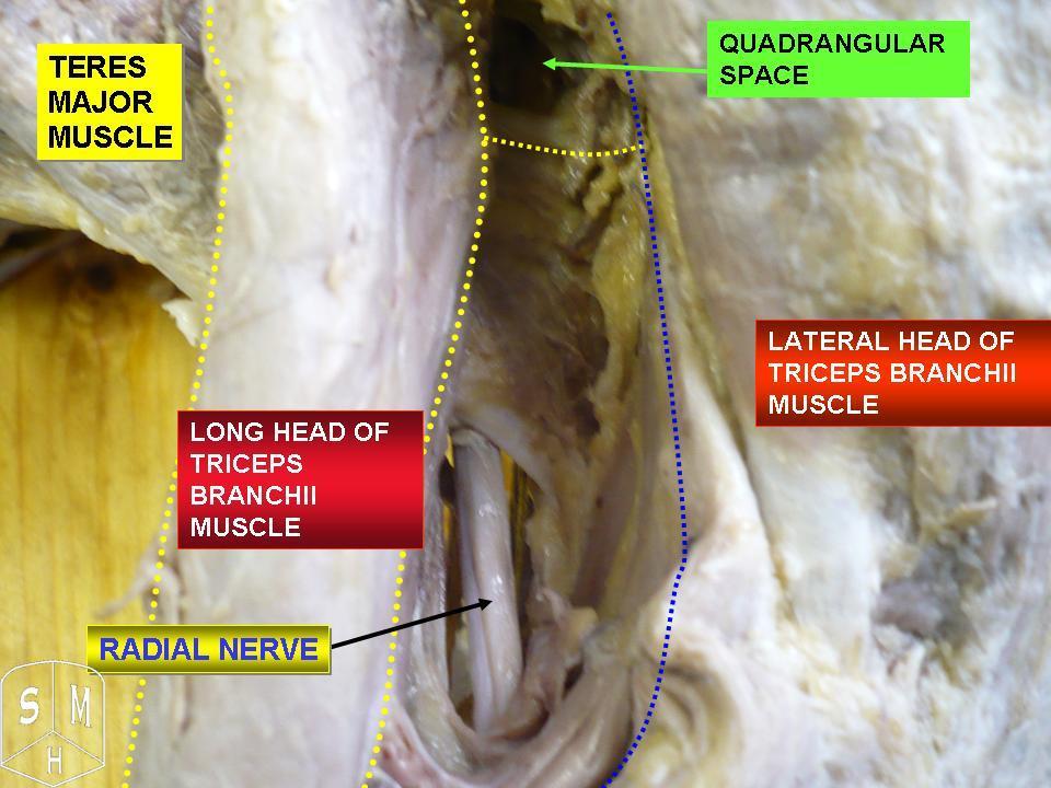
The radial nerve is a nerve in the human body that supplies the posterior portion of the upper limb. It innervates the medial and lateral heads of the triceps brachii muscle of the arm, as well as all 12 muscles in the Posterior compartment of the forearm, posterior osteofascial compartment of the forearm and the associated joints and overlying skin. It originates from the brachial plexus, carrying fibers from the posterior roots of spinal nerves C5, C6, C7, C8 and T1. The radial nerve and its branches provide Motor neuron, motor innervation to the dorsal arm muscles (the triceps brachii and the anconeus) and the extrinsic extensors of the wrists and hands; it also provides cutaneous Nerve supply to the skin, sensory innervation to most of the back of the hand, except for the back of the little finger and adjacent half of the ring finger (which are innervated by the ulnar nerve). The radial nerve divides into a deep branch, which becomes the posterior interosseous nerve, and a su ... [...More Info...] [...Related Items...] OR: [Wikipedia] [Google] [Baidu] |
Superficial Branch Of Radial Nerve
The superficial branch of the radial nerve passes along the front of the radial side of the forearm to the commencement of its lower third. It is a sensory nerve. It lies at first slightly lateral to the radial artery, concealed beneath the brachioradialis. In the middle third of the forearm, it lies behind the same muscle, close to the lateral side of the artery. It quits the artery about 7 cm. above the wrist, passes beneath the tendon of the Brachioradialis, and, piercing the deep fascia, divides into two branches: lateral and medial. Structure Lateral branch The ''lateral branch'', the smaller, supplies the radial side of the thumb (by a digital nerve), the skin of the radial side and ball of the thumb, joining with the volar branch of the lateral antebrachial cutaneous nerve. Medial branch The ''medial branch'' communicates, above the wrist, with the dorsal branch of the lateral antebrachial cutaneous, and, on the back of the hand, with the dorsal branch of the ... [...More Info...] [...Related Items...] OR: [Wikipedia] [Google] [Baidu] |
Cephalic Vein
In human anatomy, the cephalic vein (also called the antecubital vein) is a superficial vein in the arm. It is the longest vein of the upper limb. It starts at the anatomical snuffbox from the radial end of the dorsal venous network of hand, and ascends along the radial (lateral) side of the arm before emptying into the axillary vein. At the elbow, it communicates with the basilic vein via the median cubital vein. Anatomy The cephalic vein is situated within the superficial fascia along the anterolateral surface of the biceps. Origin The cephalic vein forms at the roof of the anatomical snuffbox at the radial end of the dorsal venous network of hand. Course and relations From its origin, it ascends up the lateral aspect of the radius. Near the shoulder, the cephalic vein passes between the deltoid and pectoralis major muscles ( deltopectoral groove) through the clavipectoral triangle, where it empties into the axillary vein. Anastomoses It communicates wit ... [...More Info...] [...Related Items...] OR: [Wikipedia] [Google] [Baidu] |
Lateral Cutaneous Nerve Of Forearm
The lateral cutaneous nerve of forearm (or lateral antebrachial cutaneous nerve) is a sensory nerve representing the continuation of the musculocutaneous nerve beyond the lateral edge of the tendon of the biceps brachii muscle. The lateral cutaneous nerve provides sensory innervation to the skin of the lateral forearm. It pierces the deep fascia of forearm to enter the subcutaneous compartment before splitting into a volar branch and a dorsal branch. Anatomy Course and relations It passes behind the cephalic vein and divides opposite the elbow-joint into a volar branch and a dorsal branch. Branches Volar branch The volar branch (ramus volaris; anterior branch) descends along the radial border of the forearm to the wrist, and supplies the skin over the lateral half of its volar surface. At the wrist-joint it is placed in front of the radial artery, and some filaments, piercing the deep fascia, accompany that vessel to the dorsal surface of the carpus. The nerve the ... [...More Info...] [...Related Items...] OR: [Wikipedia] [Google] [Baidu] |
Superficial Branch Of Radial Nerve
The superficial branch of the radial nerve passes along the front of the radial side of the forearm to the commencement of its lower third. It is a sensory nerve. It lies at first slightly lateral to the radial artery, concealed beneath the brachioradialis. In the middle third of the forearm, it lies behind the same muscle, close to the lateral side of the artery. It quits the artery about 7 cm. above the wrist, passes beneath the tendon of the Brachioradialis, and, piercing the deep fascia, divides into two branches: lateral and medial. Structure Lateral branch The ''lateral branch'', the smaller, supplies the radial side of the thumb (by a digital nerve), the skin of the radial side and ball of the thumb, joining with the volar branch of the lateral antebrachial cutaneous nerve. Medial branch The ''medial branch'' communicates, above the wrist, with the dorsal branch of the lateral antebrachial cutaneous, and, on the back of the hand, with the dorsal branch of the ... [...More Info...] [...Related Items...] OR: [Wikipedia] [Google] [Baidu] |
Deep Palmar Arch
The deep palmar arch (deep volar arch) is an arterial network found in the palm. It is usually primarily formed from the terminal part of the radial artery. The ulnar artery also contributes through an anastomosis. This is in contrast to the superficial palmar arch, which is formed predominantly by the ulnar artery. Structure The deep palmar arch is usually primarily formed from the radial artery. The ulnar artery also contributes through an anastomosis. The deep palmar arch lies upon the bases of the metacarpal bones and on the interossei of the hand. It is deep to the oblique head of the adductor pollicis muscle, the flexor tendons of the fingers, and the lumbricals of the hand. Alongside of it, but running in the opposite direction—toward the radial side of the hand—is the deep branch of the ulnar nerve. The superficial palmar arch is more distally located than the deep palmar arch. If one were to fully extend the thumb and draw a line from the distal border of the thu ... [...More Info...] [...Related Items...] OR: [Wikipedia] [Google] [Baidu] |
Superficial Palmar Arch
The superficial palmar arch is formed predominantly by the ulnar artery, with a contribution from the superficial palmar branch of the radial artery. However, in some individuals the contribution from the radial artery might be absent, and instead anastomoses with either the princeps pollicis artery, the radialis indicis artery, or the median artery, the former two of which are branches from the radial artery. Alternative names for this arterial arch are: superficial volar arch, superficial ulnar arch, arcus palmaris superficialis, or arcus volaris superficialis.Again, ''palmar'' and ''volar'' may be used synonymously, but ''arcus volaris superficialis'' does not occur in the TA, and can therefore be considered deprecated. The arch passes across the palm in a curve (Boeckel's line) with its convexity downward, With the thumb fully extended, the superficial palmar arch would lie approximately 1 cm from a line drawn between the first web space to the hook of hamate (Kapla ... [...More Info...] [...Related Items...] OR: [Wikipedia] [Google] [Baidu] |
Metacarpals
In human anatomy, the metacarpal bones or metacarpus, also known as the "palm bones", are the appendicular skeleton, appendicular bones that form the intermediate part of the hand between the phalanges (fingers) and the carpal bones (wrist, wrist bones), which joint, articulate with the forearm. The metacarpal bones are homologous to the metatarsal bones in the foot. Structure The metacarpals form a transverse arch to which the rigid row of distal carpal bones are fixed. The peripheral metacarpals (those of the thumb and little finger) form the sides of the cup of the palmar gutter and as they are brought together they deepen this concavity. The index metacarpal is the most firmly fixed, while the thumb metacarpal articulates with the trapezium and acts independently from the others. The middle metacarpals are tightly united to the carpus by intrinsic interlocking bone elements at their bases. The ring metacarpal is somewhat more mobile while the fifth metacarpal is semi-indepen ... [...More Info...] [...Related Items...] OR: [Wikipedia] [Google] [Baidu] |
Radial Artery
In human anatomy, the radial artery is the main artery of the lateral aspect of the forearm. Structure The radial artery arises from the bifurcation of the brachial artery in the antecubital fossa. It runs distally on the anterior part of the forearm. There, it serves as a landmark for the division between the anterior compartment of the forearm, anterior and posterior compartment of the forearm, posterior compartments of the forearm, with the posterior compartment beginning just lateral to the artery. The artery winds laterally around the wrist, passing through the anatomical snuff box and between the heads of the first dorsal interossei of the hand, dorsal interosseous muscle. It passes anteriorly between the heads of the adductor pollicis, and becomes the deep palmar arch, which joins with the deep branch of the ulnar artery. Along its course, it is accompanied by a similarly named vein, the radial vein. Branches The named branches of the radial artery may be divided int ... [...More Info...] [...Related Items...] OR: [Wikipedia] [Google] [Baidu] |


