|
öÝ1-adrenergic Receptor
alpha-1 (öÝ1) adrenergic are G protein-coupled receptors (GPCRs) associated with the Gq heterotrimeric G protein. öÝ1-adrenergic receptors are subdivided into three highly homologous subtypes, i.e., öÝ1A-, öÝ1B-, and öÝ1D-adrenergic receptor subtypes. There is no öÝ1C receptor. At one time, there was a subtype known as öÝ1C, but it was found to be identical to the previously discovered öÝ1A receptor subtype. To avoid confusion, naming was continued with the letter D. Catecholamines like norepinephrine (noradrenaline) and epinephrine (adrenaline) signal through the öÝ1-adrenergic receptors in the central and peripheral nervous systems. The crystal structure of the öÝ1B-adrenergic receptor subtype has been determined in complex with the inverse agonist (+)-cyclazosin. Effects The öÝ1-adrenergic receptor has several general functions in common with the öÝ2-adrenergic receptor, but also has specific effects of its own. öÝ1-receptors primarily mediate smooth muscle contracti ... [...More Info...] [...Related Items...] OR: [Wikipedia] [Google] [Baidu] [Amazon] |
G Protein-coupled Receptors
G protein-coupled receptors (GPCRs), also known as seven-(pass)-transmembrane domain receptors, 7TM receptors, heptahelical receptors, serpentine receptors, and G protein-linked receptors (GPLR), form a large protein family, group of evolutionarily related proteins that are cell surface receptors that detect molecules outside the cell (biology), cell and activate cellular responses. They are coupled with G proteins. They pass through the cell membrane seven times in the form of six loops (three extracellular loops interacting with ligand molecules, three intracellular loops interacting with G proteins, an N-terminus, N-terminal extracellular region and a C-terminal intracellular region) of amino acid residues, which is why they are sometimes referred to as seven-transmembrane receptors. Text was copied from this source, which is available under Attribution 2.5 Generic (CC BY 2.5) licence/ref> Ligands can bind either to the extracellular N-terminus and loops (e.g. glutama ... [...More Info...] [...Related Items...] OR: [Wikipedia] [Google] [Baidu] [Amazon] |
Skin
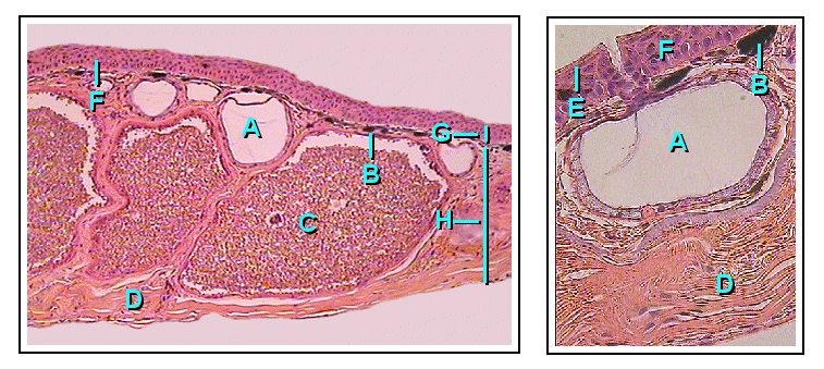
Skin is the layer of usually soft, flexible outer tissue covering the body of a vertebrate animal, with three main functions: protection, regulation, and sensation. Other animal coverings, such as the arthropod exoskeleton, have different developmental origin, structure and chemical composition. The adjective cutaneous means "of the skin" (from Latin ''cutis'' 'skin'). In mammals, the skin is an organ of the integumentary system made up of multiple layers of ectodermal tissue and guards the underlying muscles, bones, ligaments, and internal organs. Skin of a different nature exists in amphibians, reptiles, and birds. Skin (including cutaneous and subcutaneous tissues) plays crucial roles in formation, structure, and function of extraskeletal apparatus such as horns of bovids (e.g., cattle) and rhinos, cervids' antlers, giraffids' ossicones, armadillos' osteoderm, and os penis/ os clitoris. All mammals have some hair on their skin, even marine mammals like whales, ... [...More Info...] [...Related Items...] OR: [Wikipedia] [Google] [Baidu] [Amazon] |
Bronchioles
The bronchioles ( ) are the smaller branches of the bronchial airways in the lower respiratory tract. They include the terminal bronchioles, and finally the respiratory bronchioles that mark the start of the respiratory zone delivering air to the gas exchanging units of the alveoli. The bronchioles no longer contain the cartilage that is found in the bronchi, or glands in their submucosa. Structure The pulmonary lobule is the portion of the lung ventilated by one bronchiole. Bronchioles are approximately 1 mm or less in diameter and their walls consist of ciliated cuboidal epithelium and a layer of smooth muscle. Bronchioles divide into even smaller bronchioles, called ''terminal'', which are 0.5 mm or less in diameter. Terminal bronchioles in turn divide into smaller respiratory bronchioles which divide into alveolar ducts. Terminal bronchioles mark the end of the conducting division of air flow in the respiratory system while respiratory bronchioles are the be ... [...More Info...] [...Related Items...] OR: [Wikipedia] [Google] [Baidu] [Amazon] |
Urethral Sphincter
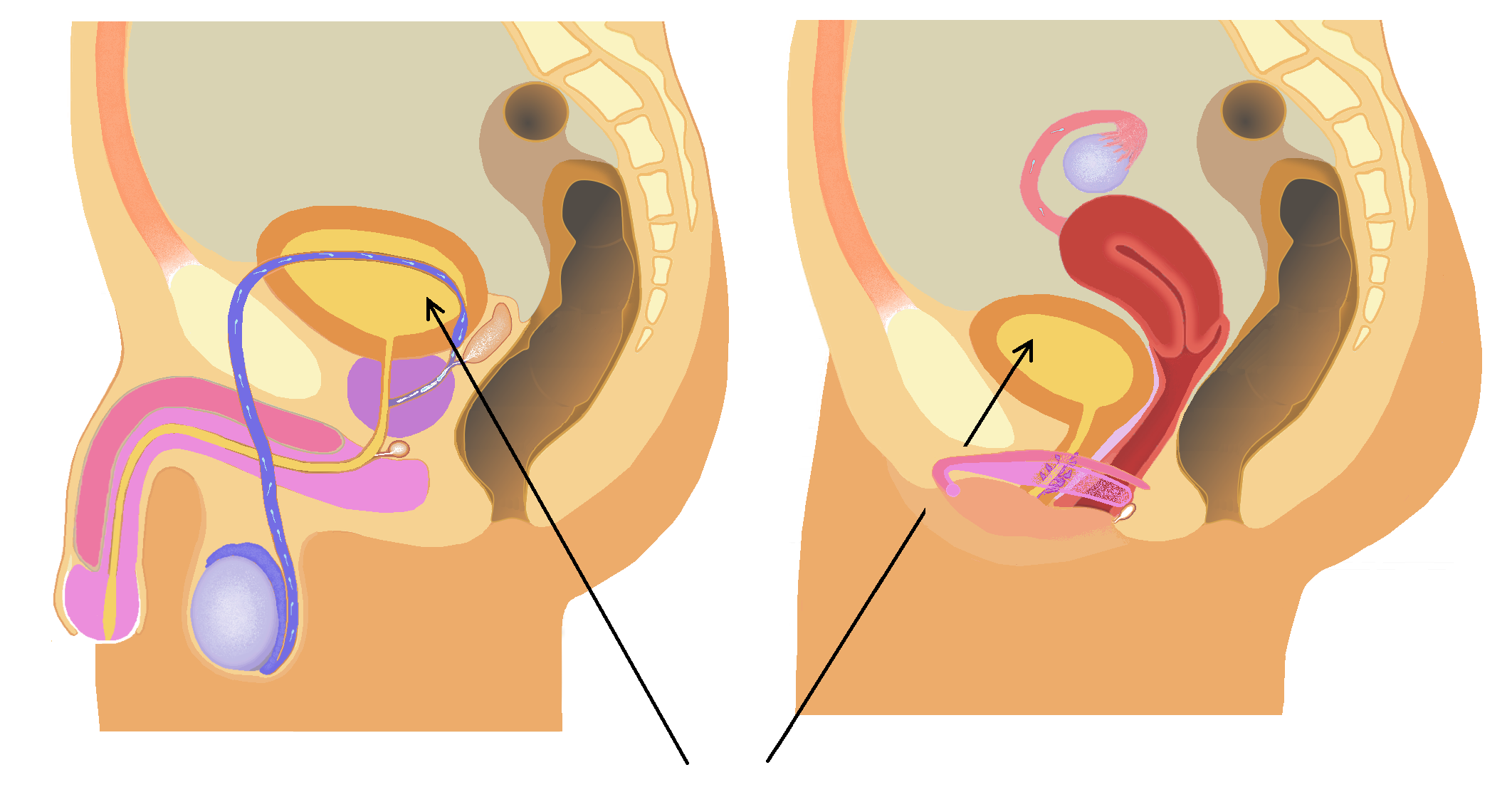
The urethral sphincters are two muscles used to control the exit of urine in the urinary bladder through the urethra. The two muscles are either the external sphincter muscle of male urethra, male or external sphincter muscle of female urethra, female external urethral sphincter and the internal urethral sphincter. When either of these muscles contracts, the urethra is sealed shut. The external urethral sphincter originates at the ischiopubic ramus and inserts into the intermeshing muscle fibers from the other side. It is controlled by the deep perineal branch of the pudendal nerve. Activity in the nerve fibers constricts the urethra. * The internal sphincter muscle of urethra: located at the bladder's inferior end and the urethra's proximal end at the junction of the urethra with the urinary bladder. The internal sphincter is a continuation of the detrusor muscle and is made of smooth muscle, therefore it is under Smooth muscle tissue, involuntary or autonomic nervous system, a ... [...More Info...] [...Related Items...] OR: [Wikipedia] [Google] [Baidu] [Amazon] |
Uterus
The uterus (from Latin ''uterus'', : uteri or uteruses) or womb () is the hollow organ, organ in the reproductive system of most female mammals, including humans, that accommodates the embryonic development, embryonic and prenatal development, fetal development of one or more Fertilized egg, fertilized eggs until birth. The uterus is a hormone-responsive sex organ that contains uterine gland, glands in its endometrium, lining that secrete uterine milk for embryonic nourishment. (The term ''uterus'' is also applied to analogous structures in some non-mammalian animals.) In humans, the lower end of the uterus is a narrow part known as the Uterine isthmus, isthmus that connects to the cervix, the anterior gateway leading to the vagina. The upper end, the body of the uterus, is connected to the fallopian tubes at the uterine horns; the rounded part, the fundus, is above the openings to the fallopian tubes. The connection of the uterine cavity with a fallopian tube is called the utero ... [...More Info...] [...Related Items...] OR: [Wikipedia] [Google] [Baidu] [Amazon] |
Ureter
The ureters are tubes composed of smooth muscle that transport urine from the kidneys to the urinary bladder. In an adult human, the ureters typically measure 20 to 30 centimeters in length and about 3 to 4 millimeters in diameter. They are lined with urothelial cells, a form of transitional epithelium, and feature an extra layer of smooth muscle in the lower third to aid in peristalsis. The ureters can be affected by a number of diseases, including urinary tract infections and kidney stone. is when a ureter is narrowed, due to for example chronic inflammation. Congenital abnormalities that affect the ureters can include the development of two ureters on the same side or abnormally placed ureters. Additionally, reflux of urine from the bladder back up the ureters is a condition commonly seen in children. The ureters have been identified for at least two thousand years, with the word "ureter" stemming from the stem relating to urinating and seen in written records since at ... [...More Info...] [...Related Items...] OR: [Wikipedia] [Google] [Baidu] [Amazon] |
Micturition
Urination is the release of urine from the bladder through the urethra in placental mammals, or through the cloaca in other vertebrates. It is the urinary system's form of excretion. It is also known medically as micturition, voiding, uresis, or, rarely, emiction, and known colloquially by various names including peeing, weeing, pissing, and euphemistically number one. The process of urination is under voluntary control in healthy humans and other animals, but may occur as a reflex in infants, some elderly individuals, and those with neurological injury. It is normal for adult humans to urinate up to seven times during the day. In some animals, in addition to expelling waste material, urination can mark territory or express submissiveness. Physiologically, urination involves coordination between the central, autonomic, and somatic nervous systems. Brain centres that regulate urination include the pontine micturition center, periaqueductal gray, and the cerebral cortex. ... [...More Info...] [...Related Items...] OR: [Wikipedia] [Google] [Baidu] [Amazon] |
Beta-2 Adrenergic Receptor
The beta-2 adrenergic receptor (öý2 adrenoreceptor), also known as ADRB2, is a cell membrane-spanning beta-adrenergic receptor that binds epinephrine (adrenaline), a hormone and neurotransmitter whose signaling, via adenylate cyclase stimulation through trimeric Gs proteins, increases cAMP, and, via downstream L-type calcium channel interaction, mediates physiologic responses such as smooth muscle relaxation and bronchodilation. Robert Lefkowitz and Brian Kobilka's study of the beta-2 adrenergic receptor as a model system earned them the 2012 Nobel Prize in Chemistry "for studies of G-protein-coupled receptors". The official symbol for the human gene encoding the öý2 adrenoreceptor is ''ADRB2''. Gene The gene is intronless. Different polymorphic forms, point mutations, and/or downregulation of this gene are associated with nocturnal asthma, obesity and type 2 diabetes. Structure The 3D crystallographic structure (see figure and links to the right) of the öý2-adrener ... [...More Info...] [...Related Items...] OR: [Wikipedia] [Google] [Baidu] [Amazon] |
Urinary Bladder
The bladder () is a hollow organ in humans and other vertebrates that stores urine from the Kidney (vertebrates), kidneys. In placental mammals, urine enters the bladder via the ureters and exits via the urethra during urination. In humans, the bladder is a distensible organ that sits on the pelvic floor. The typical adult human bladder will hold between 300 and (10 and ) before the urge to empty occurs, but can hold considerably more. The Latin phrase for "urinary bladder" is ''vesica urinaria'', and the term ''vesical'' or prefix ''vesico-'' appear in connection with associated structures such as vesical veins. The modern Latin word for "bladder" ã ''cystis'' ã appears in associated terms such as cystitis (inflammation of the bladder). Structure In humans, the bladder is a hollow muscular organ situated at the base of the pelvis. In gross anatomy, the bladder can be divided into a broad (base), a body, an apex, and a neck. The apex (also called the vertex) is directed ... [...More Info...] [...Related Items...] OR: [Wikipedia] [Google] [Baidu] [Amazon] |
Fight-or-flight Response
The fight-or-flight or the fight-flight-freeze-or-fawn (also called hyperarousal or the acute stress response) is a physiological reaction that occurs in response to a perceived harmful event, attack, or threat to survival. It was first described by Walter Bradford Cannon in 1915. His theory states that animals react to threats with a general discharge of the sympathetic nervous system, preparing the animal for fighting or fleeing. More specifically, the adrenal medulla produces a hormonal cascade that results in the secretion of catecholamines, especially norepinephrine and epinephrine. The hormones estrogen, testosterone, and cortisol, as well as the neurotransmitters dopamine and serotonin, also affect how organisms react to stress. The hormone osteocalcin might also play a part. This response is recognised as the first stage of the general adaptation syndrome that regulates stress responses among vertebrates and other organisms. Name Originally understood as the "figh ... [...More Info...] [...Related Items...] OR: [Wikipedia] [Google] [Baidu] [Amazon] |
Brain
The brain is an organ (biology), organ that serves as the center of the nervous system in all vertebrate and most invertebrate animals. It consists of nervous tissue and is typically located in the head (cephalization), usually near organs for special senses such as visual perception, vision, hearing, and olfaction. Being the most specialized organ, it is responsible for receiving information from the sensory nervous system, processing that information (thought, cognition, and intelligence) and the coordination of motor control (muscle activity and endocrine system). While invertebrate brains arise from paired segmental ganglia (each of which is only responsible for the respective segmentation (biology), body segment) of the ventral nerve cord, vertebrate brains develop axially from the midline dorsal nerve cord as a brain vesicle, vesicular enlargement at the rostral (anatomical term), rostral end of the neural tube, with centralized control over all body segments. All vertebr ... [...More Info...] [...Related Items...] OR: [Wikipedia] [Google] [Baidu] [Amazon] |
Renal Artery
The renal arteries are paired arteries that supply the kidneys with blood. Each is directed across the crus of the diaphragm, so as to form nearly a right angle. The renal arteries carry a large portion of total blood flow to the kidneys. Up to a third of total cardiac output can pass through the renal arteries to be filtered by the kidneys. Structure The renal arteries normally arise at a 90ô¯ angle off of the left interior side of the abdominal aorta, immediately below the superior mesenteric artery. They have a radius of approximately 0.25 cm, 0.26 cm at the root. The measured mean diameter can differ depending on the imaging method used. For example, the diameter was found to be 5.04 ôÝ 0.74 mm using ultrasound but 5.68 ôÝ 1.19 mm using angiography. Due to the anatomical position of the aorta, the inferior vena cava, and the kidneys, the right renal artery is normally longer than the left renal artery. * The right passes behind the inferior vena cava, ... [...More Info...] [...Related Items...] OR: [Wikipedia] [Google] [Baidu] [Amazon] |


