|
α-smooth Muscle Actin
ACTA2 (actin alpha 2) is an actin protein with several aliases including alpha-actin, alpha-actin-2, aortic smooth muscle or alpha smooth muscle actin (α-SMA, SMactin, alpha-SM-actin, ASMA). Actins are a Protein family, family of Globular protein, globular multi-functional proteins that form microfilaments. ACTA2 is one of 6 different actin isoforms and is involved in the contractility, contractile apparatus of smooth muscle. ACTA2 (as with all the actins) is extremely Conserved sequence, highly conserved and found in nearly all mammals. In humans, ACTA2 is encoded by the ''ACTA2'' gene located on 10q22-q24. Mutations in this gene cause a variety of vascular diseases, such as thoracic aortic disease, coronary artery disease, stroke, Moyamoya disease, and multisystemic smooth muscle dysfunction syndrome. ACTA2 (commonly referred to as alpha-smooth muscle actin or α-SMA) is often used as a marker of myofibroblast formation. Studies have shown that ACTA2 is associated with TGF ... [...More Info...] [...Related Items...] OR: [Wikipedia] [Google] [Baidu] |
Actin
Actin is a family of globular multi-functional proteins that form microfilaments in the cytoskeleton, and the thin filaments in muscle fibrils. It is found in essentially all eukaryotic cells, where it may be present at a concentration of over 100 ÎĽM; its mass is roughly 42 kDa, with a diameter of 4 to 7 nm. An actin protein is the monomeric subunit of two types of filaments in cells: microfilaments, one of the three major components of the cytoskeleton, and thin filaments, part of the contractile apparatus in muscle cells. It can be present as either a free monomer called G-actin (globular) or as part of a linear polymer microfilament called F-actin (filamentous), both of which are essential for such important cellular functions as the mobility and contraction of cells during cell division. Actin participates in many important cellular processes, including muscle contraction, cell motility, cell division and cytokinesis, vesicle and organelle mov ... [...More Info...] [...Related Items...] OR: [Wikipedia] [Google] [Baidu] |
Thoracic Aortic Disease
A thoracic aortic aneurysm is an aortic aneurysm that presents primarily in the thorax. A thoracic aortic aneurysm is the "ballooning" of the upper aspect of the aorta, above the diaphragm. Untreated or unrecognized they can be fatal due to dissection or "popping" of the aneurysm leading to nearly instant death. Thoracic aneurysms are less common than an abdominal aortic aneurysm. However, a syphilitic aneurysm is more likely to be a thoracic aortic aneurysm than an abdominal aortic aneurysm. This condition is commonly treated via a specialized multidisciplinary approach with both vascular surgeons and cardiac surgeons. Presentation Complications The principal causes of death due to thoracic aneurysmal disease are dissection and rupture. Once rupture occurs, the mortality rate is 50–80%. Most deaths in patients with Marfan syndrome are the result of aortic disease. Causes There are a number of causes, Aneurysms in patients younger than 40 usually involve the ascending aort ... [...More Info...] [...Related Items...] OR: [Wikipedia] [Google] [Baidu] |
Hepatic Stellate Cell
Hepatic stellate cells (HSC), also known as perisinusoidal cells or Ito cells (earlier ''lipocytes'' or ''fat-storing cells''), are pericytes found in the perisinusoidal space of the liver, also known as the space of Disse (a small area between the sinusoids and hepatocytes). The stellate cell is the major cell type involved in liver fibrosis, which is the formation of scar tissue in response to liver damage; in addition these cells store and concentrate vitamin A. Structure Hepatic stellate cells can be selectively stained with gold chloride, but their distinguishing feature in routine histological preparations is the presence of multiple lipid droplets in their cytoplasm. Cytoglobin expression has been shown to be a specific marker with which hepatic stellate cells can be distinguished from portal myofibroblasts in the damaged human liver. In murine (rats, mice) liver, reelin expressed by Ito cells has been shown to be a reliable marker in discerning them from other myo ... [...More Info...] [...Related Items...] OR: [Wikipedia] [Google] [Baidu] |
TGF-β Pathway
The transforming growth factor beta (TGFβ) signaling pathway is involved in many cellular processes in both the adult organism and the developing embryo including cell growth, cell differentiation, cell migration, apoptosis, cellular homeostasis and other cellular functions. The pathway is also involved in multiple physiological processes such as regulation of the immune system, the vascular system and embryonic development. The TGFβ signaling pathways are conserved. In spite of the wide range of cellular processes that the TGFβ signaling pathway regulates, the process is relatively simple. TGFβ superfamily ligands bind to a type II receptor, which recruits and phosphorylates a type I receptor. The type I receptor then phosphorylates receptor-regulated SMADs (R-SMADs) which can now bind the coSMAD SMAD4. R-SMAD/coSMAD complexes accumulate in the nucleus where they act as transcription factors and participate in the regulation of target gene expression. Mechanism Ligand binding ... [...More Info...] [...Related Items...] OR: [Wikipedia] [Google] [Baidu] |
Myofibroblast
A myofibroblast is a cell phenotype that was first described as being in a state between a fibroblast and a smooth muscle cell. Structure Myofibroblasts are contractile web-like fusiform cells that are identifiable by their expression of α-smooth muscle actin within their cytoplasmic stress fibers. In the gastrointestinal and genitourinary tracts, myofibroblasts are found subepithelially in mucosal surfaces. Here they not only act as a regulator of the shape of the crypts and villi, but also act as stem-niche cells in the intestinal crypts and as parts of atypical antigen-presenting cells. They have both support as well as paracrine function in most places. Location Myofibroblasts were first identified in granulation tissue during skin wound healing. Typically, these cells are found in granulation tissue, scar tissue (fibrosis) and the stroma of tumours. They also line the gastrointestinal tract, wherein they regulate the shapes of crypts and villi. Markers Myofibroblas ... [...More Info...] [...Related Items...] OR: [Wikipedia] [Google] [Baidu] |
Multisystemic Smooth Muscle Dysfunction Syndrome
Multisystemic smooth muscle dysfunction syndrome (MSMDS) is a genetic disorder caused by R179 missense mutations in the ACTA2 gene. Initially described as a case report in 1999, it was characterized in 2010 as a syndrome of congenital mydriasis, patent ductus arteriosus, and aneurysmal arterial disease—in particular aortic and thoracic aneurysms. The disorder has variable penetrance, ranging from severely symptomatic and fatal in early neonatal period to a more benign and manageable course with surgical intervention. Signs and symptoms Signs and symptoms are usually detectable prenatally or shortly after birth. In its severe manifestations, MSMDS has been associated with prune belly sequence. In less severe forms, the earliest signs of MSMDS are congenital fixed mydriasis (can be misdiagnosed as partial aniridia Aniridia is a condition characterized by the absence or near absence of the iris, the colored, muscular ring in the eye that controls the size of the pupil and regu ... [...More Info...] [...Related Items...] OR: [Wikipedia] [Google] [Baidu] |
Moyamoya Disease
Moyamoya disease is a disease in which certain arteries in the brain are constricted. Blood flow is blocked by constriction and blood clots (thrombosis). A collateral circulation develops around the blocked vessels to compensate for the blockage, but the collateral vessels are small, weak, and prone to bleeding, aneurysm, and thrombosis. On a conventional angiography, these collateral vessels have the appearance of a "puff of smoke", described as in Japanese language, Japanese. When moyamoya is diagnosed by itself, with no underlying correlational conditions, it is diagnosed as moyamoya disease. This is also the case when the arterial constriction and collateral circulation are bilateral. Moyamoya syndrome is unilateral arterial constriction, or occurs when one of the several specified conditions is also present. This may also be considered as moyamoya being secondary to the primary condition. Mainly, occlusion of the distal internal carotid artery occurs. On angiography, a "puff ... [...More Info...] [...Related Items...] OR: [Wikipedia] [Google] [Baidu] |
Stroke
Stroke is a medical condition in which poor cerebral circulation, blood flow to a part of the brain causes cell death. There are two main types of stroke: brain ischemia, ischemic, due to lack of blood flow, and intracranial hemorrhage, hemorrhagic, due to bleeding. Both cause parts of the brain to stop functioning properly. Signs and symptoms of stroke may include an hemiplegia, inability to move or feel on one side of the body, receptive aphasia, problems understanding or expressive aphasia, speaking, dizziness, or homonymous hemianopsia, loss of vision to one side. Signs and symptoms often appear soon after the stroke has occurred. If symptoms last less than 24 hours, the stroke is a transient ischemic attack (TIA), also called a mini-stroke. subarachnoid hemorrhage, Hemorrhagic stroke may also be associated with a thunderclap headache, severe headache. The symptoms of stroke can be permanent. Long-term complications may include pneumonia and Urinary incontinence, loss of b ... [...More Info...] [...Related Items...] OR: [Wikipedia] [Google] [Baidu] |
Coronary Artery Disease
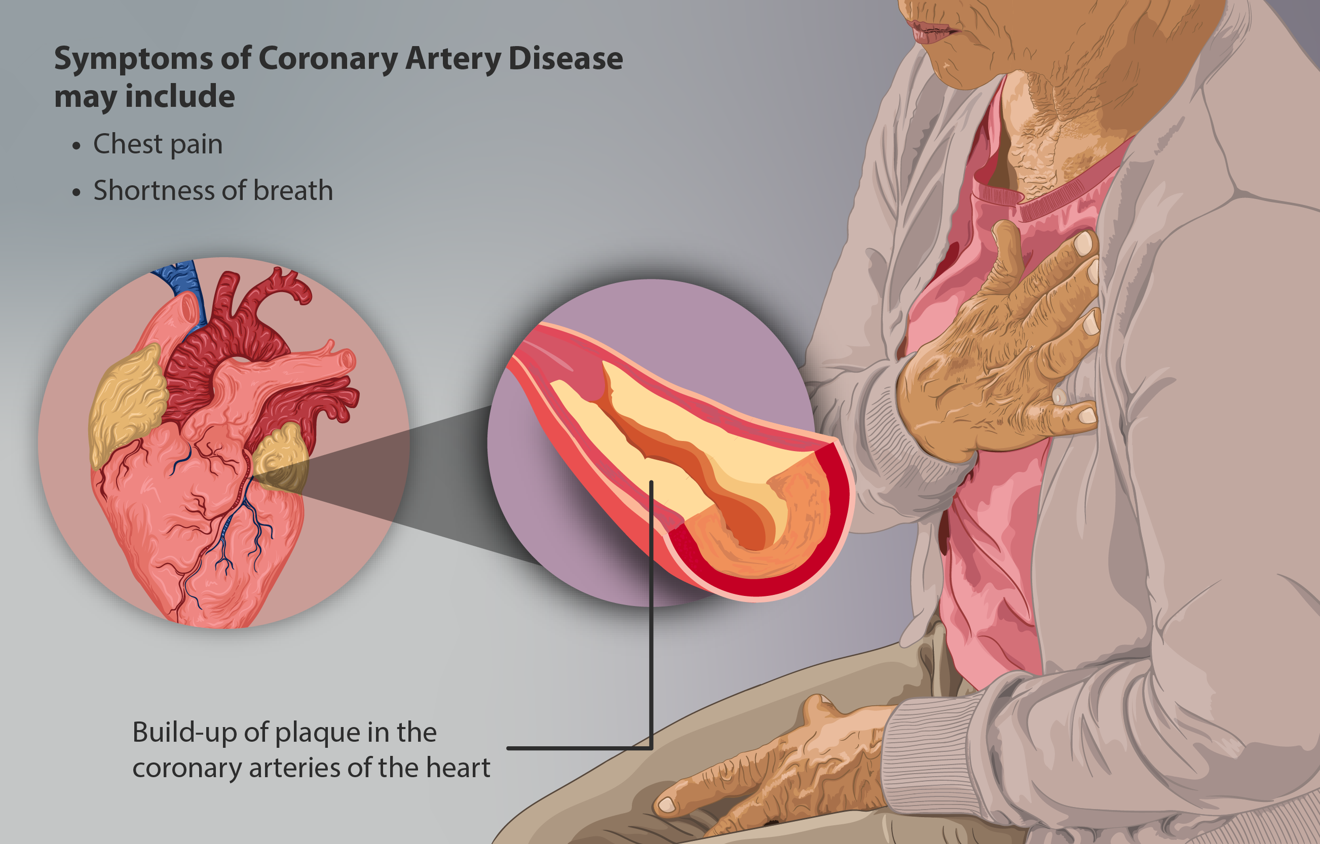
Coronary artery disease (CAD), also called coronary heart disease (CHD), or ischemic heart disease (IHD), is a type of cardiovascular disease, heart disease involving Ischemia, the reduction of blood flow to the cardiac muscle due to a build-up of atheromatous plaque in the Coronary arteries, arteries of the heart. It is the most common of the cardiovascular diseases. CAD can cause stable angina, unstable angina, myocardial ischemia, and myocardial infarction. A common symptom is angina, which is chest pain or discomfort that may travel into the shoulder, arm, back, neck, or jaw. Occasionally it may feel like heartburn. In stable angina, symptoms occur with exercise or emotional Psychological stress, stress, last less than a few minutes, and improve with rest. Shortness of breath may also occur and sometimes no symptoms are present. In many cases, the first sign is a Myocardial infarction, heart attack. Other complications include heart failure or an Heart arrhythmia, abnormal h ... [...More Info...] [...Related Items...] OR: [Wikipedia] [Google] [Baidu] |
Gene
In biology, the word gene has two meanings. The Mendelian gene is a basic unit of heredity. The molecular gene is a sequence of nucleotides in DNA that is transcribed to produce a functional RNA. There are two types of molecular genes: protein-coding genes and non-coding genes. During gene expression (the synthesis of Gene product, RNA or protein from a gene), DNA is first transcription (biology), copied into RNA. RNA can be non-coding RNA, directly functional or be the intermediate protein biosynthesis, template for the synthesis of a protein. The transmission of genes to an organism's offspring, is the basis of the inheritance of phenotypic traits from one generation to the next. These genes make up different DNA sequences, together called a genotype, that is specific to every given individual, within the gene pool of the population (biology), population of a given species. The genotype, along with environmental and developmental factors, ultimately determines the phenotype ... [...More Info...] [...Related Items...] OR: [Wikipedia] [Google] [Baidu] |
Protein
Proteins are large biomolecules and macromolecules that comprise one or more long chains of amino acid residue (biochemistry), residues. Proteins perform a vast array of functions within organisms, including Enzyme catalysis, catalysing metabolic reactions, DNA replication, Cell signaling, responding to stimuli, providing Cytoskeleton, structure to cells and Fibrous protein, organisms, and Intracellular transport, transporting molecules from one location to another. Proteins differ from one another primarily in their sequence of amino acids, which is dictated by the Nucleic acid sequence, nucleotide sequence of their genes, and which usually results in protein folding into a specific Protein structure, 3D structure that determines its activity. A linear chain of amino acid residues is called a polypeptide. A protein contains at least one long polypeptide. Short polypeptides, containing less than 20–30 residues, are rarely considered to be proteins and are commonly called pep ... [...More Info...] [...Related Items...] OR: [Wikipedia] [Google] [Baidu] |
Conserved Sequence
In evolutionary biology, conserved sequences are identical or similar sequences in nucleic acids ( DNA and RNA) or proteins across species ( orthologous sequences), or within a genome ( paralogous sequences), or between donor and receptor taxa ( xenologous sequences). Conservation indicates that a sequence has been maintained by natural selection. A highly conserved sequence is one that has remained relatively unchanged far back up the phylogenetic tree, and hence far back in geological time. Examples of highly conserved sequences include the RNA components of ribosomes present in all domains of life, the homeobox sequences widespread amongst eukaryotes, and the tmRNA in bacteria. The study of sequence conservation overlaps with the fields of genomics, proteomics, evolutionary biology, phylogenetics, bioinformatics and mathematics. History The discovery of the role of DNA in heredity, and observations by Frederick Sanger of variation between animal insulins in 194 ... [...More Info...] [...Related Items...] OR: [Wikipedia] [Google] [Baidu] |


