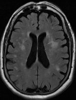
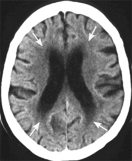
White matter hyperintensity on:
[Wikipedia]
[Google]
[Amazon]


 Leukoaraiosis is a particular abnormal change in appearance of
 White matter hyperintensities can be caused by a variety of factors, including


 Leukoaraiosis is a particular abnormal change in appearance of
 white matter
White matter refers to areas of the central nervous system (CNS) that are mainly made up of myelinated axons, also called tracts. Long thought to be passive tissue, white matter affects learning and brain functions, modulating the distribution ...
near the lateral ventricles
The lateral ventricles are the two largest ventricles of the brain and contain cerebrospinal fluid (CSF). Each cerebral hemisphere contains a lateral ventricle, known as the left or right ventricle, respectively.
Each lateral ventricle resemble ...
. It is often seen in aged individuals, but sometimes in young adults. On MRI
Magnetic resonance imaging (MRI) is a medical imaging technique used in radiology to form pictures of the anatomy and the physiological processes of the body. MRI scanners use strong magnetic fields, magnetic field gradients, and radio waves ...
, leukoaraiosis changes appear as white matter hyperintensities
A hyperintensity or T2 hyperintensity is an area of high intensity on types of magnetic resonance imaging (MRI) scans of the brain of a human or of another mammal that reflect lesions produced largely by demyelination and axonal loss. These smal ...
(WMHs) in T2 FLAIR images. On CT scans, leukoaraiosis appears as hypodense
Radiodensity (or radiopacity) is opacity to the radio wave and X-ray portion of the electromagnetic spectrum: that is, the relative inability of those kinds of electromagnetic radiation to pass through a particular material. Radiolucency or hypo ...
periventricular white-matter lesions.
The term "leukoaraiosis" was coined in 1986 by Hachinski, Potter, and Merskey as a descriptive term for rarefaction
Rarefaction is the reduction of an item's density, the opposite of compression. Like compression, which can travel in waves (sound waves, for instance), rarefaction waves also exist in nature. A common rarefaction wave is the area of low relativ ...
("araiosis") of the white matter, showing up as decreased density on CT and increased signal intensity on T2/FLAIR sequences (white matter hyperintensities) performed as part of MRI brain scans.
These white matter changes are also commonly referred to as periventricular white matter disease, or white matter hyperintensities (WMH), due to their bright white appearance on T2 MRI scans. Many patients can have leukoaraiosis without any associated clinical abnormality. However, underlying vascular mechanisms are suspected to be the cause of the imaging findings. Hypertension
Hypertension (HTN or HT), also known as high blood pressure (HBP), is a long-term medical condition in which the blood pressure in the arteries is persistently elevated. High blood pressure usually does not cause symptoms. Long-term high bl ...
, smoking, diabetes
Diabetes, also known as diabetes mellitus, is a group of metabolic disorders characterized by a high blood sugar level ( hyperglycemia) over a prolonged period of time. Symptoms often include frequent urination, increased thirst and increased ap ...
, hyperhomocysteinemia
Hyperhomocysteinemia is a medical condition characterized by an abnormally high level of homocysteine in the blood, conventionally described as above 15 μmol/L.
As a consequence of the biochemical reactions in which homocysteine is involved ...
, and heart diseases are all risk factors for leukoaraiosis.
Leukoaraiosis has been reported to be an initial stage of Binswanger's disease
Binswanger's disease, also known as subcortical leukoencephalopathy and subcortical arteriosclerotic encephalopathy, is a form of small-vessel vascular dementia caused by damage to the white brain matter. White matter atrophy can be caused by man ...
but this evolution does not always happen.
Causes
 White matter hyperintensities can be caused by a variety of factors, including
 ischemia
Ischemia or ischaemia is a restriction in blood supply to any tissue, muscle group, or organ of the body, causing a shortage of oxygen that is needed for cellular metabolism (to keep tissue alive). Ischemia is generally caused by problems wi ...
, micro-hemorrhages
Bleeding, hemorrhage, haemorrhage or blood loss, is blood escaping from the circulatory system from damaged blood vessels. Bleeding can occur internally, or externally either through a natural opening such as the mouth, nose, ear, urethra, ...
, gliosis
Gliosis is a nonspecific reactive change of glial cells in response to damage to the central nervous system (CNS). In most cases, gliosis involves the proliferation or hypertrophy of several different types of glial cells, including astrocytes, ...
, damage to small blood vessel walls, breaches of the barrier between the cerebrospinal fluid
Cerebrospinal fluid (CSF) is a clear, colorless body fluid found within the tissue that surrounds the brain and spinal cord of all vertebrates.
CSF is produced by specialised ependymal cells in the choroid plexus of the ventricles of the bra ...
and the brain, or loss and deformation of the myelin sheath
Myelin is a lipid-rich material that surrounds nerve cell axons (the nervous system's "wires") to insulate them and increase the rate at which electrical impulses (called action potentials) are passed along the axon. The myelinated axon can be l ...
. Multiple small vessel infarcts in the subcortical white matter can cause the condition, often the result of chronic hypertension leading to lipohyalinosis Lipohyalinosis is a cerebral small vessel disease affecting the small arteries, arterioles or capillaries in the brain. Originally defined by C. Miller Fisher as 'segmental arteriolar wall disorganisation', it is characterized by vessel wall thick ...
of the small vessels. Patients may develop cognitive impairment and dementia.
Special cases
* Ischaemic leukoaraiosis has been defined as the leukoaraiosis present after astroke
A stroke is a medical condition in which poor blood flow to the brain causes cell death. There are two main types of stroke: ischemic, due to lack of blood flow, and hemorrhagic, due to bleeding. Both cause parts of the brain to stop functionin ...
.
* Diabetes
Diabetes, also known as diabetes mellitus, is a group of metabolic disorders characterized by a high blood sugar level ( hyperglycemia) over a prolonged period of time. Symptoms often include frequent urination, increased thirst and increased ap ...
-associated leukoaraiosis has been reportedMaldjian JA, Whitlow CT, Saha BN, Kota G, Vandergriff C, Davenport EM, Divers J, Freedman BI, Bowden DW. "Automated White Matter Total Lesion Volume Segmentation in Diabetes". ''AJNR Am J Neuroradiol.'' 2013 Jul 18
* CuRRL syndrome: increased Cup: Disc Ratio, Retinal GanglionCell Complex thinning, Radial Peripapillary Capillary Network Density Reduction and Leukoaraiosis
* CADASIL
CADASIL or CADASIL syndrome, involving cerebral autosomal dominant arteriopathy with subcortical infarcts and leukoencephalopathy, is the most common form of hereditary stroke disorder, and is thought to be caused by mutations of the ''Notch 3'' ge ...
is a hereditary cerebrovascular disorder associated with T2-hyperintense white matter lesions that have a greater extent and earlier age of onset than age-related leukoaraiosis.
See also
*Hyperintensities
A hyperintensity or T2 hyperintensity is an area of high intensity on types of magnetic resonance imaging (MRI) scans of the brain of a human or of another mammal that reflect lesions produced largely by demyelination and axonal loss. These smal ...
* Binswanger's disease
Binswanger's disease, also known as subcortical leukoencephalopathy and subcortical arteriosclerotic encephalopathy, is a form of small-vessel vascular dementia caused by damage to the white brain matter. White matter atrophy can be caused by man ...
* Demyelinating disease
A demyelinating disease is any disease of the nervous system in which the myelin sheath of neurons is damaged. This damage impairs the conduction of signals in the affected nerves. In turn, the reduction in conduction ability causes deficiency i ...
References
Further reading
* * * {{DEFAULTSORT:Idiopathic Inflammatory Demyelinating Diseases Cerebrum