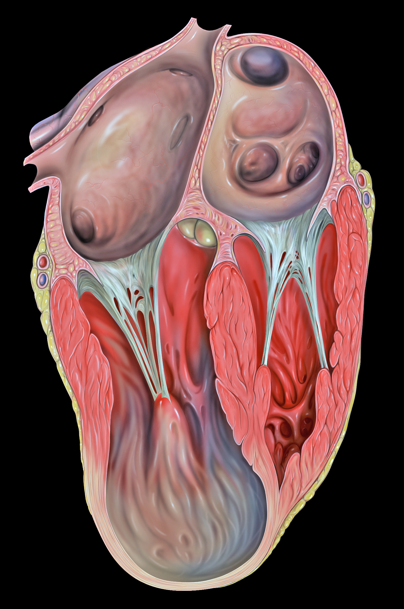
Ventricular aneurysm on:
[Wikipedia]
[Google]
[Amazon]
Ventricular aneurysms are one of the many complications that may occur after a

 When a person visits the hospital or doctor with other symptoms, especially with a history of heart problems, they will normally be required to undergo an electrocardiogram, which monitors electrical activity within the heart and shows abnormalities when a cardiac aneurysm is present. It can also appear as a bulge on a chest x-ray, and a more accurate diagnosis will then be made using an echocardiogram, which uses ultrasound to 'photograph' the heart and how it functions while it beats.
File:UOTW 57 - Ultrasound of the Week 1.webm, Left ventricular aneurysm as seen on ultrasound
File:UOTW 57 - Ultrasound of the Week 2.webm, Left ventricular aneurysm as seen on ultrasound
heart attack
A myocardial infarction (MI), commonly known as a heart attack, occurs when blood flow decreases or stops to the coronary artery of the heart, causing damage to the heart muscle. The most common symptom is chest pain or discomfort which ma ...
. The word aneurysm refers to a bulge or 'pocketing' of the wall or lining of a vessel commonly occurring in the blood vessels at the base of the septum, or within the aorta. In the heart, they usually arise from a patch of weakened tissue in a ventricular wall, which swells into a bubble filled with blood. This, in turn, may block the passageways leading out of the heart, leading to severely constricted blood flow to the body. Ventricular aneurysms can be fatal. They are usually non-rupturing because they are lined by scar tissue.
A left ventricular aneurysm can be associated with ST elevation.
Signs and symptoms
Ventricular aneurysms usually grow at a very slow pace, but can still pose problems. Usually, this type of aneurysm grows in theleft ventricle
A ventricle is one of two large chambers toward the bottom of the heart that collect and expel blood towards the peripheral beds within the body and lungs. The blood pumped by a ventricle is supplied by an atrium, an adjacent chamber in the uppe ...
. This bubble has the potential to block blood flow to the rest of the body, and thus limit the patient's stamina. In other cases, a similarly developed pseudoaneurysm
A pseudoaneurysm, also known as a false aneurysm, is a locally contained hematoma outside an artery or heart due to damage to the vessel wall. The injury goes through all the three layers of the arterial wall causing a leak, which is contained b ...
("false aneurysm") may burst, sometimes resulting in the death of the patient. Also, blood clots may form on the inside of ventricular aneurysms, and form embolism
An embolism is the lodging of an embolus, a blockage-causing piece of material, inside a blood vessel. The embolus may be a blood clot (thrombus), a fat globule (fat embolism), a bubble of air or other gas ( gas embolism), amniotic fluid (am ...
s. If such a clot escapes from the aneurysm, it will be moved in the circulation throughout the body. If it gets stuck inside a blood vessel, it may cause ischemia
Ischemia or ischaemia is a restriction in blood supply to any tissue, muscle group, or organ of the body, causing a shortage of oxygen that is needed for cellular metabolism (to keep tissue alive). Ischemia is generally caused by problems w ...
in a limb
Limb may refer to:
Science and technology
* Limb (anatomy), an appendage of a human or animal
*Limb, a large or main branch of a tree
*Limb, in astronomy, the curved edge of the apparent disk of a celestial body, e.g. lunar limb
*Limb, in botany, ...
, a painful condition that can lead to reduced movement and tissue death in the limb. Alternatively, if a clot blocks a vessel going to the brain, it can cause a stroke. In certain cases, ventricular aneurysms cause ventricular failure or arrythmia. At this stage, treatment is necessary.
Causes
Ventricular aneurysms are usually complications resulting from aheart attack
A myocardial infarction (MI), commonly known as a heart attack, occurs when blood flow decreases or stops to the coronary artery of the heart, causing damage to the heart muscle. The most common symptom is chest pain or discomfort which ma ...
. When the heart muscle (cardiac muscle
Cardiac muscle (also called heart muscle, myocardium, cardiomyocytes and cardiac myocytes) is one of three types of vertebrate muscle tissues, with the other two being skeletal muscle and smooth muscle. It is an involuntary, striated muscle ...
) partially dies during a heart attack, a layer of muscle may survive, and, being severely weakened, start to become an aneurysm. Blood may flow into the surrounding dead muscle and inflate the weakened flap of muscle into a bubble. It may also be congenital.
Diagnosis

 When a person visits the hospital or doctor with other symptoms, especially with a history of heart problems, they will normally be required to undergo an electrocardiogram, which monitors electrical activity within the heart and shows abnormalities when a cardiac aneurysm is present. It can also appear as a bulge on a chest x-ray, and a more accurate diagnosis will then be made using an echocardiogram, which uses ultrasound to 'photograph' the heart and how it functions while it beats.
Differential diagnosis
It should also not be confused with apseudoaneurysm
A pseudoaneurysm, also known as a false aneurysm, is a locally contained hematoma outside an artery or heart due to damage to the vessel wall. The injury goes through all the three layers of the arterial wall causing a leak, which is contained b ...
, coronary artery aneurysm
Coronary artery aneurysm is an abnormal dilatation of part of the coronary artery. This rare disorder occurs in about 0.3–4.9% of patients who undergo coronary angiography.
Signs and symptoms
Causes
Acquired causes include atherosclerosis in a ...
or a myocardial rupture
Myocardial rupture is a laceration of the ventricles or atria of the heart, of the interatrial or interventricular septum, or of the papillary muscles. It is most commonly seen as a serious sequela of an acute myocardial infarction (heart at ...
(which involves a hole in the wall, not just a bulge.)
Cardiac Diverticulum
Cardiac
The heart is a muscular organ in most animals. This organ pumps blood through the blood vessels of the circulatory system. The pumped blood carries oxygen and nutrients to the body, while carrying metabolic waste such as carbon dioxide to t ...
diverticulum or ventricular diverticulum is defined as a congenital
A birth defect, also known as a congenital disorder, is an abnormal condition that is present at birth regardless of its cause. Birth defects may result in disabilities that may be physical, intellectual, or developmental. The disabilities can ...
malformation of the fibrous or muscular part of the heart which is only visible during chest x-rays or during an echocardiogram
An echocardiography, echocardiogram, cardiac echo or simply an echo, is an ultrasound of the heart.
It is a type of medical imaging of the heart, using standard ultrasound or Doppler ultrasound.
Echocardiography has become routinely used in th ...
reading. This should not be confused with ventricular diverticulum, as the latter is a sub type derived from the latter in congenital cases. it is usually asymptomatic and is only detected using imaging. Fibrous diverticulum is characterised by a calcification if present at the tip ( apex) or a thrombi that may detaches to form an emboli. Muscular diverticulum is characterised by appendix forming at the ether of the ventricles. it is a rare anomaly and can be diagnosed prenatal. Diagnosis is usually done by a chest X-ray and silhouette is viewed around the heart. Echocardiogram reading present a similar picture to ventricular aneurysm
An aneurysm is an outward bulging, likened to a bubble or balloon, caused by a localized, abnormal, weak spot on a blood vessel wall. Aneurysms may be a result of a hereditary condition or an acquired disease. Aneurysms can also be a nidus ( ...
s on the ST segment. Management is dependent on the situation presented and the severity of the case. Usually, surgical resection is advised but in prenatal cases, due to combination with other cardiac abnormalities, especially in latter trimesters, but pericardiocentesis
Pericardiocentesis (PCC), also called pericardial tap, is a medical procedure where fluid is aspirated from the pericardium (the sac enveloping the heart).
Anatomy and Physiology
The pericardium is a fibrous sac surrounding the heart composed o ...
is useful technique to reduce pleural effusion or/ and secondary disorders.
Treatment
Some people live with this type of aneurysm for many years without any specific treatment. Treatment is limited to surgery ( ventricular reduction) for this defect of the heart. However, surgery is not required in most cases but, limiting the patient's physical activity levels to lower the risk of making the aneurysm bigger is advised. Also, ACE Inhibitors seem to prevent Left Ventricular remodeling and aneurysm formation. Blood thinning agents may be given to help reduce the likelihood of blood thickening and clots forming, along with the use of drugs to correct the irregular rhythm of the heart (seen on the electrocardiogram)See also
*Coronary artery aneurysm
Coronary artery aneurysm is an abnormal dilatation of part of the coronary artery. This rare disorder occurs in about 0.3–4.9% of patients who undergo coronary angiography.
Signs and symptoms
Causes
Acquired causes include atherosclerosis in a ...
References
Further reading
* *External links
{{Heart diseases Heart diseases