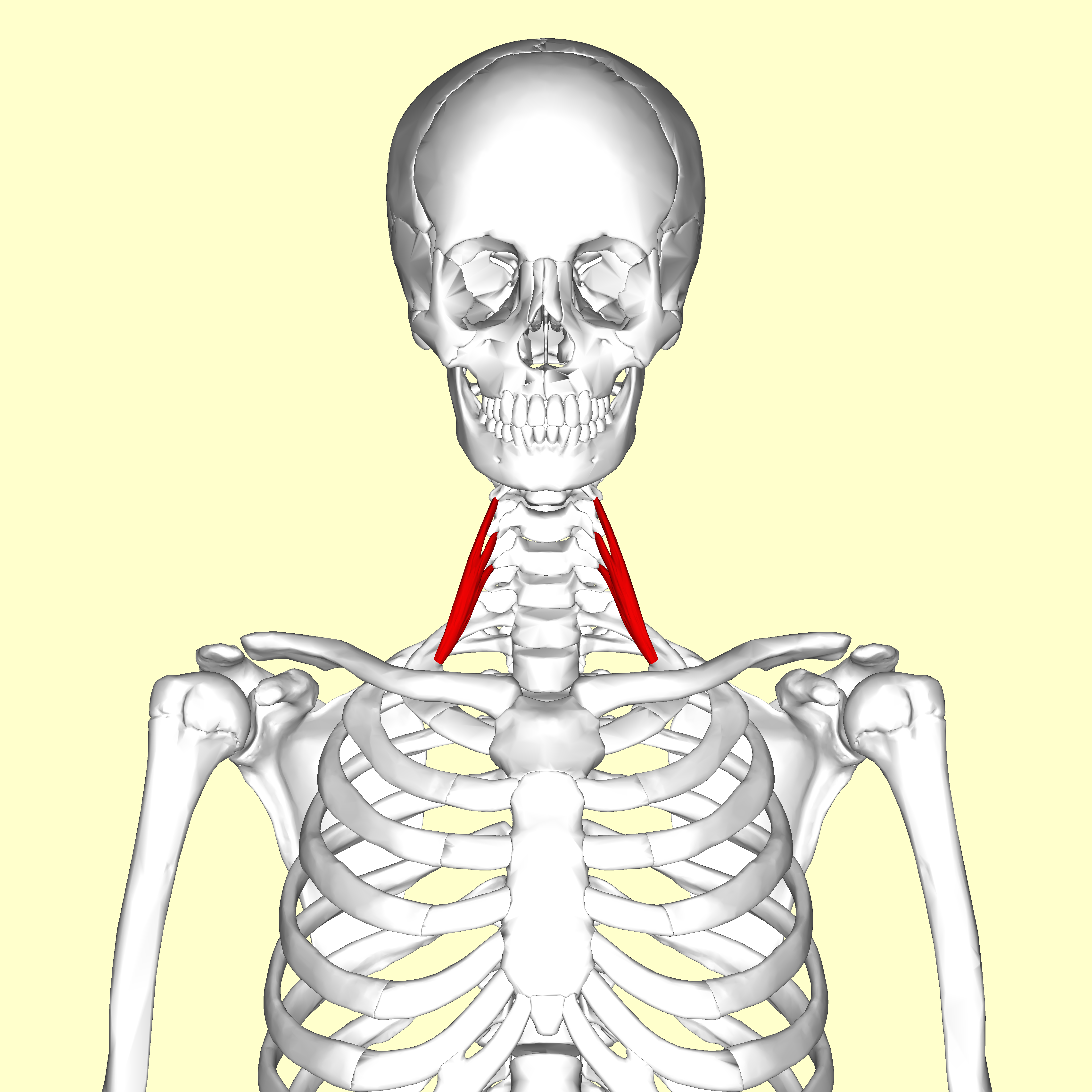
Scalenus posterior on:
[Wikipedia]
[Google]
[Amazon]
The scalene muscles are a group of three pairs of


File:Musculi coli base.svg, Musculi colli base
File:Slide1ABBA.JPG, Scalene muscles. Muscles of the neck. Lateral view.
File:Slide2ABBA.JPG, Scalene muscles. Muscles of the neck. Lateral view.
File:Gray384.png, Section of the neck at about the level of the sixth cervical vertebra. Showing the arrangement of the
muscle
Skeletal muscles (commonly referred to as muscles) are organs of the vertebrate muscular system and typically are attached by tendons to bones of a skeleton. The muscle cells of skeletal muscles are much longer than in the other types of muscl ...
s in the lateral neck
The neck is the part of the body on many vertebrates that connects the head with the torso. The neck supports the weight of the head and protects the nerves that carry sensory and motor information from the brain down to the rest of the body. In ...
, namely the anterior scalene, middle scalene, and posterior scalene. They are innervated by the third to the eight cervical spinal nerves (C3-C8).
The anterior and middle scalene muscles lift the first rib and bend the neck
The neck is the part of the body on many vertebrates that connects the head with the torso. The neck supports the weight of the head and protects the nerves that carry sensory and motor information from the brain down to the rest of the body. In ...
to the same side; the posterior scalene lifts the second rib and tilts the neck to the same side.
The muscles are named .
Structure
The scalene muscles originate from thetransverse processes
The spinal column, a defining synapomorphy shared by nearly all vertebrates,Hagfish are believed to have secondarily lost their spinal column is a moderately flexible series of vertebrae (singular vertebra), each constituting a characteristic i ...
from the cervical vertebrae
In tetrapods, cervical vertebrae (singular: vertebra) are the vertebrae of the neck, immediately below the skull. Truncal vertebrae (divided into thoracic and lumbar vertebrae in mammals) lie caudal (toward the tail) of cervical vertebrae. In ...
of C2 to C7 and insert onto the first and second rib
In vertebrate anatomy, ribs ( la, costae) are the long curved bones which form the rib cage, part of the axial skeleton. In most tetrapods, ribs surround the chest, enabling the lungs to expand and thus facilitate breathing by expanding the ches ...
s.
Anterior scalene
The anterior scalene muscle ( la, scalenus anterior), lies deeply at the side of the neck, behind the sternocleidomastoid muscle. It arises from the anterior tubercles of thetransverse processes
The spinal column, a defining synapomorphy shared by nearly all vertebrates,Hagfish are believed to have secondarily lost their spinal column is a moderately flexible series of vertebrae (singular vertebra), each constituting a characteristic i ...
of the third, fourth, fifth, and sixth cervical vertebrae
In tetrapods, cervical vertebrae (singular: vertebra) are the vertebrae of the neck, immediately below the skull. Truncal vertebrae (divided into thoracic and lumbar vertebrae in mammals) lie caudal (toward the tail) of cervical vertebrae. In ...
, and descending, almost vertically, is inserted by a narrow, flat tendon into the scalene tubercle on the inner border of the first rib, and into the ridge on the upper surface of the second rib in front of the subclavian groove. It is supplied by the anterior ramus of cervical nerve
A spinal nerve is a mixed nerve, which carries motor, sensory, and autonomic signals between the spinal cord and the body. In the human body there are 31 pairs of spinal nerves, one on each side of the vertebral column. These are grouped into the ...
5 and 6.
Middle scalene
The middle scalene, ( la, scalenus medius), is the largest and longest of the three scalene muscles. The middle scalene arises from the posterior tubercles of the transverse processes of the lower six cervical vertebrae. It descends along the side of the vertebral column to insert by a broad attachment into the upper surface of the first rib, posterior to the subclavian groove. The brachial plexus and thesubclavian artery
In human anatomy, the subclavian arteries are paired major arteries of the upper thorax, below the clavicle. They receive blood from the aortic arch. The left subclavian artery supplies blood to the left arm and the right subclavian artery supplie ...
pass anterior to it.

Posterior scalene
The posterior scalene, ( la, scalenus posterior) is the smallest and most deeply seated of the scalene muscles. It arises, by two or three separate tendons, from the posterior tubercles of the transverse processes of the lower two or three cervical vertebrae, and is inserted by a thin tendon into the outer surface of the second rib, behind the attachment of the anterior scalene. It is supplied by cervical nerves C5, C6 and C7. It is occasionally blended with the middle scalene.Variation
A fourth muscle, the scalenus minimus (Sibson's muscle), is sometimes present behind the lower portion of the anterior scalene.Function
The anterior and middle scalene muscles lift the first rib and bend theneck
The neck is the part of the body on many vertebrates that connects the head with the torso. The neck supports the weight of the head and protects the nerves that carry sensory and motor information from the brain down to the rest of the body. In ...
to the same side as the acting muscle; the posterior scalene lifts the second rib and tilts the neck to the same side.
Because they elevate the upper ribs, they also act as accessory muscles of respiration, along with the sternocleidomastoids.
Relations
The scalene muscles have an important relationship to other structures in the neck. The brachial plexus andsubclavian artery
In human anatomy, the subclavian arteries are paired major arteries of the upper thorax, below the clavicle. They receive blood from the aortic arch. The left subclavian artery supplies blood to the left arm and the right subclavian artery supplie ...
pass between the anterior and middle scalenes. The subclavian vein
The subclavian vein is a paired large vein, one on either side of the body, that is responsible for draining blood from the upper extremities, allowing this blood to return to the heart. The left subclavian vein plays a key role in the absorption ...
and phrenic nerve
The phrenic nerve is a mixed motor/sensory nerve which originates from the C3-C5 spinal nerves in the neck. The nerve is important for breathing because it provides exclusive motor control of the diaphragm, the primary muscle of respiration. In ...
pass anteriorly to the anterior scalene as the muscle crosses over the first rib. The phrenic nerve is oriented vertically as it passes in front of the anterior scalene, while the subclavian vein is oriented horizontally as it passes in front of the anterior scalene muscle.
The passing of the brachial plexus and the subclavian artery
In human anatomy, the subclavian arteries are paired major arteries of the upper thorax, below the clavicle. They receive blood from the aortic arch. The left subclavian artery supplies blood to the left arm and the right subclavian artery supplie ...
through the space of the anterior and middle scalene muscles constitute the scalene hiatus (the term "scalene fissure" is also used). The region in which this lies is referred to as the scaleotracheal fossa. It is bounded by the clavicle inferior anteriorly, the trachea medially, posteriorly by the trapezius, and anteriorly by the platysma muscle.
Clinical significance
The anterior and middle scalene muscles can be involved in certain forms of thoracic outlet syndrome as well asmyofascial pain syndrome
Myofascial pain syndrome (MPS), also known as chronic myofascial pain (CMP), is a syndrome characterized by chronic pain in multiple myofascial trigger points ("knots") and fascial (connective tissue) constrictions. It can appear in any body part ...
, the symptoms of which may mimic a spinal disc herniation of the cervical vertebrae
In tetrapods, cervical vertebrae (singular: vertebra) are the vertebrae of the neck, immediately below the skull. Truncal vertebrae (divided into thoracic and lumbar vertebrae in mammals) lie caudal (toward the tail) of cervical vertebrae. In ...
.
Since the nerves of the brachial plexus pass through the space between the anterior and middle scalene muscles, that area is sometimes targeted with the administration of regional anesthesia by an anesthesia provider. The nerve block, called an interscalene block, may be performed prior to arm or shoulder surgery.
According to the medical codes in the 2016 Procedural Coding Expert, published by the American Academy of Professional Coders
The AAPC, previously known by the full title of the American Academy of Professional Coders, is a professional association for people working in specific areas of administration within healthcare businesses in the United States. AAPC is one of a ...
, for Current Procedural Terminology
The Current Procedural Terminology (CPT) code set is a procedural code set developed by the American Medical Association (AMA). It is maintained by the CPT Editorial Panel. The CPT code set describes medical, surgical, and diagnostic services and ...
(CPT) and other medical codes, the scalenus anticus muscle can be divided by reparative or reconstructive surgery, with (# 21705) or without (# 21700) resection of the cervical rib
A cervical rib in humans is an extra rib which arises from the seventh cervical vertebra. Their presence is a congenital abnormality located above the normal first rib. A cervical rib is estimated to occur in 0.2% to 0.5% (1 in 200 to 500) of the ...
.
History
The scalenes used to be known as the ''lateral vertebral muscles''.Etymology
The muscles are named from Greek σκαληνός, or ''skalenos'', meaning uneven Mosby's Medical, Nursing & Allied Health Dictionary, Fourth Edition, Mosby-Year Book Inc., 1994, p. 1395 as the pairs are all of differing lengthAdditional images
fascia coli
The deep cervical fascia (or fascia colli in older texts) lies under cover of the platysma, and invests the muscles of the neck; it also forms sheaths for the carotid vessels, and for the structures situated in front of the vertebral column. Its a ...
.
References
{{Authority control Human anatomy Muscles of the head and neck