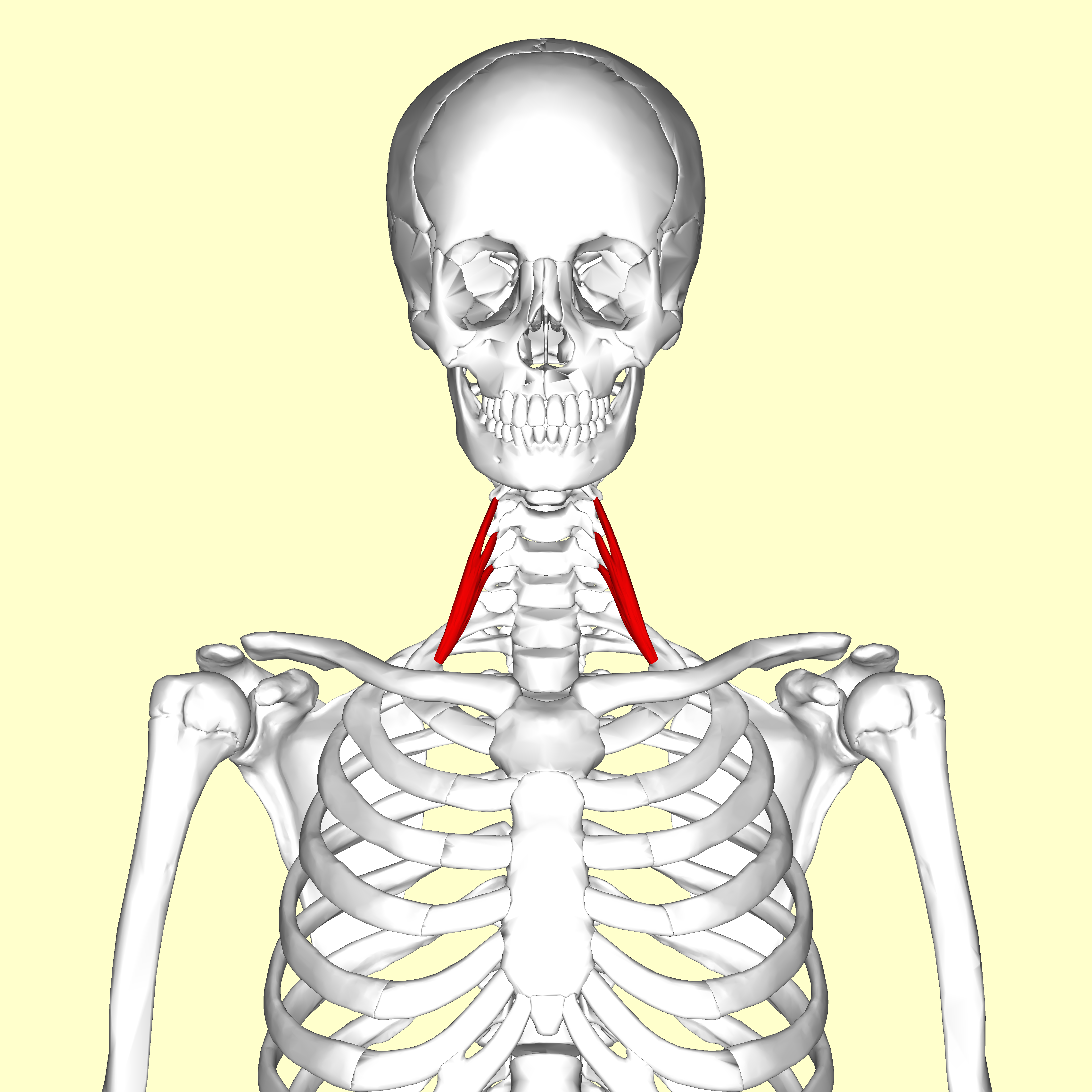
Scalenus medius on:
[Wikipedia]
[Google]
[Amazon]
The scalene muscles are a group of three pairs of muscles in the lateral neck, namely the anterior scalene, middle scalene, and posterior scalene. They are innervated by the third to the eight cervical


File:Musculi coli base.svg, Musculi colli base
File:Slide1ABBA.JPG, Scalene muscles. Muscles of the neck. Lateral view.
File:Slide2ABBA.JPG, Scalene muscles. Muscles of the neck. Lateral view.
File:Gray384.png, Section of the neck at about the level of the sixth cervical vertebra. Showing the arrangement of the fascia coli.
spinal nerves
A spinal nerve is a mixed nerve, which carries motor, sensory, and autonomic signals between the spinal cord and the body. In the human body there are 31 pairs of spinal nerves, one on each side of the vertebral column. These are grouped into the ...
(C3-C8).
The anterior and middle scalene muscles lift the first rib
The rib cage, as an enclosure that comprises the ribs, vertebral column and sternum in the thorax of most vertebrates, protects vital organs such as the heart, lungs and great vessels.
The sternum, together known as the thoracic cage, is a semi ...
and bend the neck to the same side; the posterior scalene lifts the second rib and tilts the neck to the same side.
The muscles are named .
Structure
The scalene muscles originate from the transverse processes from the cervical vertebrae of C2 to C7 and insert onto the first and second ribs.
Anterior scalene
The anterior scalene muscle ( la, scalenus anterior), lies deeply at the side of the neck, behind thesternocleidomastoid muscle
The sternocleidomastoid muscle is one of the largest and most superficial cervical muscles. The primary actions of the muscle are rotation of the head to the opposite side and flexion of the neck. The sternocleidomastoid is innervated by the access ...
. It arises from the anterior tubercles of the transverse processes of the third, fourth, fifth, and sixth cervical vertebrae, and descending, almost vertically, is inserted by a narrow, flat tendon into the scalene tubercle
The scalene tubercle is a small projection that runs along the medial border of the first rib between two grooves, which travel anteriorly for the subclavian artery and posteriorly for the subclavian vein. It projects outward medially, and is the ...
on the inner border of the first rib
The rib cage, as an enclosure that comprises the ribs, vertebral column and sternum in the thorax of most vertebrates, protects vital organs such as the heart, lungs and great vessels.
The sternum, together known as the thoracic cage, is a semi ...
, and into the ridge on the upper surface of the second rib
The rib cage, as an enclosure that comprises the ribs, vertebral column and sternum in the thorax of most vertebrates, protects vital organs such as the heart, lungs and great vessels.
The sternum, together known as the thoracic cage, is a semi ...
in front of the subclavian groove In general, Subclavian means beneath the clavicle, and it may refer to:
* Subclavian vein
* Subclavian artery
In human anatomy, the subclavian arteries are paired major arteries of the upper thorax, below the clavicle. They receive blood from t ...
. It is supplied by the anterior ramus
The ventral ramus (pl. ''rami'') (Latin for ''branch'') is the anterior division of a spinal nerve. The ventral rami supply the antero-lateral parts of the trunk and the limbs. They are mainly larger than the dorsal rami.
Shortly after a spinal n ...
of cervical nerve 5 and 6.
Middle scalene
The middle scalene, ( la, scalenus medius), is the largest and longest of the three scalene muscles. The middle scalene arises from the posterior tubercles of the transverse processes of the lower six cervical vertebrae. It descends along the side of thevertebral column
The vertebral column, also known as the backbone or spine, is part of the axial skeleton. The vertebral column is the defining characteristic of a vertebrate in which the notochord (a flexible rod of uniform composition) found in all chordate ...
to insert by a broad attachment into the upper surface of the first rib, posterior to the subclavian groove. The brachial plexus
The brachial plexus is a network () of nerves formed by the anterior rami of the lower four cervical nerves and first thoracic nerve ( C5, C6, C7, C8, and T1). This plexus extends from the spinal cord, through the cervicoaxillary canal in t ...
and the subclavian artery pass anterior to it.

Posterior scalene
The posterior scalene, ( la, scalenus posterior) is the smallest and most deeply seated of the scalene muscles. It arises, by two or three separate tendons, from the posterior tubercles of the transverse processes of the lower two or three cervical vertebrae, and is inserted by a thin tendon into the outer surface of the second rib, behind the attachment of the anterior scalene. It is supplied by cervical nerves C5, C6 and C7. It is occasionally blended with the middle scalene.Variation
A fourth muscle, the scalenus minimus (Sibson's muscle), is sometimes present behind the lower portion of the anterior scalene.Function
The anterior and middle scalene muscles lift thefirst rib
The rib cage, as an enclosure that comprises the ribs, vertebral column and sternum in the thorax of most vertebrates, protects vital organs such as the heart, lungs and great vessels.
The sternum, together known as the thoracic cage, is a semi ...
and bend the neck to the same side as the acting muscle; the posterior scalene lifts the second rib and tilts the neck to the same side.
Because they elevate the upper ribs, they also act as accessory muscles of respiration
The muscles of respiration are the muscles that contribute to inhalation and exhalation, by aiding in the expansion and contraction of the thoracic cavity. The diaphragm and, to a lesser extent, the intercostal muscles drive respiration during q ...
, along with the sternocleidomastoids
The sternocleidomastoid muscle is one of the largest and most superficial cervical muscles. The primary actions of the muscle are rotation of the head to the opposite side and flexion of the neck. The sternocleidomastoid is innervated by the access ...
.
Relations
The scalene muscles have an important relationship to other structures in the neck. Thebrachial plexus
The brachial plexus is a network () of nerves formed by the anterior rami of the lower four cervical nerves and first thoracic nerve ( C5, C6, C7, C8, and T1). This plexus extends from the spinal cord, through the cervicoaxillary canal in t ...
and subclavian artery pass between the anterior and middle scalenes. The subclavian vein and phrenic nerve pass anteriorly to the anterior scalene as the muscle crosses over the first rib. The phrenic nerve is oriented vertically as it passes in front of the anterior scalene, while the subclavian vein is oriented horizontally as it passes in front of the anterior scalene muscle.
The passing of the brachial plexus
The brachial plexus is a network () of nerves formed by the anterior rami of the lower four cervical nerves and first thoracic nerve ( C5, C6, C7, C8, and T1). This plexus extends from the spinal cord, through the cervicoaxillary canal in t ...
and the subclavian artery through the space of the anterior and middle scalene muscles constitute the scalene hiatus (the term "scalene fissure" is also used). The region in which this lies is referred to as the scaleotracheal fossa. It is bounded by the clavicle
The clavicle, or collarbone, is a slender, S-shaped long bone approximately 6 inches (15 cm) long that serves as a strut between the shoulder blade and the sternum (breastbone). There are two clavicles, one on the left and one on the r ...
inferior anteriorly, the trachea medially, posteriorly by the trapezius
The trapezius is a large paired trapezoid-shaped surface muscle that extends longitudinally from the occipital bone to the lower thoracic vertebrae of the spine and laterally to the spine of the scapula. It moves the scapula and supports th ...
, and anteriorly by the platysma muscle
The platysma muscle is a superficial muscle of the human neck that overlaps the sternocleidomastoid. It covers the anterior surface of the neck superficially. When it contracts, it produces a slight wrinkling of the neck, and a "bowstring" effect ...
.
Clinical significance
The anterior and middle scalene muscles can be involved in certain forms of thoracic outlet syndrome as well as myofascial pain syndrome, the symptoms of which may mimic aspinal disc herniation
Spinal disc herniation is an injury to the cushioning and connective tissue between vertebrae, usually caused by excessive strain or trauma to the spine. It may result in back pain, pain or sensation in different parts of the body, and physical ...
of the cervical vertebrae.
Since the nerves of the brachial plexus pass through the space between the anterior and middle scalene muscles, that area is sometimes targeted with the administration of regional anesthesia by an anesthesia provider. The nerve block, called an interscalene block, may be performed prior to arm or shoulder surgery.
According to the medical codes in the 2016 Procedural Coding Expert, published by the American Academy of Professional Coders, for Current Procedural Terminology (CPT) and other medical codes, the scalenus anticus muscle can be divided by reparative or reconstructive surgery, with (# 21705) or without (# 21700) resection of the cervical rib
A cervical rib in humans is an extra rib which arises from the seventh cervical vertebra. Their presence is a congenital abnormality located above the normal first rib. A cervical rib is estimated to occur in 0.2% to 0.5% (1 in 200 to 500) of th ...
.
History
The scalenes used to be known as the ''lateral vertebral muscles''.Etymology
The muscles are named fromGreek
Greek may refer to:
Greece
Anything of, from, or related to Greece, a country in Southern Europe:
*Greeks, an ethnic group.
*Greek language, a branch of the Indo-European language family.
**Proto-Greek language, the assumed last common ancestor ...
σκαληνός, or ''skalenos'', meaning unevenMosby's Medical, Nursing & Allied Health Dictionary
''Mosby's Dictionary of Medicine, Nursing & Health Professions'' is a dictionary of health related topics. The 8th edition, published in 2009, contains 2,240 pages and 2,400 colour illustrations. It includes some encyclopaedic definitions and 12 ap ...
, Fourth Edition, Mosby-Year Book Inc., 1994, p. 1395 as the pairs are all of differing length
Additional images
References
{{Authority control Human anatomy Muscles of the head and neck