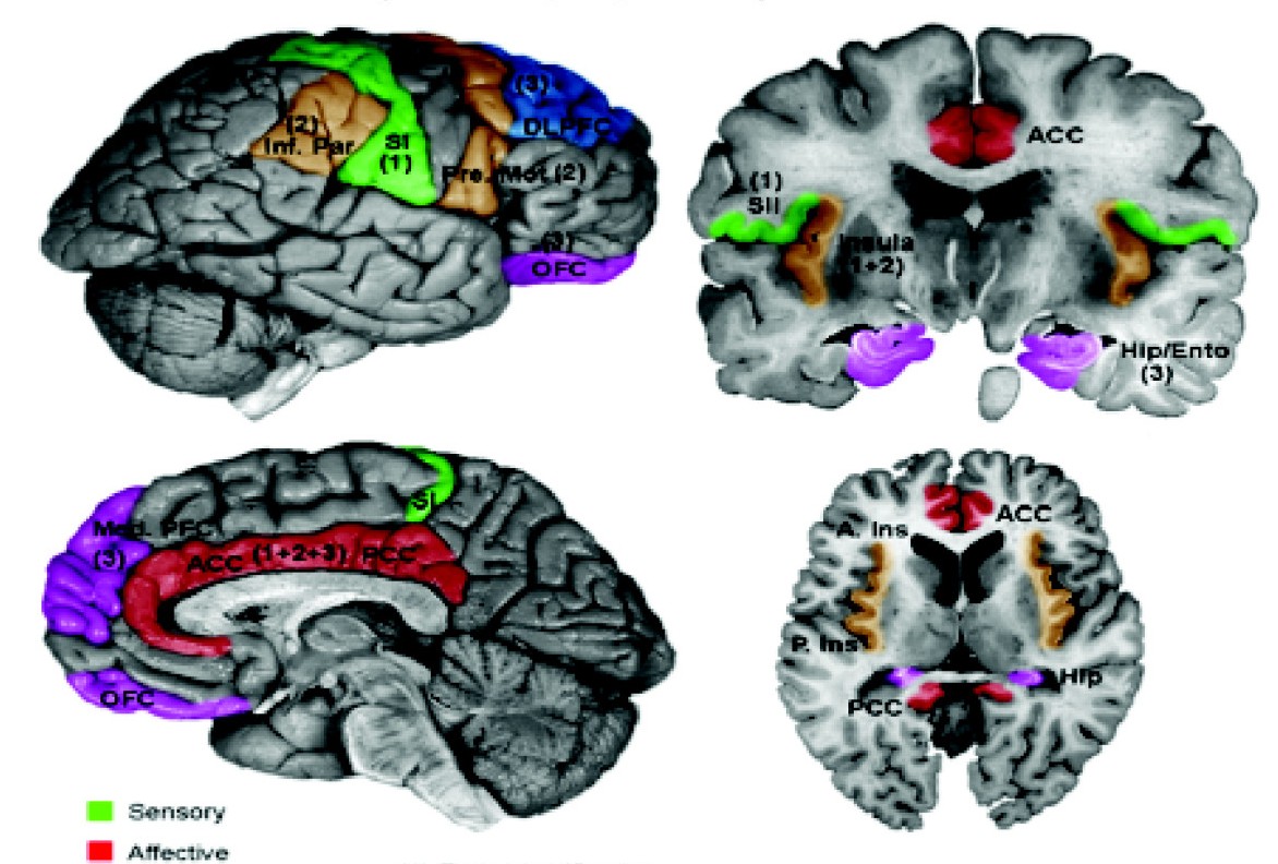
Primary somatosensory cortex on:
[Wikipedia]
[Google]
[Amazon]
In

neuroanatomy
Neuroanatomy is the study of the structure and organization of the nervous system. In contrast to animals with radial symmetry, whose nervous system consists of a distributed network of cells, animals with bilateral symmetry have segregated, defi ...
, the primary somatosensory cortex is located in the postcentral gyrus
In neuroanatomy, the postcentral gyrus is a prominent gyrus in the lateral parietal lobe of the human brain. It is the location of the primary somatosensory cortex, the main sensory receptive area for the sense of touch. Like other sensory areas, ...
of the brain
A brain is an organ that serves as the center of the nervous system in all vertebrate and most invertebrate animals. It is located in the head, usually close to the sensory organs for senses such as vision. It is the most complex organ in a ve ...
's parietal lobe
The parietal lobe is one of the four major lobes of the cerebral cortex in the brain of mammals. The parietal lobe is positioned above the temporal lobe and behind the frontal lobe and central sulcus.
The parietal lobe integrates sensory informa ...
, and is part of the somatosensory system
In physiology, the somatosensory system is the network of neural structures in the brain and body that produce the perception of touch (haptic perception), as well as temperature (thermoception), body position (proprioception), and pain. It ...
. It was initially defined from surface stimulation studies of Wilder Penfield
Wilder Graves Penfield (January 26, 1891April 5, 1976) was an American-Canadian neurosurgeon. He expanded brain surgery's methods and techniques, including mapping the functions of various regions of the brain such as the cortical homunculus. ...
, and parallel surface potential studies of Bard, Woolsey, and Marshall. Although initially defined to be roughly the same as Brodmann areas 3, 1 and 2
In neuroanatomy, the primary somatosensory cortex is located in the postcentral gyrus of the brain's parietal lobe, and is part of the somatosensory system. It was initially defined from surface stimulation studies of Wilder Penfield, and paralle ...
, more recent work by Kaas has suggested that for homogeny with other sensory fields only area 3 should be referred to as "primary somatosensory cortex", as it receives the bulk of the thalamocortical projections from the sensory input fields.
At the primary somatosensory cortex, tactile representation is orderly arranged (in an inverted fashion) from the toe (at the top of the cerebral hemisphere
The vertebrate cerebrum (brain) is formed by two cerebral hemispheres that are separated by a groove, the longitudinal fissure. The brain can thus be described as being divided into left and right cerebral hemispheres. Each of these hemispheres ...
) to mouth (at the bottom). However, some body parts may be controlled by partially overlapping regions of cortex. Each cerebral hemisphere of the primary somatosensory cortex only contains a tactile representation of the opposite (contralateral) side of the body. The amount of primary somatosensory cortex devoted to a body part is not proportional to the absolute size of the body surface, but, instead, to the relative density of cutaneous tactile receptors located on that body part. The density of cutaneous tactile receptors on a body part is generally indicative of the degree of sensitivity of tactile stimulation experienced at said body part. For this reason, the human lips
The lips are the visible body part at the mouth of many animals, including humans. Lips are soft, movable, and serve as the opening for food intake and in the articulation of sound and speech. Human lips are a tactile sensory organ, and can be ...
and hands have a larger representation than other body parts.
Structure

Brodmann areas 3, 1 and 2
Brodmann areas 3, 1, and 2 make up the primary somatosensory cortex of the humanbrain
A brain is an organ that serves as the center of the nervous system in all vertebrate and most invertebrate animals. It is located in the head, usually close to the sensory organs for senses such as vision. It is the most complex organ in a ve ...
(or S1). Because Brodmann sliced the brain
A brain is an organ that serves as the center of the nervous system in all vertebrate and most invertebrate animals. It is located in the head, usually close to the sensory organs for senses such as vision. It is the most complex organ in a ve ...
somewhat obliquely, he encountered area 1 first; however, from anterior to posterior, the Brodmann designations are 3, 1, and 2, respectively.
Brodmann area (BA) 3 is subdivided into areas 3a and 3b. Where BA 1 occupies the apex of the postcentral gyrus, the rostral border of BA 3a is in the nadir of the Central sulcus, and is caudally followed by BA 3b, then BA 1, with BA 2 following and ending in the nadir of the postcentral sulcus. BA 3b is now conceived as the primary somatosensory cortex because 1) it receives dense inputs from the NP nucleus of the thalamus; 2) its neurons are highly responsive to somatosensory stimuli, but not other stimuli; 3) lesions here impair somatic sensation; and 4) electrical stimulation evokes somatic sensory experience. BA 3a also receives dense input from the thalamus; however, this area is concerned with proprioception
Proprioception ( ), also referred to as kinaesthesia (or kinesthesia), is the sense of self-movement, force, and body position. It is sometimes described as the "sixth sense".
Proprioception is mediated by proprioceptors, mechanosensory neurons ...
.
Areas 1 and 2 receive dense inputs from BA 3b. The projection from 3b to 1 primarily relays texture information; the projection to area 2 emphasizes size and shape. Lesions confined to these areas produce predictable dysfunction in texture, size, and shape discrimination.
Somatosensory cortex, like other neocortex, is layered. Like other sensory cortex (i.e., visual and auditory) the thalamic inputs project into layer IV, which in turn project into other layers. As in other sensory cortices, S1 neurons are grouped together with similar inputs and responses into vertical columns that extend across cortical layers (e.g., As shown by Vernon Mountcastle, into alternating layers of slowly adapting and rapidly adapting neurons; or spatial segmentation of the vibrissae on mouse/rat cerebral cortex).
This area of cortex, as shown by Wilder Penfield
Wilder Graves Penfield (January 26, 1891April 5, 1976) was an American-Canadian neurosurgeon. He expanded brain surgery's methods and techniques, including mapping the functions of various regions of the brain such as the cortical homunculus. ...
and others, is organized somatotopically, having the pattern of a homunculus. That is, the legs and trunk fold over the midline; the arms and hands are along the middle of the area shown here; and the face is near the bottom of the figure. While it is not well-shown here, the lips and hands are enlarged on a proper homunculus
A homunculus ( , , ; "little person") is a representation of a small human being, originally depicted as small statues made out of clay. Popularized in sixteenth-century alchemy and nineteenth-century fiction, it has historically referred to the ...
, since a larger number of neuron
A neuron, neurone, or nerve cell is an electrically excitable cell that communicates with other cells via specialized connections called synapses. The neuron is the main component of nervous tissue in all animals except sponges and placozoa. ...
s in the cerebral cortex
The cerebral cortex, also known as the cerebral mantle, is the outer layer of neural tissue of the cerebrum of the brain in humans and other mammals. The cerebral cortex mostly consists of the six-layered neocortex, with just 10% consistin ...
are devoted to processing information from these areas.
The positions of Brodmann areas 3, 1, and 2 are - from the nadir of the central sulcus toward the apex of the postcentral gyrus - 3a, 3b, 1, and 2, respectively.
These areas contain cells that project to the secondary somatosensory cortex
The human secondary somatosensory cortex (S2, SII) is a region of cortex in the parietal operculum on the ceiling of the lateral sulcus.
Region S2 was first described by Adrian in 1940, who found that feeling in cats' feet was not only represe ...
.
Clinical significance
Lesions affecting the primary somatosensory cortex produce characteristic symptoms including:agraphesthesia Agraphesthesia is a disorder of directional cutaneous kinesthesia or a disorientation of the skin's sensation across its space. It is a difficulty recognizing a written number or letter traced on the skin after parietal damage.
Causes
Agraphest ...
, astereognosia
Astereognosis (or tactile agnosia if only one hand is affected) is the inability to identify an object by active touch of the hands without other sensory input, such as visual or sensory information. An individual with astereognosis is unable to i ...
, hemihypesthesia
Hemihypesthesia is a reduction in sensitivity on one side of the body. A person with this condition may not be able to perceive being lightly touched on one side, but has normal function on the other side of the body.
It can occur from damage to ...
, and loss of vibration
Vibration is a mechanical phenomenon whereby oscillations occur about an equilibrium point. The word comes from Latin ''vibrationem'' ("shaking, brandishing"). The oscillations may be periodic, such as the motion of a pendulum—or random, su ...
, proprioception
Proprioception ( ), also referred to as kinaesthesia (or kinesthesia), is the sense of self-movement, force, and body position. It is sometimes described as the "sixth sense".
Proprioception is mediated by proprioceptors, mechanosensory neurons ...
and fine touch
In physiology, the somatosensory system is the network of neural structures in the brain and body that produce the perception of touch (haptic perception), as well as temperature (thermoception), body position (proprioception), and pain. It i ...
(because the third-order neuron of the medial-lemniscal pathway cannot synapse in the cortex). It can also produce hemineglect
Hemispatial neglect is a neuropsychological condition in which, after damage to one hemisphere of the brain (e.g. after a stroke), a deficit in attention and awareness towards the side of space opposite brain damage (contralesional space) is obse ...
, if it affects the non-dominant hemisphere. Destruction of brodmann area 3, 1, and 2 results in contralateral hemihypesthesia and astereognosis.
It could also reduce nociception, thermoception
Thermoception or thermoreception is the sensation and perception of temperature, or more accurately, temperature differences inferred from heat flux. It deals with a series of events and processes required for an organism to receive a temperature s ...
, and crude touch, but, since information from the spinothalamic tract
The spinothalamic tract is a part of the anterolateral system or the ventrolateral system, a sensory pathway to the thalamus. From the ventral posterolateral nucleus in the thalamus, sensory information is relayed upward to the somatosensory co ...
is interpreted mainly by other areas of the brain (see insular cortex
The insular cortex (also insula and insular lobe) is a portion of the cerebral cortex folded deep within the lateral sulcus (the fissure separating the temporal lobe from the parietal and frontal lobes) within each hemisphere of the mammalian b ...
and cingulate gyrus
The cingulate cortex is a part of the brain situated in the medial aspect of the cerebral cortex. The cingulate cortex includes the entire cingulate gyrus, which lies immediately above the corpus callosum, and the continuation of this in the ...
), it is not as relevant as the other symptoms.
See also
* List of regions in the human brainReferences
External links
* - area 1 * - area 2 * - area 3 {{Neural tracts Cerebral cortex 01 somatosensory system Parietal lobe