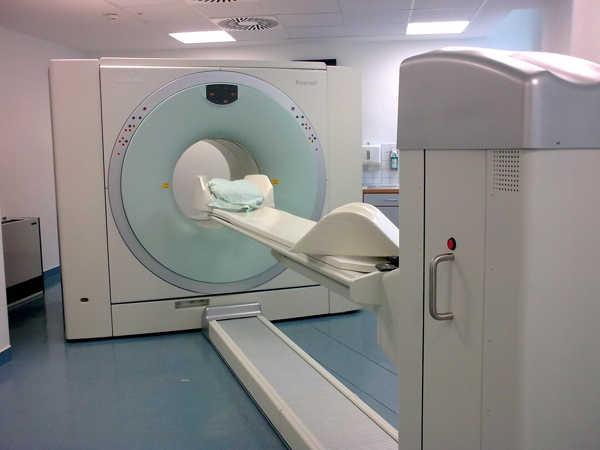
PET/CT on:
[Wikipedia]
[Google]
[Amazon]
Positron emission tomography–computed tomography (better known as PET-CT or PET/CT) is a
 The combination of PET and CT scanners was first suggested by R. Raylman in his 1991 Ph.D. thesis. The first PET-CT systems were constructed by David Townsend (at the
Human Health Campus, The official website of the International Atomic Energy Agency dedicated to Professionals in Radiation Medicine. This site is managed by the Division of Human Health, Department of Nuclear Sciences and Applications
- from Harvard Medical School
PET CT for evaluation of Lung Cancer
- from Harvard Medical School {{Medical imaging 3D nuclear medical imaging Computing in medical imaging Medical physics Neuroimaging
nuclear medicine
Nuclear medicine or nucleology is a medical specialty involving the application of radioactive substances in the diagnosis and treatment of disease. Nuclear imaging, in a sense, is " radiology done inside out" because it records radiation emi ...
technique which combines, in a single gantry, a positron emission tomography (PET) scanner and an x-ray computed tomography
An X-ray, or, much less commonly, X-radiation, is a penetrating form of high-energy electromagnetic radiation. Most X-rays have a wavelength ranging from 10 picometers to 10 nanometers, corresponding to frequencies in the range 30 ...
(CT) scanner, to acquire sequential images from both devices in the same session, which are combined into a single superposed ( co-registered) image. Thus, functional imaging obtained by PET, which depicts the spatial distribution of metabolic or biochemical activity in the body can be more precisely aligned or correlated with anatomic imaging obtained by CT scanning. Two- and three-dimensional image reconstruction may be rendered as a function of a common software
Software is a set of computer programs and associated software documentation, documentation and data (computing), data. This is in contrast to Computer hardware, hardware, from which the system is built and which actually performs the work.
...
and control system.
PET-CT has revolutionized medical diagnosis
Medical diagnosis (abbreviated Dx, Dx, or Ds) is the process of determining which disease or condition explains a person's symptoms and signs. It is most often referred to as diagnosis with the medical context being implicit. The information r ...
in many fields, by adding precision of anatomic localization to functional imaging, which was previously lacking from pure PET imaging. For example, many diagnostic imaging procedures in oncology
Oncology is a branch of medicine that deals with the study, treatment, diagnosis and prevention of cancer. A medical professional who practices oncology is an ''oncologist''. The name's etymological origin is the Greek word ὄγκος (''� ...
, surgical planning
Surgical planning is the preoperative method of pre-visualising a surgical intervention, in order to predefine the surgical steps and furthermore the bone segment navigation in the context of computer-assisted surgery.
The surgical planning is mos ...
, radiation therapy
Radiation therapy or radiotherapy, often abbreviated RT, RTx, or XRT, is a therapy using ionizing radiation, generally provided as part of cancer treatment to control or kill malignant cells and normally delivered by a linear accelerator. Radi ...
and cancer staging
Cancer staging is the process of determining the extent to which a cancer has developed by growing and spreading. Contemporary practice is to assign a number from I to IV to a cancer, with I being an isolated cancer and IV being a cancer that ha ...
have been changing rapidly under the influence of PET-CT availability, and centers have been gradually abandoning conventional PET devices and substituting them by PET-CTs. Although the combined/hybrid device is considerably more expensive, it has the advantage of providing both functions as stand-alone examinations, being, in fact, two devices in one.
The only other obstacle to the wider use of PET-CT is the difficulty and cost of producing and transporting the radiopharmaceutical
Radiopharmaceuticals, or medicinal radiocompounds, are a group of pharmaceutical drugs containing radioactive isotopes. Radiopharmaceuticals can be used as diagnostic and therapeutic agents. Radiopharmaceuticals emit radiation themselves, which ...
s used for PET imaging, which are usually extremely short-lived. For instance, the half-life
Half-life (symbol ) is the time required for a quantity (of substance) to reduce to half of its initial value. The term is commonly used in nuclear physics to describe how quickly unstable atoms undergo radioactive decay or how long stable at ...
of radioactive fluorine-18
Fluorine-18 (18F) is a fluorine radioisotope which is an important source of positrons. It has a mass of 18.0009380(6) u and its half-life is 109.771(20) minutes. It decays by positron emission 96% of the time and electron capture 4% of the time ...
(18F) used to trace glucose
Glucose is a simple sugar with the molecular formula . Glucose is overall the most abundant monosaccharide, a subcategory of carbohydrates. Glucose is mainly made by plants and most algae during photosynthesis from water and carbon dioxide, u ...
metabolism (using fluorodeoxyglucose
18F.html" ;"title="sup>18F">sup>18Fluorodeoxyglucose ( INN), or fluorodeoxyglucose F 18 (USAN and USP), also commonly called fluorodeoxyglucose and abbreviated 18F.html" ;"title="sup>18F">sup>18FDG, 2- 18F.html" ;"title="sup>18F">sup>18FDG or ...
, FDG) is only two hours. Its production requires a very expensive cyclotron
A cyclotron is a type of particle accelerator invented by Ernest O. Lawrence in 1929–1930 at the University of California, Berkeley, and patented in 1932. Lawrence, Ernest O. ''Method and apparatus for the acceleration of ions'', filed: Jan ...
as well as a production line for the radiopharmaceuticals. At least one PET-CT radiopharmaceutical is made on site from a generator: Ga-68 from a gallium-68 generator
A germanium-68/gallium-68 generator is a device used to extract the positron-emitting isotope 68Ga of gallium from a source of decaying germanium-68. The parent isotope 68Ge has a half-life of 271 days and can be easily utilized for in-hospital ...
.
PET-MRI
Positron emission tomography–magnetic resonance imaging (PET–MRI) is a hybrid imaging technology that incorporates magnetic resonance imaging (MRI) soft tissue morphological imaging and positron emission tomography (PET) functional imaging.
T ...
, like PET-CT, combines modalities to produce co-registered images.
History
 The combination of PET and CT scanners was first suggested by R. Raylman in his 1991 Ph.D. thesis. The first PET-CT systems were constructed by David Townsend (at the
 University of Geneva
The University of Geneva (French: ''Université de Genève'') is a public research university located in Geneva, Switzerland. It was founded in 1559 by John Calvin as a theological seminary. It remained focused on theology until the 17th centur ...
at the time) and Ronald Nutt (at ''CPS Innovations'' in Knoxville, TN
Knoxville is a city in and the county seat of Knox County in the U.S. state of Tennessee. As of the 2020 United States census, Knoxville's population was 190,740, making it the largest city in the East Tennessee Grand Division and the state' ...
) with help from colleagues. The first PET-CT prototype for clinical evaluation was funded by the NCI and installed at the University of Pittsburgh Medical Center
The University of Pittsburgh Medical Center (UPMC) is a $23billion integrated global nonprofit health enterprise that has 92,000 employees, 40 hospitals with more than 8,000 licensed beds, 800 clinical locations including outpatient sites and d ...
in 1998. The first commercial system reached the market by 1997, and by 2004, over 400 systems had been installed worldwide.
Procedure for FDG imaging
An example of how PET-CT works in the work-up of FDG metabolic mapping follows: :::::::::::* Before the exam, the patient fasts for at least 6 hours.NHS_Choices
:_PET_scan">NHS_Choices">NHS_Choices
:_PET_scan_Retrieved_11_November_2016. :::::::::::*_On_the_day_of_the_exam,_the_patient_rests_lying_for_a_minimum_of_15_min,_in_order_to_quiet_down_muscle.html" "title="NHS_Choices
:_PET_scan.html" ;"title="NHS Choices">NHS Choices
: PET scan">NHS Choices">NHS Choices
: PET scan Retrieved 11 November 2016. :::::::::::* On the day of the exam, the patient rests lying for a minimum of 15 min, in order to quiet down muscle">muscular Skeletal muscles (commonly referred to as muscles) are organs of the vertebrate muscular system and typically are attached by tendons to bones of a skeleton. The muscle cells of skeletal muscles are much longer than in the other types of muscle ...
activity, which might be interpreted as abnormal metabolism.
* An intravenous bolus (medicine), bolus injection of a dose of recently produced 2-FDG or 3-FDG is made, usually by arm vein. Dosage ranges from per kilogram of body weight.
* After one or two hours, the patient is placed into the PET-CT device, usually lying in a :_PET_scan">NHS_Choices">NHS_Choices
:_PET_scan_Retrieved_11_November_2016. :::::::::::*_On_the_day_of_the_exam,_the_patient_rests_lying_for_a_minimum_of_15_min,_in_order_to_quiet_down_muscle.html" "title="NHS_Choices
:_PET_scan.html" ;"title="NHS Choices">NHS Choices
: PET scan">NHS Choices">NHS Choices
: PET scan Retrieved 11 November 2016. :::::::::::* On the day of the exam, the patient rests lying for a minimum of 15 min, in order to quiet down muscle">muscular Skeletal muscles (commonly referred to as muscles) are organs of the vertebrate muscular system and typically are attached by tendons to bones of a skeleton. The muscle cells of skeletal muscles are much longer than in the other types of muscle ...
supine position
The supine position ( or ) means lying horizontally with the face and torso facing up, as opposed to the prone position, which is face down. When used in surgical procedures, it grants access to the peritoneal, thoracic and pericardium, pericardi ...
with the arms resting at the sides, or brought together above the head, depending on the main region of interest ( ROI).
* An automatic bed moves head first into the gantry, first obtaining a tomogram, also called a scout view or surview, which is a kind of whole body flat sagittal
The sagittal plane (; also known as the longitudinal plane) is an anatomical plane that divides the body into right and left sections. It is perpendicular to the transverse and coronal planes. The plane may be in the center of the body and divi ...
section, obtained with the X-ray tube fixed into the upper position.
* The operator uses the PET-CT computer console to identify the patient and examination, delimit the caudal and rostral limits of the body scan onto the scout view, selects the scanning parameters and starts the image acquisition period, which follows without human intervention.
* The patient is automatically moved head first into the CT gantry, and the x-ray tomogram is acquired.
* Now the patient is automatically moved through the PET gantry, which is mounted in parallel with the CT gantry, and the PET slices are acquired.
* The patient may now leave the device, and the PET-CT software starts reconstructing and aligning the PET and CT images.
A whole body scan, which usually is made from mid-thighs to the top of the head, takes from 5 minutes to 40 minutes depending on the acquisition protocol and technology of the equipment used. FDG imaging protocols acquires slices with a thickness of 2 to 3 mm. Hypermetabolic lesions are shown as false color
False color (or pseudo color) refers to a group of color rendering methods used to display images in color which were recorded in the visible or non-visible parts of the electromagnetic spectrum. A false-color image is an image that depicts ...
-coded pixel
In digital imaging, a pixel (abbreviated px), pel, or picture element is the smallest addressable element in a raster image, or the smallest point in an all points addressable display device.
In most digital display devices, pixels are the ...
s or voxel
In 3D computer graphics, a voxel represents a value on a regular grid in three-dimensional space. As with pixels in a 2D bitmap, voxels themselves do not typically have their position (i.e. coordinates) explicitly encoded with their values. I ...
s onto the gray-value coded CT images. Standardized Uptake Values are calculated by the software for each hypermetabolic region detected in the image. It provides a quantification of size of the lesion, since functional imaging does not provide a precise anatomical estimate of its extent. The CT can be used for that, when the lesion is also visualized in its images (this is not always the case when hypermetabolic lesions are not accompanied by anatomical changes).
FDG doses in quantities sufficient to carry out 4–5 examinations are delivered daily, twice or more per day, by the provider to the diagnostic imaging center.
For uses in image-guided radiation therapy of cancer, special fiducial marker
A fiducial marker or fiducial is an object placed in the field of view of an imaging system that appears in the image produced, for use as a point of reference or a measure. It may be either something placed into or on the imaging subject, or a m ...
s are placed in the patient's body before acquiring the PET-CT images. The slices thus acquired may be transferred digitally to a linear accelerator
A linear particle accelerator (often shortened to linac) is a type of particle accelerator that accelerates charged subatomic particles or ions to a high speed by subjecting them to a series of oscillating electric potentials along a linear ...
which is used to perform precise bombardment of the target areas using high energy photons ( radiosurgery).
See also
* Single-photon emission computed tomography *Neuroimaging
Neuroimaging is the use of quantitative (computational) techniques to study the structure and function of the central nervous system, developed as an objective way of scientifically studying the healthy human brain in a non-invasive manner. Incr ...
References
External links
Human Health Campus, The official website of the International Atomic Energy Agency dedicated to Professionals in Radiation Medicine. This site is managed by the Division of Human Health, Department of Nuclear Sciences and Applications
- from Harvard Medical School
PET CT for evaluation of Lung Cancer
- from Harvard Medical School {{Medical imaging 3D nuclear medical imaging Computing in medical imaging Medical physics Neuroimaging