Diagnostic microbiology is the study of microbial identification. Since the discovery of the
germ theory of disease
The germ theory of disease is the currently accepted scientific theory for many diseases. It states that microorganisms known as pathogens or "germs" can lead to disease. These small organisms, too small to be seen without magnification, invade h ...
, scientists have been finding ways to harvest specific organisms. Using methods such as
differential media
A growth medium or culture medium is a solid, liquid, or semi-solid designed to support the growth of a population of microorganisms or cells via the process of cell proliferation or small plants like the moss ''Physcomitrella patens''. Different ...
or
genome sequencing
Whole genome sequencing (WGS), also known as full genome sequencing, complete genome sequencing, or entire genome sequencing, is the process of determining the entirety, or nearly the entirety, of the DNA sequence of an organism's genome at a ...
, physicians and scientists can observe novel functions in organisms for more effective and accurate diagnosis of organisms. Methods used in diagnostic microbiology are often used to take advantage of a particular difference in organisms and attain information about what species it can be identified as, which is often through a reference of previous studies. New studies provide information that others can reference so that scientists can attain a basic understanding of the organism they are examining.
Aerobic vs anaerobic
Anaerobic organism
An anaerobic organism or anaerobe is any organism that does not require molecular oxygen for growth. It may react negatively or even die if free oxygen is present. In contrast, an aerobic organism (aerobe) is an organism that requires an oxygenate ...
s require an oxygen-free environment. When culturing anaerobic microbes, broths are often flushed with nitrogen gas to extinguish oxygen present, and growth can also occur on media in a chamber without oxygen present. Sodium resazurin can be added to indicate
redox
Redox (reduction–oxidation, , ) is a type of chemical reaction in which the oxidation states of substrate (chemistry), substrate change. Oxidation is the loss of Electron, electrons or an increase in the oxidation state, while reduction ...
potential. Cultures are to be incubated in an oxygen-free environment for 48 hours at 35 °C before growth is examined.

Anaerobic bacteria collection can come from a variety of sources in patient samples, including blood, bile, bone marrow,
cerebrospinal fluid
Cerebrospinal fluid (CSF) is a clear, colorless body fluid found within the tissue that surrounds the brain and spinal cord of all vertebrates.
CSF is produced by specialised ependymal cells in the choroid plexus of the ventricles of the bra ...
, direct lung aspirate,
tissue biopsies
A biopsy is a medical test commonly performed by a surgeon, interventional radiologist, or an interventional cardiologist. The process involves extraction of sample cells or tissues for examination to determine the presence or extent of a disease ...
from a normally sterile site, fluid from a normally sterile site (like a joint), dental, abscess, abdominal or pelvic abscess, knife, gunshot, or surgical wound, or severe burn.
Incubation length
Incubation times vary based upon the microbe that requires culturing. Traditional culturing techniques, for example, require less than 24 hours culture time for ''
Escherichia coli
''Escherichia coli'' (),Wells, J. C. (2000) Longman Pronunciation Dictionary. Harlow ngland Pearson Education Ltd. also known as ''E. coli'' (), is a Gram-negative, facultative anaerobic, rod-shaped, coliform bacterium of the genus ''Escher ...
'' but 6–8 weeks for successful culturing of ''
Mycobacterium tuberculosis
''Mycobacterium tuberculosis'' (M. tb) is a species of pathogenic bacteria in the family Mycobacteriaceae and the causative agent of tuberculosis. First discovered in 1882 by Robert Koch, ''M. tuberculosis'' has an unusual, waxy coating on its c ...
'' before definitive results are expressed.
A benefit of non-culture tests is that physicians and microbiologists are not handicapped by waiting periods.
Incubation follows a growth curve variable for every microorganism. Cultures follow a lag, log, stationary, and finally death phase.
The
lag phase
250px, Growth is shown as ''L'' = log(numbers) where numbers is the number of colony forming units per ml, versus ''T'' (time.)
Bacterial growth is proliferation of bacterium into two daughter cells, in a process called binary fission. Providing ...
is not well known in microbiology, but it is speculated that this phase consists of the microorganism adjusting to its environment by synthesizing proteins specific for the surrounding habitat.
The
log phase
Log most often refers to:
* Trunk (botany), the stem and main wooden axis of a tree, called logs when cut
** Logging, cutting down trees for logs
** Firewood, logs used for fuel
** Lumber or timber, converted from wood logs
* Logarithm, in mathem ...
is the period where a culture experiences logarithmic growth until nutrients become scarce. The stationary phase is when culture concentration is the highest and cells stop reproducing. When nutrients in the environment are depleting, organisms enter the death phase where toxic
metabolite
In biochemistry, a metabolite is an intermediate or end product of metabolism.
The term is usually used for small molecules. Metabolites have various functions, including fuel, structure, signaling, stimulatory and inhibitory effects on enzymes, c ...
s become abundant and nutrients are depleted to the point where cell death exceeds reproduction.
Rapid identification after culture
Automated culturing systems
Automatic cell culturing systems are becoming popular because of their ability to maintain a sterile growth environment and remove strain on the laboratory staff involving repetitive experimentation. Laboratories can also set incubation times to adjust for the lag period involved in bacterial growth.
Blood cultures
Blood culture
A blood culture is a medical laboratory test used to detect bacteria or fungi in a person's blood. Under normal conditions, the blood does not contain microorganisms: their presence can indicate a bloodstream infection such as bacteremia or f ...
s can allow for diagnostic results after culture. Recent development of DNA based
PCR diagnostics have provided faster diagnostic results as opposed to overnight biochemical tests. DNA diagnostic test can diagnose with near the same specificity as biochemical test, resulting in the same diagnostic result in 90% of cases.
Breath tests
Breath test
A breath test is a type of test performed on air generated from the act of exhalation.
Types include:
*Breathalyzer – by far the most common usage of this term relates to the legal breath test to determine if a person is driving under the inf ...
for microbial diagnosis on patients has been used in a clinical setting for bacteria, including ''
Helicobacter pylori
''Helicobacter pylori'', previously known as ''Campylobacter pylori'', is a gram-negative, microaerophilic, spiral (helical) bacterium usually found in the stomach. Its helical shape (from which the genus name, helicobacter, derives) is though ...
''. Diagnostic test using the breath of patients look for metabolites excreted that were manufactured by the infectious microorganism. ''H. pylori'' is tested by testing patients for CO
2 concentration, increased because of the organism’s ability to convert urea into other derivatives.
Conventional tests
Antibody detection
A benefit of antibody detection (
ELISA
The enzyme-linked immunosorbent assay (ELISA) (, ) is a commonly used analytical biochemistry assay, first described by Eva Engvall and Peter Perlmann in 1971. The assay uses a solid-phase type of enzyme immunoassay (EIA) to detect the presence ...
) is that protein identification on a microorganism becomes faster than a
western blot
The western blot (sometimes called the protein immunoblot), or western blotting, is a widely used analytical technique in molecular biology and immunogenetics to detect specific proteins in a sample of tissue homogenate or extract. Besides detect ...
.
Antibody
An antibody (Ab), also known as an immunoglobulin (Ig), is a large, Y-shaped protein used by the immune system to identify and neutralize foreign objects such as pathogenic bacteria and viruses. The antibody recognizes a unique molecule of the ...
detection works by attaching an indicator to an antibody with a known specificity and observing whether the antibody attaches. ELISA can also indicate viral presence and is highly specific, having a detection specificity of 10
−9-10
−12 moles per litre detection. By knowing the
epitope
An epitope, also known as antigenic determinant, is the part of an antigen that is recognized by the immune system, specifically by antibodies, B cells, or T cells. The epitope is the specific piece of the antigen to which an antibody binds. The p ...
sequence of the antibody, ELISA can also be used for antigen detection in a sample.
Histological detection and culture
Histological
Histology,
also known as microscopic anatomy or microanatomy, is the branch of biology which studies the microscopic anatomy of biological tissues. Histology is the microscopic counterpart to gross anatomy, which looks at larger structures vis ...
methods used for microbiology are useful because of their ability to quickly identify a disease present in a tissue
biopsy
A biopsy is a medical test commonly performed by a surgeon, interventional radiologist, or an interventional cardiologist. The process involves extraction of sample cells or tissues for examination to determine the presence or extent of a diseas ...
.
Rapid antigen tests
Immunofluorescence

Immunofluorescence
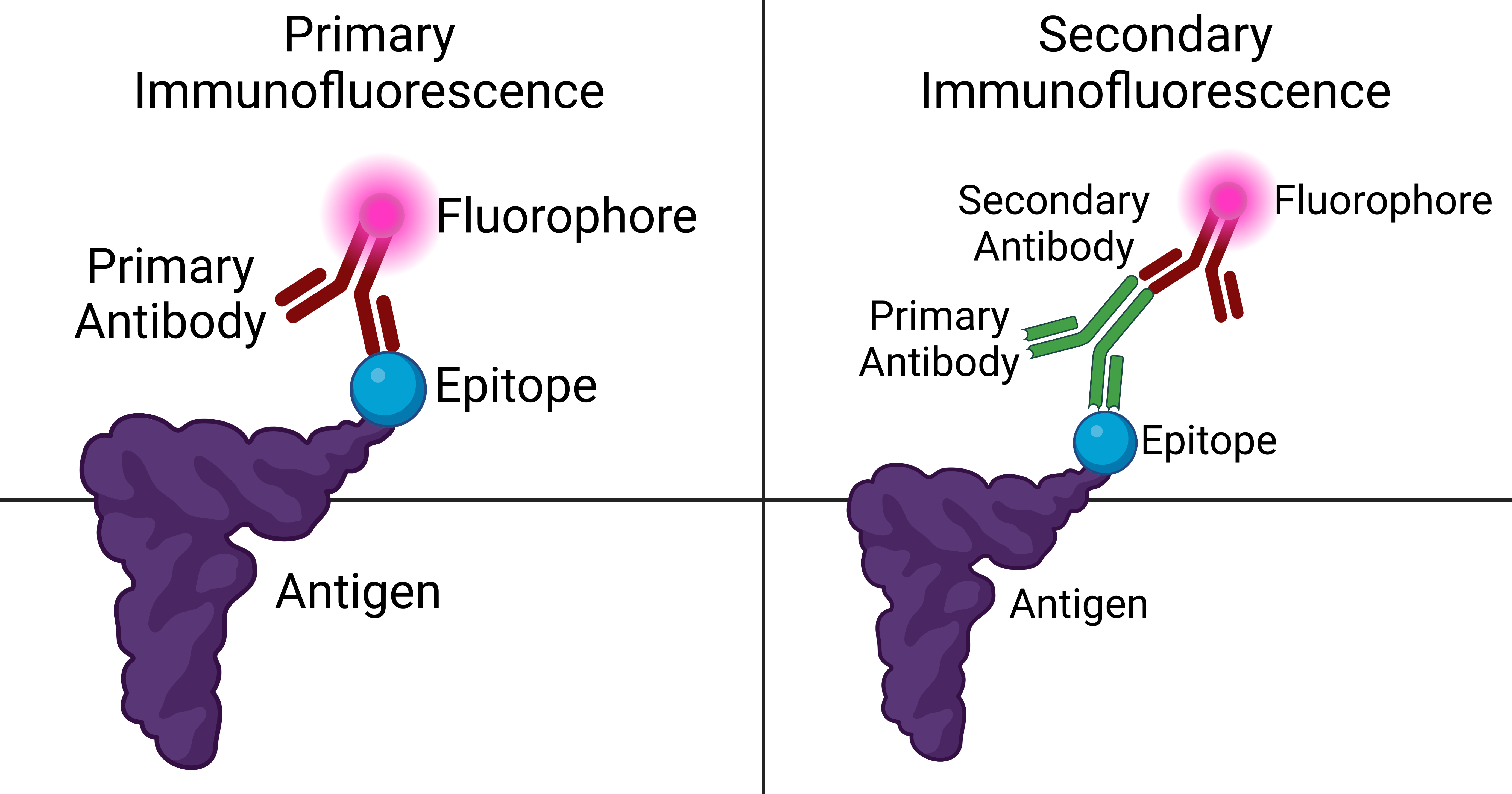
Immunofluorescence is a technique used for light microscopy with a fluorescence microscope and is used primarily on microbiological samples. This technique uses the specificity of antibodies to their antigen to target fluorescent dyes to specif ...
is performed by the production of anti-antibodies with a fluorescent molecule attached, making it a
chemiluminescent
Chemiluminescence (also chemoluminescence) is the emission of light (luminescence) as the result of a chemical reaction. There may also be limited emission of heat. Given reactants A and B, with an excited intermediate ◊,
: + -> lozenge - ...
molecule, which provides a glow when subject to ultraviolet light. Antibodies are added to a bacterial solution, providing an antigen for the binding of fluorescent anti-antibody adherence.

Mass spectrometry
MALDI-TOF (
Matrix-assisted laser desorption/ionization
In mass spectrometry, matrix-assisted laser desorption/ionization (MALDI) is an ionization technique that uses a laser energy absorbing matrix to create ions from large molecules with minimal fragmentation. It has been applied to the analysis of ...
- time of flight) is a specific type of
mass spectrometry
Mass spectrometry (MS) is an analytical technique that is used to measure the mass-to-charge ratio of ions. The results are presented as a ''mass spectrum'', a plot of intensity as a function of the mass-to-charge ratio. Mass spectrometry is use ...
that is able to identify microorganisms. A pure culture is isolated and spread directly on a stainless steel or disposable target. The cells are lysed and overlaid with a matrix, which forms protein complexes with the bacterial proteins. The MALDI fires a laser and ionizes the protein complexes, which break off and travel up the vacuum where they are detected based on mass and charge. The resulting protein spectra is compared to a known database of previously catalogued organisms, resulting in rapid diagnosis of microorganisms.
Recent studies have suggested that these tests can become specific enough to diagnose down to the sub-species level by observing novel
biomarkers
In biomedical contexts, a biomarker, or biological marker, is a measurable indicator of some biological state or condition. Biomarkers are often measured and evaluated using blood, urine, or soft tissues to examine normal biological processes, p ...
.
The MALDI-TOF identification method requires pure cultures that are less than 72 hours old. This places the organism in log phase with an abundance of ribosomal proteins, which are the most common proteins detected in the spectra. Identifications with this technology can also be impacted if the culture is exposed to cold temperatures, as this would change the typical protein distribution.
Biochemical Profile-based Microbial Identification Systems
Phenotypic tests are used to identify microbes based on metabolic and biochemical pathways present in those microbes. There are many automated and semi-automated commercial systems available. These methods can be very informative but are not as accurate as MALDI-TOF or genotypic methods.
6.5% salt broth
The 6.5%
salt broth test
Salt is a mineral composed primarily of sodium chloride (NaCl), a chemical compound belonging to the larger class of Salt (chemistry), salts; salt in the form of a natural crystallinity, crystalline mineral is known as rock salt or halite. ...
is used to analyze the tolerance level of various bacteria under halophilic conditions. This test is used because most organisms cannot survive in high salt concentrations while ''Staphylococci'', ''Enterococci'', and
''Aerococci'' are all expected to tolerate 6.5% NaCl concentrations.
Acetate utilization
The
acetate utilization test is used primarily to differentiate between ''Escherichia coli ''from members of the genus ''
Shigella
''Shigella'' is a genus of bacteria that is Gram-negative, facultative anaerobic, non-spore-forming, nonmotile, rod-shaped, and genetically closely related to ''E. coli''. The genus is named after Kiyoshi Shiga, who first discovered it in 1897. ...
''. Many of the ''Escherichia coli ''strains have the capability of the utilization of acetate for a sole carbon and energy source, while ''Shigella'' does not. Since acetate utilization results in an increase in pH, an indicator is added that changes color under conditions of acetate utilization.
ALA
An ALA (
delta-aminolevulinic acid
δ-Aminolevulinic acid (also dALA, δ-ALA, 5ALA or 5-aminolevulinic acid), an endogenous non-proteinogenic amino acid, is the first compound in the porphyrin synthesis pathway, the pathway that leads to heme in mammals, as well as chlorophyll in p ...
) test is used to test for the presence of
porphyrin
Porphyrins ( ) are a group of heterocyclic macrocycle organic compounds, composed of four modified pyrrole subunits interconnected at their α carbon atoms via methine bridges (=CH−). The parent of porphyrin is porphine, a rare chemical com ...
and
cytochrome
Cytochromes are redox-active proteins containing a heme, with a central Fe atom at its core, as a cofactor. They are involved in electron transport chain and redox catalysis. They are classified according to the type of heme and its mode of bin ...
compounds. Finding
hemin
Hemin (haemin; ferric chloride heme) is an iron-containing porphyrin with chlorine that can be formed from a heme group, such as heme B found in the hemoglobin of human blood.
Chemistry
Hemin is protoporphyrin IX containing a ferric iron (Fe3 ...
synthesis indicates that the organism is likely ''
Haemophilus
''Haemophilus'' is a genus of Gram-negative, pleomorphic, coccobacilli bacteria belonging to the family Pasteurellaceae. While ''Haemophilus'' bacteria are typically small coccobacilli, they are categorized as pleomorphic bacteria because of ...
''.
Aminopeptidase
The
aminopeptidase test analyzes bacteria for the production of the enzyme L-alanine-aminopeptidase, an enzyme found in many
gram-negative bacteria
Gram-negative bacteria are bacteria that do not retain the crystal violet stain used in the Gram staining method of bacterial differentiation. They are characterized by their cell envelopes, which are composed of a thin peptidoglycan cell wall ...
. Adding L-Alanine-4-nitroanilide hydrochloride to a bacterial culture works as an indicator, changing to a yellow color in the presence of L-alanine-aminopeptidase.
Analytical profile index
An
analytical profile index
The analytical profile index or API is a classification of bacteria based on biochemical tests, allowing fast identification. This system is developed for quick identification of clinically relevant bacteria. Because of this, only known bacteria ...
is a fast identification system based on biochemical incubation tests. Usually, this test is used to quickly diagnose clinically relevant bacteria by allowing physicians to run about 20 tests at one time.

Antibiotic disks
 Antibiotic disk
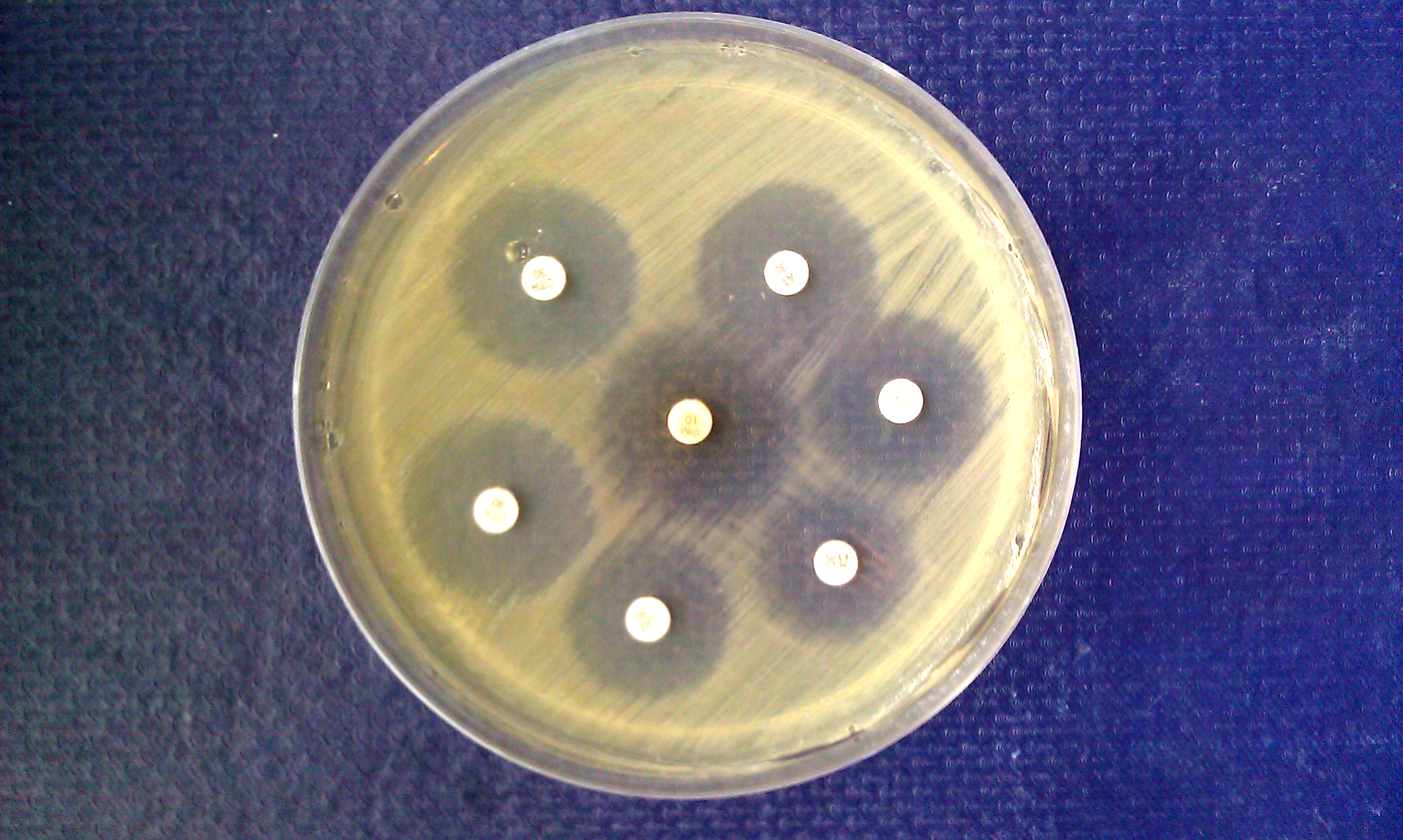
s are used to test the ability for an antibiotic to inhibit growth of a microorganism. This method, which is commonly used with
Mueller–Hinton agar
Mueller–Hinton agar is a microbiological growth medium that is commonly used for antibiotic susceptibility testing, specifically disk diffusion tests. It is also used to isolate and maintain ''Neisseria'' and ''Moraxella'' species.
It typi ...
, is used by evenly seeding bacteria over a petri dish and applying an antibiotic treated disk to the top of the agar. By observing the ring formed around the disk formed due to the lack of bacterial growth, the
zone of inhibition
The disk diffusion test (also known as the agar diffusion test, Kirby–Bauer test, disc-diffusion antibiotic susceptibility test, disc-diffusion antibiotic sensitivity test and KB test) is a culture-based microbiology assay used in diagnos ...
can be found, which is used to find the susceptibility of an organism to an antibiotic.
Bile esculin agar
Bile Esculin Agar (BEA) is a selective differential agar used to isolate and identify members of the genus ''Enterococcus'', formerly part of the "group D streptococci" (enterococci were reclassified in their own genus in 1984).
Composition and ...
The
bile esculin test is used to differentiate members of the genus ''Enterococcus'' from ''Streptococcus''.
Bile solubility
Bile solubility is used to test for ''Streptococcus Pneumoniae'' due to their unique ability to be lysed by
sodium deoxycholate. Lysis indicates ''S. Pneumoniae'' while no lysis does not.
CAMP

A
CAMP test
The CAMP test (Christie–Atkins–Munch-Peterson) is a test to identify group B β-hemolytic streptococci (''Streptococcus agalactiae'')
based on their formation of a substance (CAMP factor) that enlarges the area of hemolysis formed by the β ...
is used to differentiate between ''
Streptococcus agalactiae
''Streptococcus agalactiae'' (also known as group B streptococcus or GBS) is a gram-positive coccus (round bacterium) with a tendency to form chains (as reflected by the genus name ''Streptococcus''). It is a beta-hemolytic, catalase-negative, a ...
'' and other species of
beta-hemolytic
''Streptococcus'' is a genus of gram-positive ' (plural ) or spherical bacteria that belongs to the family Streptococcaceae, within the order Lactobacillales (lactic acid bacteria), in the phylum Bacillota. Cell division in streptococci occurs ...
''Streptococcus.'' This biochemical test uses the fact that ''Streptococcus agalactiae'' excretes a CAMP substance, making it slightly more hemolytic, which can be observed on blood agar media.
Catalase
The catalase test tests whether a microbe produces the enzyme catalase, which catalyzes the breakdown of hydrogen peroxide. Smearing a colony sample onto a glass slide and adding a solution of hydrogen peroxide (3% H
2O
2) will indicate whether the enzyme is present or not. Bubbling is a positive test while nothing happening is a negative result.
Cetrimide agar
Cetrimide agar Cetrimide agar is a type of agar used for the selective isolation of the gram-negative bacterium, ''Pseudomonas aeruginosa''.http://www.bd.com/ds/productCenter/297882.asp "Cetrimide Agar Base • Pseudosel Agar". Accessed May 3, 2008. As the name s ...
slants is a selective agar used to isolate ''
Pseudomonas aeruginosa
''Pseudomonas aeruginosa'' is a common encapsulated, gram-negative, aerobic–facultatively anaerobic, rod-shaped bacterium that can cause disease in plants and animals, including humans. A species of considerable medical importance, ''P. aerugi ...
''.
CLO tests
The
CLO test is used to diagnose ''H. Pylori'' in patient biopsies. A sample of the biopsy is places in a medium containing
urea
Urea, also known as carbamide, is an organic compound with chemical formula . This amide has two amino groups (–) joined by a carbonyl functional group (–C(=O)–). It is thus the simplest amide of carbamic acid.
Urea serves an important r ...
, which ''H. Pylori'' can use in some of its biochemical pathways. Consumption of urea indicates a positive test result.
Coagulase
The
coagulase test
Coagulase is a protein enzyme produced by several microorganisms that enables the conversion of fibrinogen to fibrin. In the laboratory, it is used to distinguish between different types of ''Staphylococcus'' isolates. Importantly, '' S. aureus'' ...
determines whether an organism can produce the enzyme coagulase, which causes the
fibrin
Fibrin (also called Factor Ia) is a fibrous, non-globular protein involved in the clotting of blood. It is formed by the action of the protease thrombin on fibrinogen, which causes it to polymerize. The polymerized fibrin, together with platele ...
to clot. Inoculating a plasma test tube with the microbe indicates whether coagulase is produced. A clot indicates the presence of coagulase, while no clot indicates the lack of coagulase.
DNA hydrolysis
DNase agar is used to test whether a microbe can produce the
exoenzyme
An exoenzyme, or extracellular enzyme, is an enzyme that is secreted by a cell and functions outside that cell. Exoenzymes are produced by both prokaryotic and eukaryotic cells and have been shown to be a crucial component of many biological pr ...
deoxyribonuclease Deoxyribonuclease (DNase, for short) refers to a group of glycoprotein endonucleases which are enzymes that catalyze the hydrolytic cleavage of phosphodiester linkages in the DNA backbone, thus degrading DNA. The role of the DNase enzyme in cells ...
(DNase), which hydrolyzes DNA.
Methyl green
Methyl green (CI 42585) is a cationic or positive charged stain, related to Ethyl Green, that has been used for staining DNA since the 19th century. It has been used for staining cell nuclei either as a part of the classical Unna-Pappenheim sta ...
is used as an indicator in the growth medium because it is a cation that provides an opaqueness to a medium with the presence of negatively charged DNA strands. When DNA is cleaved, the media becomes clear, showing the presence of DNase activity. DNA hydrolysis is tested by growing an organism on a DNase Test Agar plate (providing nutrients and DNA) and then checking the plate for hydrolysis. The agar plate has DNA-methyl green complex, and if the organism on the agar does hydrolyze DNA then the green color fades and the colony is surrounded by a colorless zone.
Gelatin
The
gelatin test is used to analyze whether a microbe can hydrolyze gelatin with the enzyme
gelatinase Gelatinases are enzymes capable of degrading gelatin.
Gelatinases are expressed in several bacteria including ''Pseudomonas aeruginosa'' and ''Serratia marcescens''.
In humans, the gelatinases are matrix metalloproteinases MMP2 and MMP9
Matrix ...
. The gelatin makes the agar solid, so if an organism can produce gelatinase and consume gelatin as an energy and carbon source, the agar will become liquid during growth.
Gonocheck II
The Gonochek II test, a commercial biochemical test, is used to differentiate between ''
Neisseria lactamica'', ''
Neisseria meningitidis
''Neisseria meningitidis'', often referred to as meningococcus, is a Gram-negative bacterium that can cause meningitis and other forms of meningococcal disease such as meningococcemia, a life-threatening sepsis. The bacterium is referred to as a ...
'', ''N. gonorrhoeae'' and ''Moraxella catarrhalis.'' The principle behind this test is to use enzymes native to the organism to create a colored product in the presence of foreign molecules. The chemical
galactosidase, turning the solution into a blue color. Gamma-glutamyl-p-nitroanilide is added to the solution to indicate whether the bacteria is ''N. meningitides,'' which hydrolyzes the molecule with the enzyme gamma-glutamylaminopeptidase, producing a yellow end-product. Prolyl-4-methoxynaphthylamide is in the solution to identify ''N. gonorrhoeae'' because of its ability to hydrolyze the molecule with the enzyme hydroxyprolylaminopeptidase, creating a red-pink derivative. ''M. catarrhalis'' contains none of these enzymes, rendering the solution colorless. This process of identification takes approximately 30 minutes in total.
Hippurate
The Hippurate diagnostic test is used to differentiate between ''Gardnerella vaginalis'', ''Campylobacter jejuni,'' ''Listeria monocytogenes'' and group B streptococci using the chemical Hippurate. The Hippurate hydrolysis pathway, capable by organisms with the necessary enzymes, produces glycine as a byproduct. Using the indicator
ninhydrin
Ninhydrin (2,2-dihydroxyindane-1,3-dione) is an organic compound with the formula C6H4(CO)2C(OH)2. It is used to detect ammonia and amines. Upon reaction with these amines, ninhydrin gets converted into deep blue or purple derivatives, which are ...
, which changes color in the presence of glycine, will display either a colorless product, a negative result, of a dark blue color, a positive result.
Indole butyrate disk
An
indole butyrate disc is used to differentiate between ''
Neisseria gonorrhoeae
''Neisseria gonorrhoeae'', also known as ''gonococcus'' (singular), or ''gonococci'' (plural), is a species of Gram-negative diplococci bacteria isolated by Albert Ludwig Sigesmund Neisser, Albert Neisser in 1879. It causes the sexually transmit ...
'' (negative result) and ''
Moraxella catarrhalis
''Moraxella catarrhalis'' is a fastidious, nonmotile, Gram-negative, aerobic, oxidase-positive diplococcus that can cause infections of the respiratory system, middle ear, eye, central nervous system, and joints of humans. It causes the infec ...
'' (positive result). This test involves a
butyrate
The conjugate acids are in :Carboxylic acids.
{{Commons category, Carboxylate ions, Carboxylate anions
Carbon compounds
Oxyanions ...
disk, which when smeared with a culture, will change color for a positive result after 5 minutes of incubation. A blue color is the result of a positive test.
Lysine iron agar slant
The
lysine iron agar slant test is used to tell whether an organism can
decarboxylate lysine
Lysine (symbol Lys or K) is an α-amino acid that is a precursor to many proteins. It contains an α-amino group (which is in the protonated form under biological conditions), an α-carboxylic acid group (which is in the deprotonated −C ...
and/or produce
hydrogen sulfide
Hydrogen sulfide is a chemical compound with the formula . It is a colorless chalcogen-hydride gas, and is poisonous, corrosive, and flammable, with trace amounts in ambient atmosphere having a characteristic foul odor of rotten eggs. The unde ...
.
Lysostaphin
The
lysostaphin test is used to differentiate between ''Staphylococcus'' and ''Micrococcus'' bacteria.
Lysostaphin
Lysostaphin (, ''glycyl-glycine endopeptidase'') is a '' Staphylococcus simulans'' metalloendopeptidasecrystal structure of lysostaphin. It can function as a bacteriocin (antimicrobial) against '' Staphylococcus aureus''.
Lysostaphin is a 27 KDa g ...
can
lyse Lyse may refer to:
* Lyse Abbey, a former Cistercian abbey in Norway
* Lyse, an alternative name of Lysebotn, Norway
* Lyse Energi, a Norwegian power company
* Łyse, Masovian Voivodeship, a village in east-central Poland
* Łyse, Podlaskie Voivode ...
''Staphylococcus,'' but ''Micrococcus'' bacteria are resistant to the chemical.
Methyl red test

The
methyl red test
Methyl red (2-(''N'',''N''-dimethyl-4-aminophenyl) azobenzenecarboxylic acid), also called C.I. Acid Red 2, is an indicator dye that turns red in acidic solutions. It is an azo dye, and is a dark red crystalline powder. Methyl red is a pH indi ...
is used to analyze whether a bacterium produces acids through sugar fermentation.
Microdase
Microdase is a modified oxidase test used to differentiate ''
Micrococcus
''Micrococcus'' (mi’ krō kŏk’ Əs) is a genus of bacteria in the Micrococcaceae family. ''Micrococcus'' occurs in a wide range of environments, including water, dust, and soil. Micrococci have Gram-positive spherical cells ranging from abo ...
'' from ''
Staphylococcus
''Staphylococcus'' is a genus of Gram-positive bacteria in the family Staphylococcaceae from the order Bacillales. Under the microscope, they appear spherical (cocci), and form in grape-like clusters. ''Staphylococcus'' species are facultative ...
'' by testing for the presence of
cytochrome c
The cytochrome complex, or cyt ''c'', is a small hemeprotein found loosely associated with the inner membrane of the mitochondrion. It belongs to the cytochrome c family of proteins and plays a major role in cell apoptosis. Cytochrome c is hig ...
. A positive result produces a dark color around the inoculant while negative result produces no color change.
Nitrite test
The
nitrite test
A nitrite test is a chemical test used to determine the presence of nitrite ion in solution.
Chemical methods Using iron(II) sulfate
A simple nitrite test can be performed by adding 4 M sulfuric acid to the sample until acidic, and then adding 0.1 ...
is commonly used to diagnose urinary tract infections by measuring the concentrations of nitrite in solution, indicating the presence of a gram-negative organism. A simple nitrite test can be performed by adding 4 M sulfuric acid to the sample until acidic, and then adding 0.1 M iron (II) sulfate to the solution. A positive test for nitrite is indicated by a dark brown solution, arising from the iron-nitric oxide complex ion.
Oxidase
The oxidase test indicates whether a microbe is aerobic. By using the chemical
N,N,N,N-tetramethyl-1,4-phenylendiamin, an electron acceptor that changes color when oxidized by
cytochrome c oxidase
The enzyme cytochrome c oxidase or Complex IV, (was , now reclassified as a translocasEC 7.1.1.9 is a large transmembrane protein complex found in bacteria, archaea, and mitochondria of eukaryotes.
It is the last enzyme in the respiratory electr ...
, one can deduce whether a microbe can perform aerobic respiration. A color change to purple indicates oxidative respiration while no color change provides evidence that the organism does not have cytochrome c oxidase.
Phenylalanine deaminase
The
phenylalanine deaminase
The enzyme phenylalanine ammonia lyase (EC 4.3.1.24) catalyzes the conversion of L-phenylalanine to ammonia and ''trans''-cinnamic acid.:
:L-phenylalanine = ''trans''-cinnamate + NH3
Phenylalanine ammonia lyase (PAL) is the first and committed ...
test is used to tell whether an organism can produce the enzyme deaminase. Deaminase is the enzyme that can deaminate the amino acid phenylalanine into the products ammonia and
phenylpyruvic acid
Phenylpyruvic acid is the organic compound with the formula C6H5CH2C(O)CO2H. It is a keto acid.
Occurrence and properties
The compound exists in equilibrium with its E- and Z-enol tautomers. It is a product from the oxidative deamination of phe ...
. The test is performed by adding phenylalanine to the growth medium and allowing growth to occur. After incubation, 10%
ferric chloride
Iron(III) chloride is the inorganic compound with the formula . Also called ferric chloride, it is a common compound of iron in the +3 oxidation state. The anhydrous compound is a crystalline solid with a melting point of 307.6 °C. The colo ...
is added to the solution, which will react with phenylpyruvic acid in solution to make a dark green color, resulting in a positive test result.
PYR
The PYR test is used to check if an organism has enzymes to
hydrolyze
Hydrolysis (; ) is any chemical reaction in which a molecule of water breaks one or more chemical bonds. The term is used broadly for substitution, elimination, and solvation reactions in which water is the nucleophile.
Biological hydrolysis ...
L-pyrrolidonyl- β-napthylamide. A positive result indicates that the organism is either group A ''
streptococcus
''Streptococcus'' is a genus of gram-positive ' (plural ) or spherical bacteria that belongs to the family Streptococcaceae, within the order Lactobacillales (lactic acid bacteria), in the phylum Bacillota. Cell division in streptococci occurs ...
'' and/or group D ''
enterococcus
''Enterococcus'' is a large genus of lactic acid bacteria of the phylum Bacillota. Enterococci are gram-positive cocci that often occur in pairs (diplococci) or short chains, and are difficult to distinguish from streptococci on physical charact ...
''.
Reverse CAMP
The
reverse CAMP test utilizes the synergetic hemolytic abilities of the CAMP factor produced by ''Streptococcus agalactiae'' with the α-toxin produced by ''
Clostridium perfringens
''Clostridium perfringens'' (formerly known as ''C. welchii'', or ''Bacillus welchii'') is a Gram-positive, rod-shaped, anaerobic, spore-forming pathogenic bacterium of the genus '' Clostridium''. ''C. perfringens'' is ever-present in nature an ...
''. Streaking these two organisms perpendicular to each other on a blood agar plate will yield a “bow-tie” clearing of the blood agar by the hemolytic capabilities of the two organisms’ toxins. Incubation requires 24 hours at 37 °C.
Simmons' citrate agar
Simmons' citrate agar is used to test whether an organism can utilize
citrate
Citric acid is an organic compound with the chemical formula HOC(CO2H)(CH2CO2H)2. It is a colorless weak organic acid. It occurs naturally in citrus fruits. In biochemistry, it is an intermediate in the citric acid cycle, which occurs in t ...
for its sole carbon source.
Spot indole

The spot indole test is used to determine if a microbe can
deaminate tryptophan
Tryptophan (symbol Trp or W)
is an α-amino acid that is used in the biosynthesis of proteins. Tryptophan contains an α-amino group, an α- carboxylic acid group, and a side chain indole, making it a polar molecule with a non-polar aromatic ...
to produce
indole
Indole is an aromatic heterocyclic organic compound with the formula C8 H7 N. It has a bicyclic structure, consisting of a six-membered benzene ring fused to a five-membered pyrrole ring. Indole is widely distributed in the natural environmen ...
. This test is performed by saturating a piece of filter paper with Indole Kovacs Reagent and scraping a portion of microbe onto the paper. A color to a pink-red color indicates a positive result while no color change indicates the lack of
tryptophanase
The enzyme tryptophanase () catalyzes the chemical reaction
:L-tryptophan + H2O \rightleftharpoons indole + pyruvate + NH3
This enzyme belongs to the family of lyases, specifically in the "catch-all" class of carbon-carbon lyases. The syst ...
.
Sulphide indole motility medium
The
sulfide indole motility medium is a three-part test for an organism’s ability to reduce sulfates, produce indoles, and motile ability.
TSI slant
The
triple sugar iron (TSI) test is a differential media used to tell whether an organism can ferment glucose, sucrose, and/or lactose and whether an organism can produce hydrogen sulfide gas.
Urea agar slant

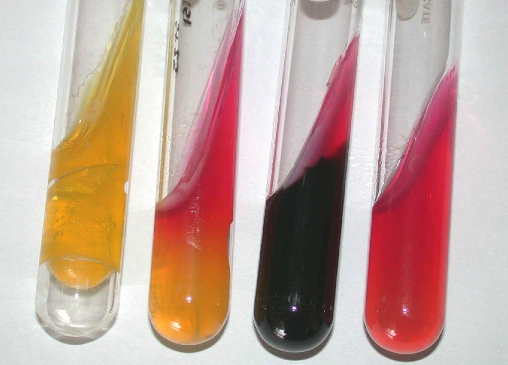
The urease agar slant is used to measure an organism’s ability to produce
urease
Ureases (), functionally, belong to the superfamily of amidohydrolases and phosphotriesterases. Ureases are found in numerous bacteria, fungi, algae, plants, and some invertebrates, as well as in soils, as a soil enzyme. They are nickel-containin ...
, an enzyme capable to digesting urea in carbon dioxide and ammonia through hydrolysis. Because ammonia is alkaline, the media contains phenol red, an indicator that changes from orange to pink when a pH increases above 8.1. When ammonia is increased to high enough concentrations, the media will change to a pink color, indicating the presence of urease production.
Voges–Proskauer test
The
Voges-Proskauer test detects whether a bacterium is producing the product
acetoin
Acetoin, also known as 3-hydroxybutanone or acetyl methyl carbinol, is an organic compound with the formula CH3CH(OH)C(O)CH3. It is a colorless liquid with a pleasant, buttery odor. It is chiral. The form produced by bacteria is (''R'')-acetoin. ...
from the digestion of glucose.
Cellular fatty acid based identification
Mycolic acid analysis
Mycolic acid
Mycolic acids are long fatty acids found in the cell walls of the Mycolata taxon, a group of bacteria that includes ''Mycobacterium tuberculosis'', the causative agent of the disease tuberculosis. They form the major component of the cell wall of ...
analysis has been an evolving field of study for
gas-liquid chromatography
Gas chromatography (GC) is a common type of chromatography used in analytical chemistry for separating and analyzing compounds that can be vaporized without decomposition. Typical uses of GC include testing the purity of a particular substance, ...
, as it offers a solution to slow growth rates in ''
Mycobacterium
''Mycobacterium'' is a genus of over 190 species in the phylum Actinomycetota, assigned its own family, Mycobacteriaceae. This genus includes pathogens known to cause serious diseases in mammals, including tuberculosis ('' M. tuberculosis'') and ...
.'' Mycolic acid is a fatty acid found in the disease
tuberculosis
Tuberculosis (TB) is an infectious disease usually caused by '' Mycobacterium tuberculosis'' (MTB) bacteria. Tuberculosis generally affects the lungs, but it can also affect other parts of the body. Most infections show no symptoms, in ...
, offering a chemical target for diagnosticians to look for.
Nucleic acid extraction techniques
Cesium chloride / Ethidium bromide density gradient centrifugation

With high speed
buoyant density ultracentrifugation
Buoyant density centrifugation (also isopycnic centrifugation or equilibrium density-gradient centrifugation) uses the concept of buoyancy to separate molecules in solution by their differences in density.
Implementation
Historically a caesium c ...
, a density gradient is created with
caesium chloride
Caesium chloride or cesium chloride is the inorganic compound with the formula Cs Cl. This colorless salt is an important source of caesium ions in a variety of niche applications. Its crystal structure forms a major structural type where each ...
in water. DNA will go to the density that reflects its own, and
ethidium bromide
Ethidium bromide (or homidium bromide, chloride salt homidium chloride) is an intercalating agent commonly used as a fluorescent tag (nucleic acid stain) in molecular biology laboratories for techniques such as agarose gel electrophoresis. It i ...
is then added to enhance the visuals the nucleic acid band provides.
Magnetic bead method
New extraction techniques have been developed using
magnetic bead
Magnetism is the class of physical attributes that are mediated by a magnetic field, which refers to the capacity to induce attractive and repulsive phenomena in other entities. Electric currents and the magnetic moments of elementary particle ...
s for the purification of nucleic acids by taking advantage of the charged and polymeric nature of long strand of DNA. Beads are both uncoated to increase surface are and yield, while others are more selective by being coated with functional groups that interact with the
polymer
A polymer (; Greek '' poly-'', "many" + ''-mer'', "part")
is a substance or material consisting of very large molecules called macromolecules, composed of many repeating subunits. Due to their broad spectrum of properties, both synthetic a ...
s present in microbes. One common method is to use polyethylene glycol to drive DNA binding to the magnetic beads. The molecular weight and concentration of the PEG will control what molecular weight DNA binds.
Phenol–chloroform extraction
Phenol-Chloroform extraction is a liquid-liquid method used by biochemists to separate nucleic acids from proteins and lipids after cells have been lysed. This method has fallen out of favor with scientists and microbiologists as there are easier methods available which require less hazardous chemicals.
Solid phase extraction
Solid phase extraction
Solid-phase extraction (SPE) is an extractive technique by which compounds that are dissolved or suspended in a liquid mixture are separated from other compounds in the mixture according to their physical and chemical properties. Analytical labor ...
which separates long polymers like DNA from other substances found in the cells. This is similar to magnetic beads, where the solid phase is fixed and selectively binds a cellular component, allowing for its isolation.
Methods with electrophoretic outputs
Gel electrophoresis
Gel electrophoresis is a method for separation and analysis of biomacromolecules ( DNA, RNA, proteins, etc.) and their fragments, based on their size and charge. It is used in clinical chemistry to separate proteins by charge or size (IEF ...
is a technique to separate
macromolecule
A macromolecule is a very large molecule important to biophysical processes, such as a protein or nucleic acid. It is composed of thousands of covalently bonded atoms. Many macromolecules are polymers of smaller molecules called monomers. The ...
s by taking advantage of the charge on many of the molecules found in nucleic acids and protein. This is also the key method for Sanger sequencing. Fluorescent-labeled DNA fragments move through a polymer and are separated with one base precision. A laser excites the fluorescent tag and is captured by a camera. The result is an electropherogram which reads the DNA sequence.
Restriction enzyme based
Optical mapping
Optical mapping Optical mapping is a technique for constructing ordered, genome-wide, high-resolution restriction maps from single, stained molecules of DNA, called "optical maps". By mapping the location of restriction enzyme sites along the unknown DNA of an orga ...
is a technique using multiple restriction enzymes to create a genomic “barcode” which can be referenced back to diagnose an unknown microbe.
Pulsed-field gel electrophoresis
Pulsed-field gel electrophoresis
Pulsed field gel electrophoresis is a technique used for the separation of large DNA molecules by applying to a gel matrix an electric field that periodically changes direction.
Historical background
Standard gel electrophoresis techniques for ...
is a technique used to separate large DNA in an electric field that periodically changes direction. By cutting segments of the DNA with
restriction enzyme
A restriction enzyme, restriction endonuclease, REase, ENase or'' restrictase '' is an enzyme that cleaves DNA into fragments at or near specific recognition sites within molecules known as restriction sites. Restriction enzymes are one class o ...
s, pulse-field can be used to separate out the segments of DNA.
Restriction enzymes then gel electrophoresis
Restriction enzymes are first used to recognize and then cut specific nucleic acid sequences. These cut pieces of DNA can be run through a gel electrophoresis to allow diagnostics of the organism by referencing back to previous gel electrophoresis results.
Ribotyping
Ribotyping is a rapid automated method for microbial diagnostics, testing for rRNA in bacteria using restriction enzyme digestion and Southern blot technology.
PCR-based
Multiple loci VNTR analysis
Multiple-locus VNTR analysis is a test used to detect
variable number tandem repeats, which act as a
DNA fingerprint
DNA profiling (also called DNA fingerprinting) is the process of determining an individual's DNA characteristics. DNA analysis intended to identify a species, rather than an individual, is called DNA barcoding.
DNA profiling is a forensic tec ...
in microbial diagnostics.
DNA sequence-based methods
Multi-locus sequence typing
Multilocus sequence typing
Multilocus sequence typing (MLST) is a technique in molecular biology for the typing of multiple loci, using DNA sequences of internal fragments of multiple housekeeping genes to characterize isolates of microbial species.
The first MLST scheme ...
(MLST) is the sequencing of numerous loci to diagnose an organism by comparing DNA sequences to a database of known organisms. This method is often used to compare isolates or strains of the same species to see if they are indistinguishable or different from each other. This is common for tracking food-borne illnesses and public health outbreaks. Most MLST assays are published in scientific journals so consistent methods are used worldwide. There are also public databases available for tracking and comparisons.
Single-locus sequence typing
Single-locus sequence typing (SLST) is the sequencing of a single locus of an organism to produce data that can be used for strain-level comparisons between isolates of the same species.
Genotypic identifications
For bacterial identifications, microbiologists sequence the 16S rRNA gene and for fungal identifications, sequence the ITS regions. Both regions are part of the ribosomal operon so they are well-conserved but provide enough variation to allow for speciation. Accurate identifications require high quality sequence data, a robust data analysis, and a broad microbial database of known organisms. It is also useful to use a Neighbor Joining tree or some other phylogenetic approach to make the identification.
Whole genome sequencing (WGS)
Whole genome sequencing
Whole genome sequencing (WGS), also known as full genome sequencing, complete genome sequencing, or entire genome sequencing, is the process of determining the entirety, or nearly the entirety, of the DNA sequence of an organism's genome at a s ...
and genomics applications can be used for large-scale alignment and comparative analysis with both bacteria and fungi. WGS can be used to diagnose, identify, or characterize an organism down to the individual base pairs by sequencing the entire genome.
WGS can also be used to compare the genomes or average nucleotide identity (ANI) of the shared genes between two strains and can be a robust way to compare genetic relatedness and if often used for investigating organisms involved in foodborne illness and other outbreaks.
References
{{Clinical microbiology techniques
Clinical pathology
Applied microbiology
 Anaerobic bacteria collection can come from a variety of sources in patient samples, including blood, bile, bone marrow,

 Antibiotic disks are used to test the ability for an antibiotic to inhibit growth of a microorganism. This method, which is commonly used with
  The
The  The urease agar slant is used to measure an organism’s ability to produce
  With high speed