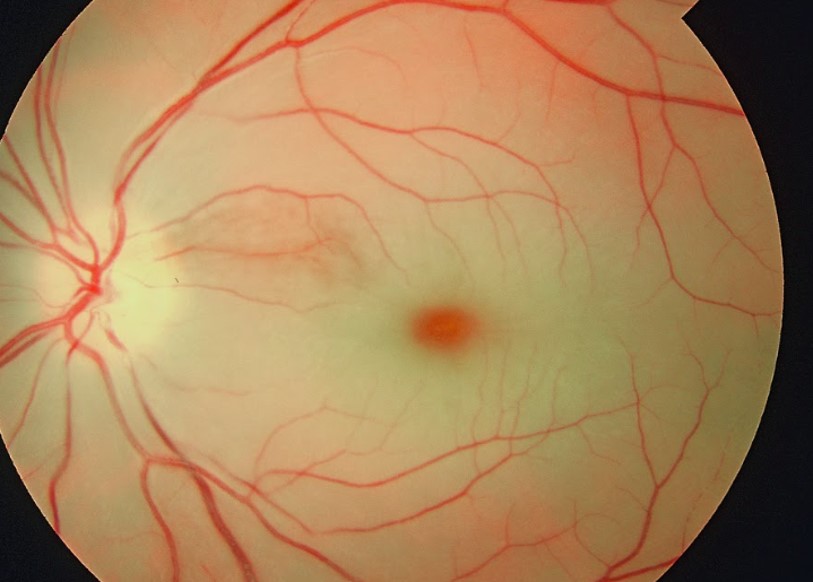
Central retinal artery occlusion on:
[Wikipedia]
[Google]
[Amazon]
Central retinal artery occlusion (CRAO) is a disease of the eye where the flow of blood through the
 Central retinal artery occlusion is characterized by painless, acute vision loss in one eye. Upon fundoscopic exam, one would expect to find:

 One diagnostic method for the confirmation of CRAO is
central retinal artery
The central retinal artery (retinal artery) branches off the ophthalmic artery, running inferior to the optic nerve within its dural sheath to the eyeball.
Structure
The central retinal artery pierces the eyeball close to the optic nerve, sendin ...
is blocked (occluded). There are several different causes of this occlusion; the most common is carotid artery atherosclerosis
Atherosclerosis is a pattern of the disease arteriosclerosis in which the wall of the artery develops abnormalities, called lesions. These lesions may lead to narrowing due to the buildup of atheromatous plaque. At onset there are usually no s ...
.
Signs and symptoms
 Central retinal artery occlusion is characterized by painless, acute vision loss in one eye. Upon fundoscopic exam, one would expect to find:
 cherry-red spot
A cherry-red spot is a finding in the macula of the eye in a variety of lipid storage disorders and in central retinal artery occlusion.
It describes the appearance of a small circular choroid shape as seen through the fovea centralis.
Its appe ...
(90%) (a morphologic description in which the normally red background of the choroid is sharply outlined by the swollen opaque retina in the central retina), retinal opacity in the posterior pole (58%), pallor (39%), retinal arterial attenuation (32%), and optic disk edema (22%). During later stages of onset, one may also find plaques, emboli
An embolism is the lodging of an embolus, a blockage-causing piece of material, inside a blood vessel. The embolus may be a blood clot (thrombus), a fat globule ( fat embolism), a bubble of air or other gas (gas embolism), amniotic fluid ( amni ...
, and optic atrophy Optic neuropathy is damage to the optic nerve from any cause. The optic nerve is a bundle of millions of fibers in the retina that sends visual signals to the brain.
Damage and death of these nerve cells, or neurons, leads to characteristic featu ...
.
Diagnosis

 One diagnostic method for the confirmation of CRAO is
 Fluorescein angiography
Fluorescein angiography (FA), fluorescent angiography (FAG), or fundus fluorescein angiography (FFA) is a technique for examining the circulation of the retina and choroid (parts of the fundus) using a fluorescent dye and a specialized camera. S ...
, it is used to examine the retinal artery filling time after the fluorescein dye is injected into the peripheral venous system
Veins are blood vessels in humans and most other animals that carry blood towards the heart. Most veins carry deoxygenated blood from the tissues back to the heart; exceptions are the pulmonary and umbilical veins, both of which carry oxygenated ...
. In an eye with CRAO some branches of the retinal artery may not fill or the time it takes for the branches of the retinal artery to fill will be increased, which is visualized by the leading edge of the fluorescein moving slower than normal through the retinal artery branches to the edges of the retina. Fluorescein angiography can also be used to determine the extent of the occlusion as well as classify it into one of four types non-arteritic CRAO, non-arteritic CRAO with cilioretinal artery sparing, transient non-arteritic CRAO and arteritic CRAO. Optical coherence tomography (OCT) may also be used to confirm the diagnosis of CRAO.
Causes
CRAO can be classified based on it pathogenesis, as arteritic versus non-arteritic. Non-arteritic CRAO is most commonly caused by anembolus
An embolus (; plural emboli; from the Greek ἔμβολος "wedge", "plug") is an unattached mass that travels through the bloodstream and is capable of creating blockages. When an embolus occludes a blood vessel, it is called an embolism or emb ...
and occlusion at the narrowest part of the carotid retinal artery due to plaques in the carotid artery resulting in carotid retinal artery atherosclerosis
Atherosclerosis is a pattern of the disease arteriosclerosis in which the wall of the artery develops abnormalities, called lesions. These lesions may lead to narrowing due to the buildup of atheromatous plaque. At onset there are usually no s ...
. Further causes of non-arteritic CRAO may include vasculitis
Vasculitis is a group of disorders that destroy blood vessels by inflammation. Both arteries and veins are affected. Lymphangitis (inflammation of lymphatic vessels) is sometimes considered a type of vasculitis. Vasculitis is primarily caused ...
and chronic systemic autoimmune diseases. Arteritic CRAO is most commonly caused by giant cell arteritis
Giant cell arteritis (GCA), also called temporal arteritis, is an inflammatory autoimmune disease of large blood vessels. Symptoms may include headache, pain over the temples, flu-like symptoms, double vision, and difficulty opening the mouth. ...
. Other causes can include dissecting aneurysms
An aneurysm is an outward bulging, likened to a bubble or balloon, caused by a localized, abnormal, weak spot on a blood vessel wall. Aneurysms may be a result of a hereditary condition or an acquired disease. Aneurysms can also be a nidus ( ...
and arterial spasms, and as a complication of patient positioning causing external compression of the eye compressing flow to the central retinal artery (e.g. in spine surgeries in the prone position).Central and branch retinal artery occlusion. Uptodate.com. Mar 14, 2012.
Mechanism
Theophthalmic artery
The ophthalmic artery (OA) is an artery of the head. It is the first branch of the internal carotid artery distal to the cavernous sinus. Branches of the ophthalmic artery supply all the structures in the orbit around the eye, as well as some s ...
branches off into the central retinal artery
The central retinal artery (retinal artery) branches off the ophthalmic artery, running inferior to the optic nerve within its dural sheath to the eyeball.
Structure
The central retinal artery pierces the eyeball close to the optic nerve, sendin ...
which travels with the optic nerve
In neuroanatomy, the optic nerve, also known as the second cranial nerve, cranial nerve II, or simply CN II, is a paired cranial nerve that transmits visual information from the retina to the brain. In humans, the optic nerve is derived fro ...
until it enters the eye. This central retinal artery provides nutrients to the retina
The retina (from la, rete "net") is the innermost, light-sensitive layer of tissue of the eye of most vertebrates and some molluscs. The optics of the eye create a focused two-dimensional image of the visual world on the retina, which then ...
of the eye, more specifically the inner retina and the surface of the optic nerve. Variations, such as branch retinal artery occlusion
Branch retinal artery occlusion (BRAO) is a rare retinal vascular disorder in which one of the branches of the central retinal artery is obstructed.
Presentation
Abrupt painless loss of vision in the visual field corresponding to territory of the ...
, can also occur. Central retinal artery occlusion is most often due to emboli blocking the artery and therefore prevents the artery from delivering nutrients to most of the retina. These emboli originate from the carotid arteries most of the time but in 25% of cases, this is due to plaque build-up in the ophthalmic artery. The most frequent site of blockage is at the most narrow part of the artery which is where the artery pierces the dura covering the optic nerve. Some people have cilioretinal arterial branches, which may or may not be included in the blocked portion.
Treatment
While no treatment has been clearly demonstrated to be benefit for CRAO in large systematic reviews of randomized clinical trials, many of the following are frequently used: * Loweringintraocular pressure
Intraocular pressure (IOP) is the fluid pressure inside the eye. Tonometry is the method eye care professionals use to determine this. IOP is an important aspect in the evaluation of patients at risk of glaucoma. Most tonometers are calibrated t ...
;
* Dilating the CRA;
* Increasing oxygenation;
* Isovolemic hemodilution;
* Anticoagulation;
* Dislodging or fragmenting thrombus or embolus;
* Thrombolysis
Thrombolysis, also called fibrinolytic therapy, is the breakdown (lysis) of blood clots formed in blood vessels, using medication. It is used in ST elevation myocardial infarction, stroke, and in cases of severe venous thromboembolism (massive ...
; and
* Hyperbaric oxygen
Hyperbaric medicine is medical treatment in which an ambient pressure greater than sea level atmospheric pressure is a necessary component. The treatment comprises hyperbaric oxygen therapy (HBOT), the medical use of oxygen at an ambient pressure ...
.
To achieve the best outcome for a person with CRAO, it is important to identify the condition in a timely manner and to refer to the appropriate specialist.
Prognosis
The artery can re-canalize over time and the edema can clear. However, optic atrophy leads to permanent loss of vision. Irreversible damage to neural tissue can occur after approximately 15 minutes of complete blockage to the central retinal artery, but this time may vary between people. Two thirds of people experience 20/400 vision while only one in six will experience 20/40 vision or better.Kunimoto, Dr., Lecture, Vascular diseases of the retina, AT Still University SOMA, October 2012Epidemiology
The incidence of CRAO is approximately 1 in 100,000 people in the general population. Risk factors for CRAO include the following: being over 50 years of age, male gender, smoking, hypertension, tranexamic acid,diabetes mellitus
Diabetes, also known as diabetes mellitus, is a group of metabolic disorders characterized by a high blood sugar level ( hyperglycemia) over a prolonged period of time. Symptoms often include frequent urination, increased thirst and increased ...
, dyslipidemia
Dyslipidemia is an abnormal amount of lipids (e.g. triglycerides, cholesterol and/or fat phospholipids) in the blood. Dyslipidemia is a risk factor for the development of atherosclerotic cardiovascular disease ( ASCVD). ASCVD includes coronary ar ...
, angina, valvular disease, transient hemiparesis
Hemiparesis, or unilateral paresis, is weakness of one entire side of the body ('' hemi-'' means "half"). Hemiplegia is, in its most severe form, complete paralysis of half of the body. Hemiparesis and hemiplegia can be caused by different medi ...
, cancer, hypercoagulable
Thrombophilia (sometimes called hypercoagulability or a prothrombotic state) is an abnormality of blood coagulation that increases the risk of thrombosis (blood clots in blood vessels). Such abnormalities can be identified in 50% of people who ...
blood conditions, lupus, or a family history of cerebrovascular or cardiovascular issues. Additional risk factors include endocarditis
Endocarditis is an inflammation of the inner layer of the heart, the endocardium. It usually involves the heart valves. Other structures that may be involved include the interventricular septum, the chordae tendineae, the mural endocardium, or the ...
, atrial myxoma, inflammatory diseases of the blood vessels, and predisposition to forming blood clots.
See also
*Central retinal vein occlusion
Central retinal vein occlusion, also CRVO, is when the central retinal vein becomes occluded, usually through thrombosis. The central retinal vein is the venous equivalent of the central retinal artery and both may become occluded. Since the centra ...
* Branch retinal artery occlusion
Branch retinal artery occlusion (BRAO) is a rare retinal vascular disorder in which one of the branches of the central retinal artery is obstructed.
Presentation
Abrupt painless loss of vision in the visual field corresponding to territory of the ...
* Branch retinal vein occlusion
Branch retinal vein occlusion is a common retinal vascular disease of the elderly. It is caused by the occlusion of one of the branches of central retinal vein.
Signs and symptoms
Patients with branch retinal vein occlusion usually have a sudden ...
* Amaurosis fugax
Amaurosis fugax (Greek ''amaurosis'' meaning ''darkening'', ''dark'', or ''obscure'', Latin '' fugax'' meaning ''fleeting'') is a painless temporary loss of vision in one or both eyes.
Signs and symptoms
The experience of amaurosis fugax is clas ...
* Ocular ischemic syndrome
Ocular ischemic syndrome is the constellation of ocular signs and symptoms secondary to severe, chronic arterial hypoperfusion to the eye. Amaurosis fugax is a form of acute vision loss caused by reduced blood flow to the eye; it may be a warn ...
References
External links
{{Eye pathology Eye Disorders of choroid and retina