|
Myofilament
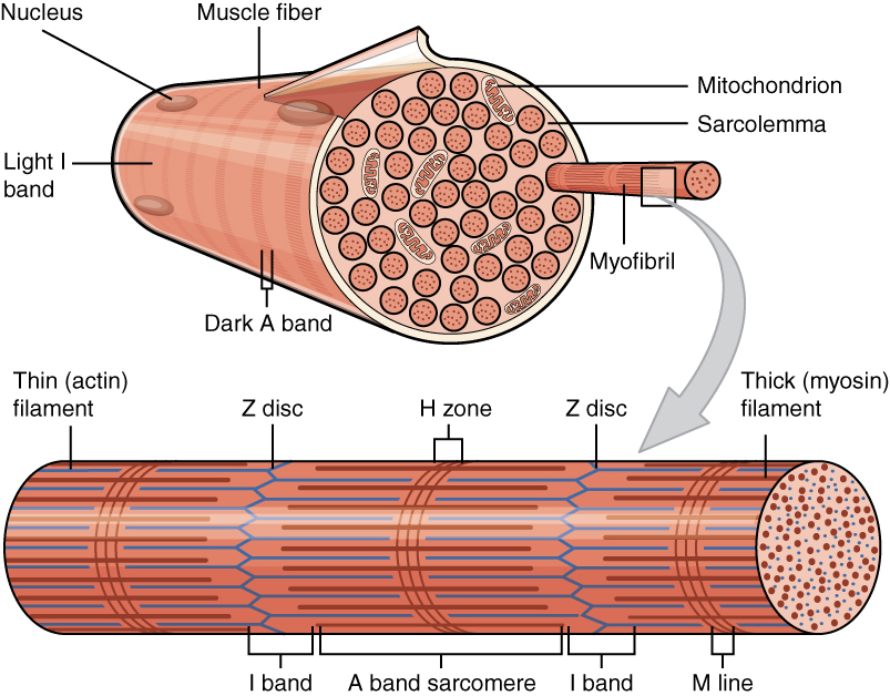
Myofilaments are the three protein filaments of myofibrils in muscle cells. The main proteins involved are myosin, actin, and titin. Myosin and actin are the ''contractile proteins'' and titin is an elastic protein. The myofilaments act together in muscle contraction, and in order of size are a thick one of mostly myosin, a thin one of mostly actin, and a very thin one of mostly titin. Types of muscle tissue are striated skeletal muscle and cardiac muscle, obliquely striated muscle (found in some invertebrates), and non-striated smooth muscle. Various arrangements of myofilaments create different muscles. Striated muscle has transverse bands of filaments. In obliquely striated muscle, the filaments are staggered. Smooth muscle has irregular arrangements of filaments. Structure There are three different types of myofilaments: thick, thin, and elastic filaments. *Thick filaments consist primarily of a type of myosin, a motor protein – myosin II. Each thick filament is approxi ... [...More Info...] [...Related Items...] OR: [Wikipedia] [Google] [Baidu] |
Muscle Contraction
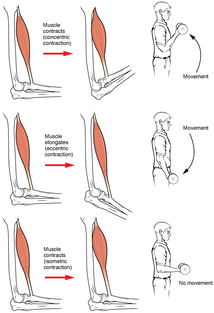
Muscle contraction is the activation of tension-generating sites within muscle cells. In physiology, muscle contraction does not necessarily mean muscle shortening because muscle tension can be produced without changes in muscle length, such as when holding something heavy in the same position. The termination of muscle contraction is followed by muscle relaxation, which is a return of the muscle fibers to their low tension-generating state. For the contractions to happen, the muscle cells must rely on the interaction of two types of filaments which are the thin and thick filaments. Thin filaments are two strands of actin coiled around each, and thick filaments consist of mostly elongated proteins called myosin. Together, these two filaments form myofibrils which are important organelles in the skeletal muscle system. Muscle contraction can also be described based on two variables: length and tension. A muscle contraction is described as isometric if the muscle tension changes ... [...More Info...] [...Related Items...] OR: [Wikipedia] [Google] [Baidu] |
Myofibril
A myofibril (also known as a muscle fibril or sarcostyle) is a basic rod-like organelle of a muscle cell. Skeletal muscles are composed of long, tubular cells known as muscle fibers, and these cells contain many chains of myofibrils. Each myofibril has a diameter of 1–2 micrometres. They are created during embryonic development in a process known as myogenesis. Myofibrils are composed of long proteins including actin, myosin, and titin, and other proteins that hold them together. These proteins are organized into thick, thin, and elastic myofilaments, which repeat along the length of the myofibril in sections or units of contraction called sarcomeres. Muscles contract by sliding the thick myosin, and thin actin myofilaments along each other. Structure Each myofibril has a diameter of between 1 and 2 micrometres (μm). The filaments of myofibrils, myofilaments, consist of three types, thick, thin, and elastic filaments. *Thin filaments consist primarily of the pro ... [...More Info...] [...Related Items...] OR: [Wikipedia] [Google] [Baidu] |
Myosin Head
The myosin head is the part of the thick myofilament made up of myosin that acts in muscle contraction, by sliding over thin myofilaments of actin. Myosin is the major component of the thick filaments and most myosin molecules are composed of a head, neck, and tail domain; the myosin head binds to thin filamentous actin, and uses ATP hydrolysis to generate force and "walk" along the thin filament. Myosin exists as a hexamer of two heavy chains, two alkali light chains, and two regulatory light chains. The heavy chain can be subdivided into the globular head at the N-terminal and the coiled-coil rod-like tail at the C-terminal, although some forms have a globular region in their C-terminal. There are many cell-specific isoforms of myosin heavy chains, coded for by a multi-gene family. Myosin interacts with actin to convert chemical energy, in the form of ATP, to mechanical energy. The 3-D structure of the head portion of myosin has been determined and a model for actin-myosin comp ... [...More Info...] [...Related Items...] OR: [Wikipedia] [Google] [Baidu] |
Sarcolemma
The sarcolemma (''sarco'' (from ''sarx'') from Greek; flesh, and ''lemma'' from Greek; sheath) also called the myolemma, is the cell membrane surrounding a skeletal muscle fiber or a cardiomyocyte. It consists of a lipid bilayer and a thin outer coat of polysaccharide material (glycocalyx) that contacts the basement membrane. The basement membrane contains numerous thin collagen fibrils and specialized proteins such as laminin that provide a scaffold to which the muscle fiber can adhere. Through transmembrane proteins in the plasma membrane, the actin skeleton inside the cell is connected to the basement membrane and the cell's exterior. At each end of the muscle fiber, the surface layer of the sarcolemma fuses with a tendon fiber, and the tendon fibers, in turn, collect into bundles to form the muscle tendons that adhere to bones. The sarcolemma generally maintains the same function in muscle cells as the plasma membrane does in other eukaryote cells. It acts as a barrier betwe ... [...More Info...] [...Related Items...] OR: [Wikipedia] [Google] [Baidu] |
Synaptic Cleft
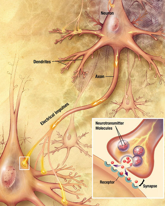
Chemical synapses are biological junctions through which neurons' signals can be sent to each other and to non-neuronal cells such as those in muscles or glands. Chemical synapses allow neurons to form circuits within the central nervous system. They are crucial to the biological computations that underlie perception and thought. They allow the nervous system to connect to and control other systems of the body. At a chemical synapse, one neuron releases neurotransmitter molecules into a small space (the synaptic cleft) that is adjacent to another neuron. The neurotransmitters are contained within small sacs called synaptic vesicles, and are released into the synaptic cleft by exocytosis. These molecules then bind to neurotransmitter receptors on the postsynaptic cell. Finally, the neurotransmitters are cleared from the synapse through one of several potential mechanisms including enzymatic degradation or re-uptake by specific transporters either on the presynaptic cell or ... [...More Info...] [...Related Items...] OR: [Wikipedia] [Google] [Baidu] |
Acetylcholine
Acetylcholine (ACh) is an organic chemical that functions in the brain and body of many types of animals (including humans) as a neurotransmitter. Its name is derived from its chemical structure: it is an ester of acetic acid and choline. Parts in the body that use or are affected by acetylcholine are referred to as cholinergic. Substances that increase or decrease the overall activity of the cholinergic system are called cholinergics and anticholinergics, respectively. Acetylcholine is the neurotransmitter used at the neuromuscular junction—in other words, it is the chemical that motor neurons of the nervous system release in order to activate muscles. This property means that drugs that affect cholinergic systems can have very dangerous effects ranging from paralysis to convulsions. Acetylcholine is also a neurotransmitter in the autonomic nervous system, both as an internal transmitter for the sympathetic nervous system and as the final product released by the par ... [...More Info...] [...Related Items...] OR: [Wikipedia] [Google] [Baidu] |
Neurotransmitter
A neurotransmitter is a signaling molecule secreted by a neuron to affect another cell across a synapse. The cell receiving the signal, any main body part or target cell, may be another neuron, but could also be a gland or muscle cell. Neurotransmitters are released from synaptic vesicles into the synaptic cleft where they are able to interact with neurotransmitter receptors on the target cell. The neurotransmitter's effect on the target cell is determined by the receptor it binds. Many neurotransmitters are synthesized from simple and plentiful precursors such as amino acids, which are readily available and often require a small number of biosynthetic steps for conversion. Neurotransmitters are essential to the function of complex neural systems. The exact number of unique neurotransmitters in humans is unknown, but more than 100 have been identified. Common neurotransmitters include glutamate, GABA, acetylcholine, glycine and norepinephrine. Mechanism and cycle ... [...More Info...] [...Related Items...] OR: [Wikipedia] [Google] [Baidu] |
Motor Neuron
A motor neuron (or motoneuron or efferent neuron) is a neuron whose cell body is located in the motor cortex, brainstem or the spinal cord, and whose axon (fiber) projects to the spinal cord or outside of the spinal cord to directly or indirectly control effector organs, mainly muscles and glands. There are two types of motor neuron – upper motor neurons and lower motor neurons. Axons from upper motor neurons synapse onto interneurons in the spinal cord and occasionally directly onto lower motor neurons. The axons from the lower motor neurons are efferent nerve fibers that carry signals from the spinal cord to the effectors. Types of lower motor neurons are alpha motor neurons, beta motor neurons, and gamma motor neurons. A single motor neuron may innervate many muscle fibres and a muscle fibre can undergo many action potentials in the time taken for a single muscle twitch. Innervation takes place at a neuromuscular junction and twitches can become superimposed as a ... [...More Info...] [...Related Items...] OR: [Wikipedia] [Google] [Baidu] |
Axon Terminal
Axon terminals (also called synaptic boutons, terminal boutons, or end-feet) are distal terminations of the telodendria (branches) of an axon. An axon, also called a nerve fiber, is a long, slender projection of a nerve cell, or neuron, that conducts electrical impulses called action potentials away from the neuron's cell body, or soma, in order to transmit those impulses to other neurons, muscle cells or glands. Neurons are interconnected in complex arrangements, and use electrochemical signals and neurotransmitter chemicals to transmit impulses from one neuron to the next; axon terminals are separated from neighboring neurons by a small gap called a synapse, across which impulses are sent. The axon terminal, and the neuron from which it comes, is sometimes referred to as the "presynaptic" neuron. Nerve impulse release Neurotransmitters are packaged into synaptic vesicles that cluster beneath the axon terminal membrane on the presynaptic side of a synapse. The axonal terminals ... [...More Info...] [...Related Items...] OR: [Wikipedia] [Google] [Baidu] |
Striated Muscle
Striations means a series of ridges, furrows or linear marks, and is used in several ways: * Glacial striation * Striation (fatigue), in material * Striation (geology), a ''striation'' as a result of a geological fault * Striation Valley, in Antarctica * In hyperbolic geometry, a ''striation'' is a reflection across two parallel mirrors. * In anatomy, striated muscle * Striations can be found in certain glasses. These have been caused by turbulent flow during teeming (pouring) of the glass. * Striations can be observed in clouds In meteorology, a cloud is an aerosol consisting of a visible mass of miniature liquid drop (liquid), droplets, ice crystals, frozen crystals, or other particulates, particles suspended in the atmosphere of a planetary body or similar space. .... See Barber's pole. * Ballistic fingerprinting {{disambiguation ... [...More Info...] [...Related Items...] OR: [Wikipedia] [Google] [Baidu] |
Sarcomere
A sarcomere (Greek σάρξ ''sarx'' "flesh", μέρος ''meros'' "part") is the smallest functional unit of striated muscle tissue. It is the repeating unit between two Z-lines. Skeletal muscles are composed of tubular muscle cells (called muscle fibers or myofibers) which are formed during embryonic myogenesis. Muscle fibers contain numerous tubular myofibrils. Myofibrils are composed of repeating sections of sarcomeres, which appear under the microscope as alternating dark and light bands. Sarcomeres are composed of long, fibrous proteins as filaments that slide past each other when a muscle contracts or relaxes. The costamere is a different component that connects the sarcomere to the sarcolemma. Two of the important proteins are myosin, which forms the thick filament, and actin, which forms the thin filament. Myosin has a long, fibrous tail and a globular head, which binds to actin. The myosin head also binds to ATP, which is the source of energy for muscle movement. ... [...More Info...] [...Related Items...] OR: [Wikipedia] [Google] [Baidu] |
Troponin
image:Troponin Ribbon Diagram.png, 400px, Ribbon representation of the human cardiac troponin core complex (52 kDa core) in the calcium-saturated form. Blue = troponin C; green = troponin I; magenta = troponin T.; ; rendered with PyMOL Troponin, or the troponin complex, is a complex of three regulatory proteins (troponin C, troponin I, and troponin T) that are integral to muscle contraction in skeletal muscle and cardiac muscle, but not smooth muscle. Measurements of cardiac-specific troponins I and T are extensively used as diagnostic and prognostic indicators in the management of myocardial infarction and acute coronary syndrome. Blood troponin levels may be used as a diagnostic marker for stroke or other myocardial injury that is ongoing, although the sensitivity of this measurement is low. Function Troponin is attached to the protein tropomyosin and lies within the groove between actin filaments in muscle tissue. In a relaxed muscle, tropomyosin blocks the attachment site fo ... [...More Info...] [...Related Items...] OR: [Wikipedia] [Google] [Baidu] |


