|
Venule
A venule is a very small blood vessel in the microcirculation that allows blood to return from the capillary beds to drain into the larger blood vessels, the veins. Venules range from 7μm to 1mm in diameter. Veins contain approximately 70% of total blood volume, while about 25% is contained in the venules. Many venules unite to form a vein. Structure Venule walls have three layers: An inner endothelium composed of squamous endothelial cells that act as a membrane, a middle layer of muscle and elastic tissue and an outer layer of fibrous connective tissue. The middle layer is poorly developed so that venules have thinner walls than arterioles. They are porous so that fluid and blood cells can move easily from the bloodstream through their walls. Short portal venules between the neural and anterior pituitary lobes provide an avenue for rapid hormonal exchange via the blood. Specifically within and between the pituitary lobes is anatomical evidence for confluent interlobe ve ... [...More Info...] [...Related Items...] OR: [Wikipedia] [Google] [Baidu] |
Microcirculation
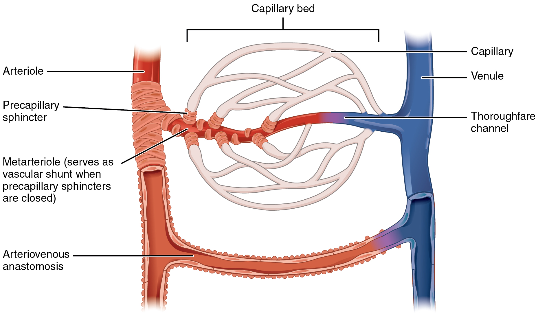
The microcirculation is the circulation of the blood in the smallest blood vessels, the microvessels of the microvasculature present within organ tissues. The microvessels include terminal arterioles, metarterioles, capillaries, and venules. Arterioles carry oxygenated blood to the capillaries, and blood flows out of the capillaries through venules into veins. In addition to these blood vessels, the microcirculation also includes lymphatic capillaries and collecting ducts. The main functions of the microcirculation are the delivery of oxygen and nutrients and the removal of carbon dioxide (CO2). It also serves to regulate blood flow and tissue perfusion thereby affecting blood pressure and responses to inflammation which can include edema (swelling). Most vessels of the microcirculation are lined by flattened cells of the endothelium and many of them are surrounded by contractile cells called pericytes. The endothelium provides a smooth surface for the flow of blood and regu ... [...More Info...] [...Related Items...] OR: [Wikipedia] [Google] [Baidu] |
High Endothelial Venules
High endothelial venules (HEV) are specialized post-capillary venous swellings characterized by plump endothelial cells as opposed to the usual thinner endothelial cells found in regular venules. HEVs enable lymphocytes circulating in the blood to directly enter a lymph node (by crossing through the HEV). Table 14-1 In humans, HEVs are found in all secondary lymphoid organs (with the exception of spleen, where blood exits through open arterioles and enters the red pulp), including hundreds of lymph nodes dispersed in the body, tonsils and adenoids in the pharynx, Peyer's patches (PIs) in the small intestine, appendix, and small aggregates of lymphoid tissue in the stomach and large intestine. In contrast to the endothelial cells from other vessels, the high endothelial cells of HEVs have a distinctive appearance, consisting of a cuboidal morphology and with various receptors to interact with leukocytes (express specialized ligands for lymphocytes and are able to support high levels ... [...More Info...] [...Related Items...] OR: [Wikipedia] [Google] [Baidu] |
Capillary
A capillary is a small blood vessel from 5 to 10 micrometres (μm) in diameter. Capillaries are composed of only the tunica intima, consisting of a thin wall of simple squamous endothelial cells. They are the smallest blood vessels in the body: they convey blood between the arterioles and venules. These microvessels are the site of exchange of many substances with the interstitial fluid surrounding them. Substances which cross capillaries include water, oxygen, carbon dioxide, urea, glucose, uric acid, lactic acid and creatinine. Lymph capillaries connect with larger lymph vessels to drain lymphatic fluid collected in the microcirculation. During early embryonic development, new capillaries are formed through vasculogenesis, the process of blood vessel formation that occurs through a '' de novo'' production of endothelial cells that then form vascular tubes. The term '' angiogenesis'' denotes the formation of new capillaries from pre-existing blood vessels and already ... [...More Info...] [...Related Items...] OR: [Wikipedia] [Google] [Baidu] |
Vein
Veins are blood vessels in humans and most other animals that carry blood towards the heart. Most veins carry deoxygenated blood from the tissues back to the heart; exceptions are the pulmonary and umbilical veins, both of which carry oxygenated blood to the heart. In contrast to veins, arteries carry blood away from the heart. Veins are less muscular than arteries and are often closer to the skin. There are valves (called ''pocket valves'') in most veins to prevent backflow. Structure Veins are present throughout the body as tubes that carry blood back to the heart. Veins are classified in a number of ways, including superficial vs. deep, pulmonary vs. systemic, and large vs. small. * Superficial veins are those closer to the surface of the body, and have no corresponding arteries. * Deep veins are deeper in the body and have corresponding arteries. * Perforator veins drain from the superficial to the deep veins. These are usually referred to in the lower limbs and feet. * ... [...More Info...] [...Related Items...] OR: [Wikipedia] [Google] [Baidu] |
Arteriole
An arteriole is a small-diameter blood vessel in the microcirculation that extends and branches out from an artery and leads to capillaries. Arterioles have muscular walls (usually only one to two layers of smooth muscle cells) and are the primary site of vascular resistance. The greatest change in blood pressure and velocity of blood flow occurs at the transition of arterioles to capillaries.This function is extremely important because it prevents the thin, one-layer capillaries from exploding upon pressure. The arterioles achieve this decrease in pressure, as they are the site with the highest resistance (a large contributor to total peripheral resistance) which translates to a large decrease in the pressure. Structure Microanatomy In a healthy vascular system the endothelium lines all blood-contacting surfaces, including arteries, arterioles, veins, venules, capillaries, and heart chambers. This healthy condition is promoted by the ample production of nitric oxide by the en ... [...More Info...] [...Related Items...] OR: [Wikipedia] [Google] [Baidu] |
Portal Vessel
The portal vein or hepatic portal vein (HPV) is a blood vessel that carries blood from the gastrointestinal tract, gallbladder, pancreas and spleen to the liver. This blood contains nutrients and toxins extracted from digested contents. Approximately 75% of total liver blood flow is through the portal vein, with the remainder coming from the hepatic artery proper. The blood leaves the liver to the heart in the hepatic veins. The portal vein is not a true vein, because it conducts blood to capillary beds in the liver and not directly to the heart. It is a major component of the hepatic portal system, one of only two portal venous systems in the body – with the hypophyseal portal system being the other. The portal vein is usually formed by the confluence of the superior mesenteric, splenic veins, inferior mesenteric, left, right gastric veins and the pancreatic vein. Conditions involving the portal vein cause considerable illness and death. An important example of such a c ... [...More Info...] [...Related Items...] OR: [Wikipedia] [Google] [Baidu] |
Blood Vessel
The blood vessels are the components of the circulatory system that transport blood throughout the human body. These vessels transport blood cells, nutrients, and oxygen to the tissues of the body. They also take waste and carbon dioxide away from the tissues. Blood vessels are needed to sustain life, because all of the body's tissues rely on their functionality. There are five types of blood vessels: the arteries, which carry the blood away from the heart; the arterioles; the capillaries, where the exchange of water and chemicals between the blood and the tissues occurs; the venules; and the veins, which carry blood from the capillaries back towards the heart. The word ''vascular'', meaning relating to the blood vessels, is derived from the Latin ''vas'', meaning vessel. Some structures – such as cartilage, the epithelium, and the lens and cornea of the eye – do not contain blood vessels and are labeled ''avascular''. Etymology * artery: late Middle English; from L ... [...More Info...] [...Related Items...] OR: [Wikipedia] [Google] [Baidu] |
Arterioles
An arteriole is a small-diameter blood vessel in the microcirculation that extends and branches out from an artery and leads to capillaries. Arterioles have muscular walls (usually only one to two layers of smooth muscle cells) and are the primary site of vascular resistance. The greatest change in blood pressure and velocity of blood flow occurs at the transition of arterioles to capillaries.This function is extremely important because it prevents the thin, one-layer capillaries from exploding upon pressure. The arterioles achieve this decrease in pressure, as they are the site with the highest resistance (a large contributor to total peripheral resistance) which translates to a large decrease in the pressure. Structure Microanatomy In a healthy vascular system the endothelium lines all blood-contacting surfaces, including arteries, arterioles, veins, venules, capillaries, and heart chambers. This healthy condition is promoted by the ample production of nitric oxide by the en ... [...More Info...] [...Related Items...] OR: [Wikipedia] [Google] [Baidu] |
Surface Chemistry Of Microvasculature
Microvasculature comprises the microvessels – venules and capillaries of the microcirculation, with a maximum average diameter of 0.3 millimeters. As the vessels decrease in size, they increase their surface-area-to-volume ratio. This allows surface properties to play a significant role in the function of the vessel. Diffusion occurs through the walls of the vessels due to a concentration gradient, allowing the necessary exchange of ions, molecules, or blood cells. The permeability of a capillary wall is determined by the type of capillary and the surface of the endothelial cells. A continuous, tightly spaced endothelial cell lining only permits the diffusion of small molecules. Larger molecules and blood cells require adequate space between cells or holes in the lining. The high resistivity of a cellular membrane prevents the diffusion of ions without a membrane transport protein. The hydrophobicity of an endothelial cell surface determines whether water or lipophilic molecules w ... [...More Info...] [...Related Items...] OR: [Wikipedia] [Google] [Baidu] |
Blood
Blood is a body fluid in the circulatory system of humans and other vertebrates that delivers necessary substances such as nutrients and oxygen to the cells, and transports metabolic waste products away from those same cells. Blood in the circulatory system is also known as ''peripheral blood'', and the blood cells it carries, ''peripheral blood cells''. Blood is composed of blood cells suspended in blood plasma. Plasma, which constitutes 55% of blood fluid, is mostly water (92% by volume), and contains proteins, glucose, mineral ions, hormones, carbon dioxide (plasma being the main medium for excretory product transportation), and blood cells themselves. Albumin is the main protein in plasma, and it functions to regulate the colloidal osmotic pressure of blood. The blood cells are mainly red blood cells (also called RBCs or erythrocytes), white blood cells (also called WBCs or leukocytes) and platelets (also called thrombocytes). The most abundant cells in vertebrate blood a ... [...More Info...] [...Related Items...] OR: [Wikipedia] [Google] [Baidu] |
Fibrous Connective Tissue
Connective tissue is one of the four primary types of animal tissue, along with epithelial tissue, muscle tissue, and nervous tissue. It develops from the mesenchyme derived from the mesoderm the middle embryonic germ layer. Connective tissue is found in between other tissues everywhere in the body, including the nervous system. The three meninges, membranes that envelop the brain and spinal cord are composed of connective tissue. Most types of connective tissue consists of three main components: elastic and collagen fibers, ground substance, and cells. Blood, and lymph are classed as specialized fluid connective tissues that do not contain fiber. All are immersed in the body water. The cells of connective tissue include fibroblasts, adipocytes, macrophages, mast cells and leucocytes. The term "connective tissue" (in German, ''Bindegewebe'') was introduced in 1830 by Johannes Peter Müller. The tissue was already recognized as a distinct class in the 18th century. Types File:I ... [...More Info...] [...Related Items...] OR: [Wikipedia] [Google] [Baidu] |


