|
Stapedius
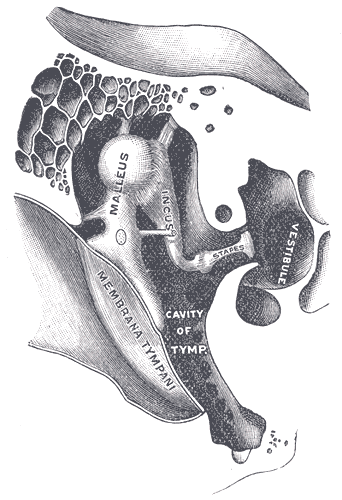
The stapedius is the smallest skeletal muscle in the human body. At just over one millimeter in length, its purpose is to stabilize the smallest bone in the body, the stapes or strirrup bone of the middle ear. Structure The stapedius emerges from a pinpoint foramen or opening in the apex of the pyramidal eminence (a hollow, cone-shaped prominence in the posterior wall of the tympanic cavity), and inserts into the neck of the stapes. Nerve supply The stapedius is supplied by the nerve to stapedius, a branch of the facial nerve. Function The stapedius dampens the vibrations of the stapes by pulling on the neck of that bone. As one of the muscles involved in the acoustic reflex it prevents excess movement of the stapes, helping to control the amplitude of sound waves from the general external environment to the inner ear. Clinical significance Paralysis of the stapedius allows wider oscillation of the stapes, resulting in heightened reaction of the auditory ossicles ... [...More Info...] [...Related Items...] OR: [Wikipedia] [Google] [Baidu] |
Acoustic Reflex
The acoustic reflex (also known as the stapedius reflex, stapedial reflex, auditory reflex, middle-ear-muscle reflex (MEM reflex, MEMR), attenuation reflex, cochleostapedial reflex or intra-aural reflex) is an involuntary muscle contraction that occurs in the middle ear in response to loud sound stimuli or when the person starts to vocalize. When presented with an intense sound stimulus, the stapedius and tensor tympani muscles of the ossicles contract. The stapedius stiffens the ossicular chain by pulling the stapes (stirrup) of the middle ear away from the oval window of the cochlea and the tensor tympani muscle stiffens the ossicular chain by loading the tympanic membrane when it pulls the malleus (hammer) in toward the middle ear. The reflex decreases the transmission of vibrational energy to the cochlea, where it is converted into electrical impulses to be processed by the brain. Acoustic reflex threshold The acoustic reflex threshold (ART) is the sound pressure level (SPL) ... [...More Info...] [...Related Items...] OR: [Wikipedia] [Google] [Baidu] |
Nerve To The Stapedius
The nerve to the stapedius is a branch of the facial nerve (CN VII) which innervates the stapedius muscle. It arises from the CN VII opposite the pyramidal eminence and passes through a small canal in this eminence to reach the stapedius muscle The stapedius is the smallest skeletal muscle in the human body. At just over one millimeter in length, its purpose is to stabilize the smallest bone in the body, the stapes or strirrup bone of the middle ear. Structure The stapedius emerges from .... References External links * () Facial nerve {{Neuroanatomy-stub ... [...More Info...] [...Related Items...] OR: [Wikipedia] [Google] [Baidu] |
Tensor Tympani
The tensor tympani is a muscle within the middle ear, located in the bony canal above the bony part of the auditory tube, and connects to the malleus bone. Its role is to dampen loud sounds, such as those produced from chewing, shouting, or thunder. Because its reaction time is not fast enough, the muscle cannot protect against hearing damage caused by sudden loud sounds, like explosions or gunshots. Structure The tensor tympani is a muscle that is present in the middle ear. It arises from the cartilaginous part of the auditory tube, and the adjacent great wing of the sphenoid. It then passes through its own canal, and ends in the tympanic cavity as a slim tendon that connects to the handle of the malleus. The tendon makes a sharp bend around the ''processus cochleariformis'', part of the wall of its cavity, before it joins with the malleus. The tensor tympani receives blood from the middle meningeal artery via the superior tympanic branch. It is one of two muscles in th ... [...More Info...] [...Related Items...] OR: [Wikipedia] [Google] [Baidu] |
Nerve To Stapedius
The nerve to the stapedius is a branch of the facial nerve (CN VII) which innervates the stapedius muscle. It arises from the CN VII opposite the pyramidal eminence and passes through a small canal in this eminence to reach the stapedius muscle The stapedius is the smallest skeletal muscle in the human body. At just over one millimeter in length, its purpose is to stabilize the smallest bone in the body, the stapes or strirrup bone of the middle ear. Structure The stapedius emerges from .... References External links * () Facial nerve {{Neuroanatomy-stub ... [...More Info...] [...Related Items...] OR: [Wikipedia] [Google] [Baidu] |
Middle Ear
The middle ear is the portion of the ear medial to the eardrum, and distal to the oval window of the cochlea (of the inner ear). The mammalian middle ear contains three ossicles, which transfer the vibrations of the eardrum into waves in the fluid and membranes of the inner ear. The hollow space of the middle ear is also known as the tympanic cavity and is surrounded by the tympanic part of the temporal bone. The auditory tube (also known as the Eustachian tube or the pharyngotympanic tube) joins the tympanic cavity with the nasal cavity (nasopharynx), allowing pressure to equalize between the middle ear and throat. The primary function of the middle ear is to efficiently transfer acoustic energy from compression waves in air to fluid–membrane waves within the cochlea. Structure Ossicles The middle ear contains three tiny bones known as the ossicles: '' malleus'', '' incus'', and ''stapes''. The ossicles were given their Latin names for their distinctive shapes; they ar ... [...More Info...] [...Related Items...] OR: [Wikipedia] [Google] [Baidu] |
Hyoid Arch
The pharyngeal arches, also known as visceral arches'','' are structures seen in the embryonic development of vertebrates that are recognisable precursors for many structures. In fish, the arches are known as the branchial arches, or gill arches. In the human embryo, the arches are first seen during the fourth week of development. They appear as a series of outpouchings of mesoderm on both sides of the developing pharynx. The vasculature of the pharyngeal arches is known as the aortic arches. In fish, the branchial arches support the gills. Structure In vertebrates, the pharyngeal arches are derived from all three germ layers (the primary layers of cells that form during embryogenesis). Neural crest cells enter these arches where they contribute to features of the skull and facial skeleton such as bone and cartilage. However, the existence of pharyngeal structures before neural crest cells evolved is indicated by the existence of neural crest-independent mechanisms of phar ... [...More Info...] [...Related Items...] OR: [Wikipedia] [Google] [Baidu] |
Pyramidal Eminence
The pyramidal eminence (pyramid) is a conical projection in the middle ear. It is situated immediately behind the fenestra vestibuli (oval window), and in front of the vertical portion of the facial canal; it is hollow, and contains the stapedius muscle; its summit projects forward toward the fenestra vestibuli, and is pierced by a small aperture which transmits the tendon of the muscle. The cavity in the pyramidal eminence is prolonged downward and backward in front of the facial canal, and communicates with it by a minute aperture which transmits a twig from the facial nerve The facial nerve, also known as the seventh cranial nerve, cranial nerve VII, or simply CN VII, is a cranial nerve that emerges from the pons of the brainstem, controls the muscles of facial expression, and functions in the conveyance of taste ... to the stapedius muscle. References Auditory system {{anatomy-stub ... [...More Info...] [...Related Items...] OR: [Wikipedia] [Google] [Baidu] |
Ossicles
The ossicles (also called auditory ossicles) are three bones in either middle ear that are among the smallest bones in the human body. They serve to transmit sounds from the air to the fluid-filled labyrinth (cochlea). The absence of the auditory ossicles would constitute a moderate-to-severe hearing loss. The term "ossicle" literally means "tiny bone". Though the term may refer to any small bone throughout the body, it typically refers to the malleus, incus, and stapes (hammer, anvil, and stirrup) of the middle ear. Structure The ossicles are, in order from the eardrum to the inner ear (from superficial to deep): the malleus, incus, and stapes, terms that in Latin are translated as "the hammer, anvil, and stirrup". * The malleus ( la, "hammer") articulates with the incus through the incudomalleolar joint and is attached to the tympanic membrane ( eardrum), from which vibrational sound pressure motion is passed. * The incus ( la, "anvil") is connected to both the ... [...More Info...] [...Related Items...] OR: [Wikipedia] [Google] [Baidu] |
Hearing
Hearing, or auditory perception, is the ability to perceive sounds through an organ, such as an ear, by detecting vibrations as periodic changes in the pressure of a surrounding medium. The academic field concerned with hearing is auditory science. Sound may be heard through solid, liquid, or gaseous matter. It is one of the traditional five senses. Partial or total inability to hear is called hearing loss. In humans and other vertebrates, hearing is performed primarily by the auditory system: mechanical waves, known as vibrations, are detected by the ear and transduced into nerve impulses that are perceived by the brain (primarily in the temporal lobe). Like touch, audition requires sensitivity to the movement of molecules in the world outside the organism. Both hearing and touch are types of mechanosensation. Hearing mechanism There are three main components of the human auditory system: the outer ear, the middle ear, and the inner ear. Outer ear Th ... [...More Info...] [...Related Items...] OR: [Wikipedia] [Google] [Baidu] |
Cranial Nerve VII
The facial nerve, also known as the seventh cranial nerve, cranial nerve VII, or simply CN VII, is a cranial nerve that emerges from the pons of the brainstem, controls the muscles of facial expression, and functions in the conveyance of taste sensations from the anterior two-thirds of the tongue. The nerve typically travels from the pons through the facial canal in the temporal bone and exits the skull at the stylomastoid foramen. It arises from the brainstem from an area posterior to the cranial nerve VI (abducens nerve) and anterior to cranial nerve VIII (vestibulocochlear nerve). The facial nerve also supplies preganglionic parasympathetic fibers to several head and neck ganglia. The facial and intermediate nerves can be collectively referred to as the nervus intermediofacialis. The path of the facial nerve can be divided into six segments: # intracranial (cisternal) segment # meatal (canalicular) segment (within the internal auditory canal) # labyrinthine segment ... [...More Info...] [...Related Items...] OR: [Wikipedia] [Google] [Baidu] |
Levator Operculi
{{disambig ...
Levator muscle can refer to: * Levator scapulae muscle * Levator palpebrae superioris muscle * Levator ani * Levator labii superioris alaeque nasi muscle * Levator veli palatini * Levator muscle of thyroid gland * Levator labii superioris * Levator anguli oris The levator anguli oris (caninus) is a facial muscle of the mouth arising from the canine fossa, immediately below the infraorbital foramen. It elevates angle of mouth medially. Its fibers are inserted into the angle of the mouth, intermingli ... [...More Info...] [...Related Items...] OR: [Wikipedia] [Google] [Baidu] |


