|
Scalp
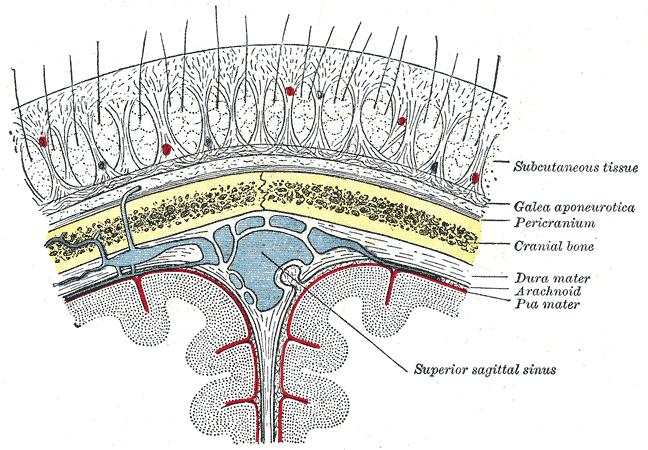
The scalp is the anatomical area bordered by the human face at the front, and by the neck at the sides and back. Structure The scalp is usually described as having five layers, which can conveniently be remembered as a mnemonic: * S: The skin on the head from which head hair grows. It contains numerous sebaceous glands and hair follicles. * C: Connective tissue. A dense subcutaneous layer of fat and fibrous tissue that lies beneath the skin, containing the nerves and vessels of the scalp. * A: The aponeurosis called epicranial aponeurosis (or galea aponeurotica) is the next layer. It is a tough layer of dense fibrous tissue which runs from the frontalis muscle anteriorly to the occipitalis posteriorly. * L: The loose areolar connective tissue layer provides an easy plane of separation between the upper three layers and the pericranium. In scalping the scalp is torn off through this layer. It also provides a plane of access in craniofacial surgery and neurosurgery. This la ... [...More Info...] [...Related Items...] OR: [Wikipedia] [Google] [Baidu] |
Scalping
Scalping is the act of cutting or tearing a part of the human scalp, with hair attached, from the head, and generally occurred in warfare with the scalp being a trophy. Scalp-taking is considered part of the broader cultural practice of the taking and display of human body parts as trophies, and may have developed as an alternative to the taking of human heads, for scalps were easier to take, transport, and preserve for subsequent display. Scalping independently developed in various cultures in both the Old and New Worlds. Europe Several human remains from the stone-age Ertebølle culture in Denmark show evidence of scalping. A man found in a grave in the Alvastra pile-dwelling in Sweden had been scalped approximately 5,000 years ago. Georg Frederici noted that “Herodotus provided the only clear and satisfactory portrayal of a scalping people in the old world” in his description of the Scythians, a nomadic people then located to the north and west of the Black Sea. Herodotus ... [...More Info...] [...Related Items...] OR: [Wikipedia] [Google] [Baidu] |
Hair Follicle
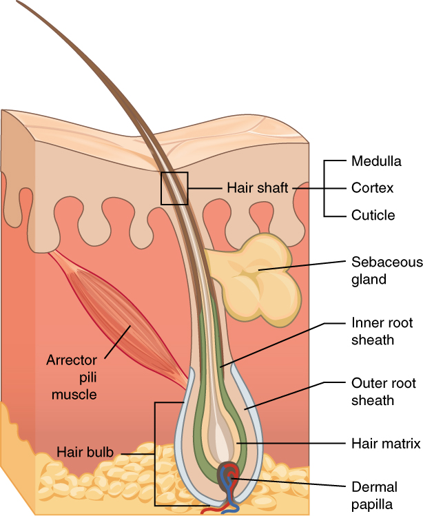
The hair follicle is an organ found in mammalian skin. It resides in the dermal layer of the skin and is made up of 20 different cell types, each with distinct functions. The hair follicle regulates hair growth via a complex interaction between hormones, neuropeptides, and immune cells. This complex interaction induces the hair follicle to produce different types of hair as seen on different parts of the body. For example, terminal hairs grow on the scalp and lanugo hairs are seen covering the bodies of fetuses in the uterus and in some newborn babies. The process of hair growth occurs in distinct sequential stages. The first stage is called ''anagen'' and is the active growth phase, ''telogen'' is the resting stage, ''catagen'' is the regression of the hair follicle phase, ''exogen'' is the active shedding of hair phase and lastly ''kenogen'' is the phase between the empty hair follicle and the growth of new hair. The function of hair in humans has long been a subject of int ... [...More Info...] [...Related Items...] OR: [Wikipedia] [Google] [Baidu] |
Occipital Vein
The occipital vein is a vein of the scalp. It originates from a plexus around the external occipital protuberance and superior nuchal line to the back part of the vertex of the skull. It usually drains into the internal jugular vein, but may also drain into the posterior auricular vein (which joins the external jugular vein). It drains part of the scalp. Structure The occipital vein is part of the scalp. It begins as a plexus at the posterior aspect of the scalp from the external occipital protuberance and superior nuchal line to the back part of the vertex of the skull. It pierces the cranial attachment of the trapezius and, dipping into the venous plexus of the suboccipital triangle, joins the deep cervical vein and the vertebral vein. Occasionally it follows the course of the occipital artery, and ends in the internal jugular vein. Alternatively, it joins the posterior auricular vein, and ends in the external jugular vein. The parietal emissary vein connects it with th ... [...More Info...] [...Related Items...] OR: [Wikipedia] [Google] [Baidu] |
Head Hair
Hair is a protein filament that grows from follicles found in the dermis. Hair is one of the defining characteristics of mammals. The human body, apart from areas of glabrous skin, is covered in follicles which produce thick terminal and fine vellus hair. Most common interest in hair is focused on hair growth, hair types, and hair care, but hair is also an important biomaterial primarily composed of protein, notably alpha-keratin. Attitudes towards different forms of hair, such as hairstyles and hair removal, vary widely across different cultures and historical periods, but it is often used to indicate a person's personal beliefs or social position, such as their age, sex, or religion. Overview The word "hair" usually refers to two distinct structures: #the part beneath the skin, called the hair follicle, or, when pulled from the skin, the bulb or root. This organ is located in the dermis and maintains stem cells, which not only re-grow the hair after it falls out, but al ... [...More Info...] [...Related Items...] OR: [Wikipedia] [Google] [Baidu] |
Greater Occipital Nerve
The greater occipital nerve is a nerve of the head. It is a spinal nerve, specifically the medial branch of the dorsal primary ramus of cervical spinal nerve 2. It arises from between the first and second cervical vertebrae, ascends, and then passes through the semispinalis muscle. It ascends further to supply the skin along the posterior part of the scalp to the vertex. It supplies sensation the scalp at the top of the head, over the ear and over the parotid glands. Structure The greater occipital nerve is the medial branch of the dorsal primary ramus of cervical spinal nerve 2. It may also involve fibres from cervical spinal nerve 3. It arises from between the first and second cervical vertebrae, along with the lesser occipital nerve. It ascends after emerging from below the suboccipital triangle beneath the obliquus capitis inferior muscle. Just below the superior nuchal ridge, it pierces the fascia. It ascends further to supply the skin along the posterior part of ... [...More Info...] [...Related Items...] OR: [Wikipedia] [Google] [Baidu] |
Occipital Artery
The occipital artery arises from the external carotid artery opposite the facial artery. Its path is below the posterior belly of digastric to the occipital region. This artery supplies blood to the back of the scalp and sternocleidomastoid muscles, and deep muscles in the back and neck. Structure At its origin, it is covered by the posterior belly of the digastricus and the stylohyoideus, and the hypoglossal nerve winds around it from behind forward; higher up, it crosses the internal carotid artery, the internal jugular vein, and the vagus and accessory nerves. It next ascends to the interval between the transverse process of the atlas and the mastoid process of the temporal bone, and passes horizontally backward, grooving the surface of the latter bone, being covered by the sternocleidomastoideus, splenius capitis, longissimus capitis, and digastricus, and resting upon the rectus capitis lateralis, the obliquus superior, and semispinalis capitis. It then changes its ... [...More Info...] [...Related Items...] OR: [Wikipedia] [Google] [Baidu] |
Lesser Occipital Nerve
The lesser occipital nerve or small occipital nerve is a cutaneous spinal nerve. It arises from second cervical (spinal) nerve (along with the greater occipital nerve). It innervates the scalp in the lateral area of the head posterior to the ear. Structure The lesser occipital nerve is one of the four cutaneous branches of the cervical plexus. Origin It arises from the (lateral branch of the ventral ramus) of cervical spinal nerve C2; it may also receive fibres from cervical spinal nerve C3. It originates between the atlas, and axis. Course and relations It curves around the accessory nerve (CN XI) to come to course anterior to it. It then curves around and ascends along the posterior border of the sternocleidomastoid muscle; rarely, it may pierce the muscle. Near the cranium, it perforates the deep fascia. It is continues upwards along the scalp posterior to the auricle. Distribution The lesser occipital nerve distributes branches to the skin. It gives off an ... [...More Info...] [...Related Items...] OR: [Wikipedia] [Google] [Baidu] |
Supratrochlear Artery
The supratrochlear artery (or frontal artery) is one of the terminal branches of the ophthalmic artery. It arises within the orbit. It exits the orbit alongside the supratrochlear nerve. It contributes arterial supply to the skin, muscles and pericranium of the forehead. Anatomy It branches from the ophthalmic artery near the trochlea of the superior oblique muscle in the orbit. Origin The supratrochlear artery branches from the ophthalmic artery in the orbit near the trochlea of the superior oblique muscle. Course After branching from the ophthalmic artery, it passes anteriorly through the superomedial orbit. It travels medial to the trochlear nerve. With the supratrochlear nerve, the supratrochlear artery exits the orbit through the supratrochlear notch (variably present), medial to the supraorbital foramen. It then ascends on the forehead. Anastomoses The supratrochlear artery anastomoses with the contralateral supratrochlear artery, and the ipsilateral supraorb ... [...More Info...] [...Related Items...] OR: [Wikipedia] [Google] [Baidu] |
Supraorbital Artery
The supraorbital artery is a branch of the ophthalmic artery. It passes anteriorly within the orbit to exit the orbit through the supraorbital foramen or notch alongside the supraorbital nerve, splitting into two terminal branches which go on to form anastomoses with arteries of the head. Structure Origin The supraorbital artery arises from the ophthalmic artery. Course and relations It travels anteriorly in the orbit by passing superior to the eye and medial to the superior rectus and levator palpebrae superioris. It then joins the supraorbital nerve to jointly pass between the periosteum of the roof of the orbit and the levator palpebrae superioris towards the supraorbital foramen or notch. After passing through the supraorbital foramen or notch, it often splits into a superficial branch and a deep branch. Distribution The supraorbital artery contributes arterial supply to: the superior rectus muscle, superior oblique muscle, levator palpebrae muscles, periorbita, t ... [...More Info...] [...Related Items...] OR: [Wikipedia] [Google] [Baidu] |
Supratrochlear Nerve
The supratrochlear nerve is a branch of the frontal nerve, itself a branch of the ophthalmic nerve (CN V1) from the trigeminal nerve (CN V). It provides sensory innervation to the skin of the forehead and the upper eyelid. Structure The supratrochlear nerve is a branch of the frontal nerve, itself a branch of the ophthalmic nerve (CN V1) from the trigeminal nerve (CN V). It is smaller than the supraorbital nerve from the frontal nerve. It branches midway between the base and apex of the orbit. It passes above the trochlea of the superior oblique muscle. It then travels anteriorly above the levator palpebrae superioris muscle. It exits the orbit through the frontal notch in the superomedial margin of the orbit. It then ascends onto the forehead beneath the corrugator supercilii muscle and frontalis muscle. It then divides into sensory branches. The supratrochlear nerve travels with the supratrochlear artery, a branch of the ophthalmic artery. Function The supratrochlear ne ... [...More Info...] [...Related Items...] OR: [Wikipedia] [Google] [Baidu] |
Galea Aponeurotica
The epicranial aponeurosis (aponeurosis epicranialis, galea aponeurotica) is an aponeurosis (a tough layer of dense fibrous tissue). It covers the upper part of the skull in humans and many other animals. Structure In humans, the epicranial aponeurosis originates from the external occipital protuberance and highest nuchal lines of the occipital bone. It merges with the occipitofrontalis muscle. In front, it forms a short and narrow prolongation between its union with the frontalis muscle (the frontal part of the occipitofrontalis muscle). On either side, the epicranial aponeurosis attaches to the anterior auricular muscles and the superior auricular muscles. Here it is less aponeurotic, and is continued over the temporal fascia to the zygomatic arch as a layer of laminated areolar tissue. It is closely connected to the integument by the firm, dense, fibro-fatty layer which forms the superficial fascia of the scalp. It is attached to the pericranium by loose cellular tissue ... [...More Info...] [...Related Items...] OR: [Wikipedia] [Google] [Baidu] |
Neurosurgery
Neurosurgery or neurological surgery, known in common parlance as brain surgery, is the medical specialty concerned with the surgical treatment of disorders which affect any portion of the nervous system including the brain, spinal cord and peripheral nervous system. Education and context In different countries, there are different requirements for an individual to legally practice neurosurgery, and there are varying methods through which they must be educated. In most countries, neurosurgeon training requires a minimum period of seven years after graduating from medical school. United States In the United States, a neurosurgeon must generally complete four years of undergraduate education, four years of medical school, and seven years of residency (PGY-1-7). Most, but not all, residency programs have some component of basic science or clinical research. Neurosurgeons may pursue additional training in the form of a fellowship after residency, or, in some cases, as a senior res ... [...More Info...] [...Related Items...] OR: [Wikipedia] [Google] [Baidu] |


