|
Immunofluorescence
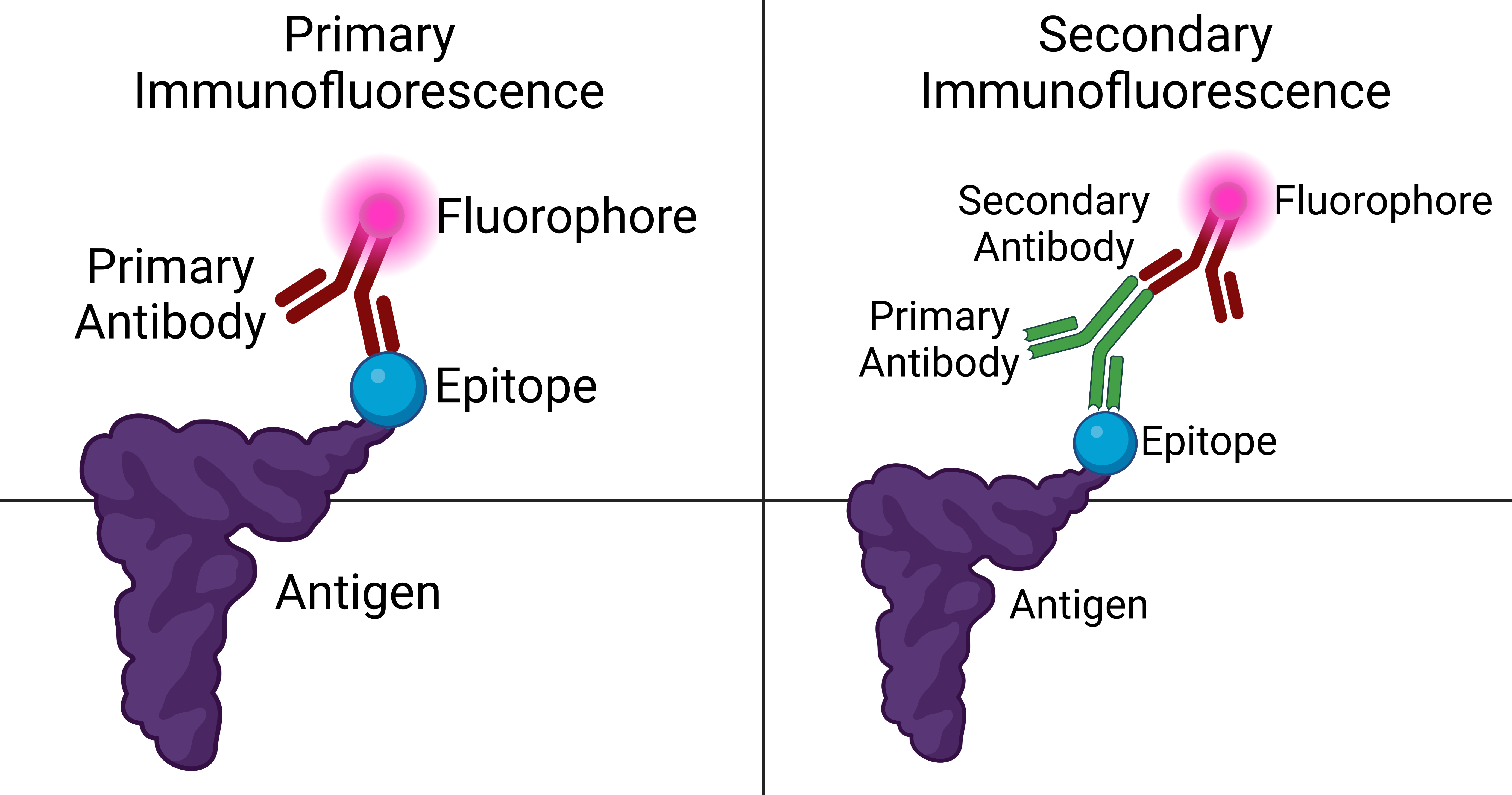
Immunofluorescence is a technique used for light microscopy with a fluorescence microscope and is used primarily on microbiological samples. This technique uses the specificity of antibodies to their antigen to target fluorescent dyes to specific biomolecule targets within a cell, and therefore allows visualization of the distribution of the target molecule through the sample. The specific region an antibody recognizes on an antigen is called an epitope. There have been efforts in epitope mapping since many antibodies can bind the same epitope and levels of binding between antibodies that recognize the same epitope can vary. Additionally, the binding of the fluorophore to the antibody itself cannot interfere with the immunological specificity of the antibody or the binding capacity of its antigen. Immunofluorescence is a widely used example of immunostaining (using antibodies to stain proteins) and is a specific example of immunohistochemistry (the use of the antibody-antige ... [...More Info...] [...Related Items...] OR: [Wikipedia] [Google] [Baidu] |
Epifluorescence Microscope
A fluorescence microscope is an optical microscope that uses fluorescence instead of, or in addition to, scattering, reflection, and attenuation or absorption, to study the properties of organic or inorganic substances. "Fluorescence microscope" refers to any microscope that uses fluorescence to generate an image, whether it is a simple set up like an epifluorescence microscope or a more complicated design such as a confocal microscope, which uses optical sectioning to get better resolution of the fluorescence image. Principle The specimen is illuminated with light of a specific wavelength (or wavelengths) which is absorbed by the fluorophores, causing them to emit light of longer wavelengths (i.e., of a different color than the absorbed light). The illumination light is separated from the much weaker emitted fluorescence through the use of a spectral emission filter. Typical components of a fluorescence microscope are a light source ( xenon arc lamp or mercury-vapor lamp ar ... [...More Info...] [...Related Items...] OR: [Wikipedia] [Google] [Baidu] |
Fluorescence Microscope
A fluorescence microscope is an optical microscope that uses fluorescence instead of, or in addition to, scattering, reflection, and attenuation or absorption, to study the properties of organic or inorganic substances. "Fluorescence microscope" refers to any microscope that uses fluorescence to generate an image, whether it is a simple set up like an epifluorescence microscope or a more complicated design such as a confocal microscope, which uses optical sectioning to get better resolution of the fluorescence image. Principle The specimen is illuminated with light of a specific wavelength (or wavelengths) which is absorbed by the fluorophores, causing them to emit light of longer wavelengths (i.e., of a different color than the absorbed light). The illumination light is separated from the much weaker emitted fluorescence through the use of a spectral emission filter. Typical components of a fluorescence microscope are a light source (xenon arc lamp or mercury-vapor lamp ar ... [...More Info...] [...Related Items...] OR: [Wikipedia] [Google] [Baidu] |
Immunohistochemistry
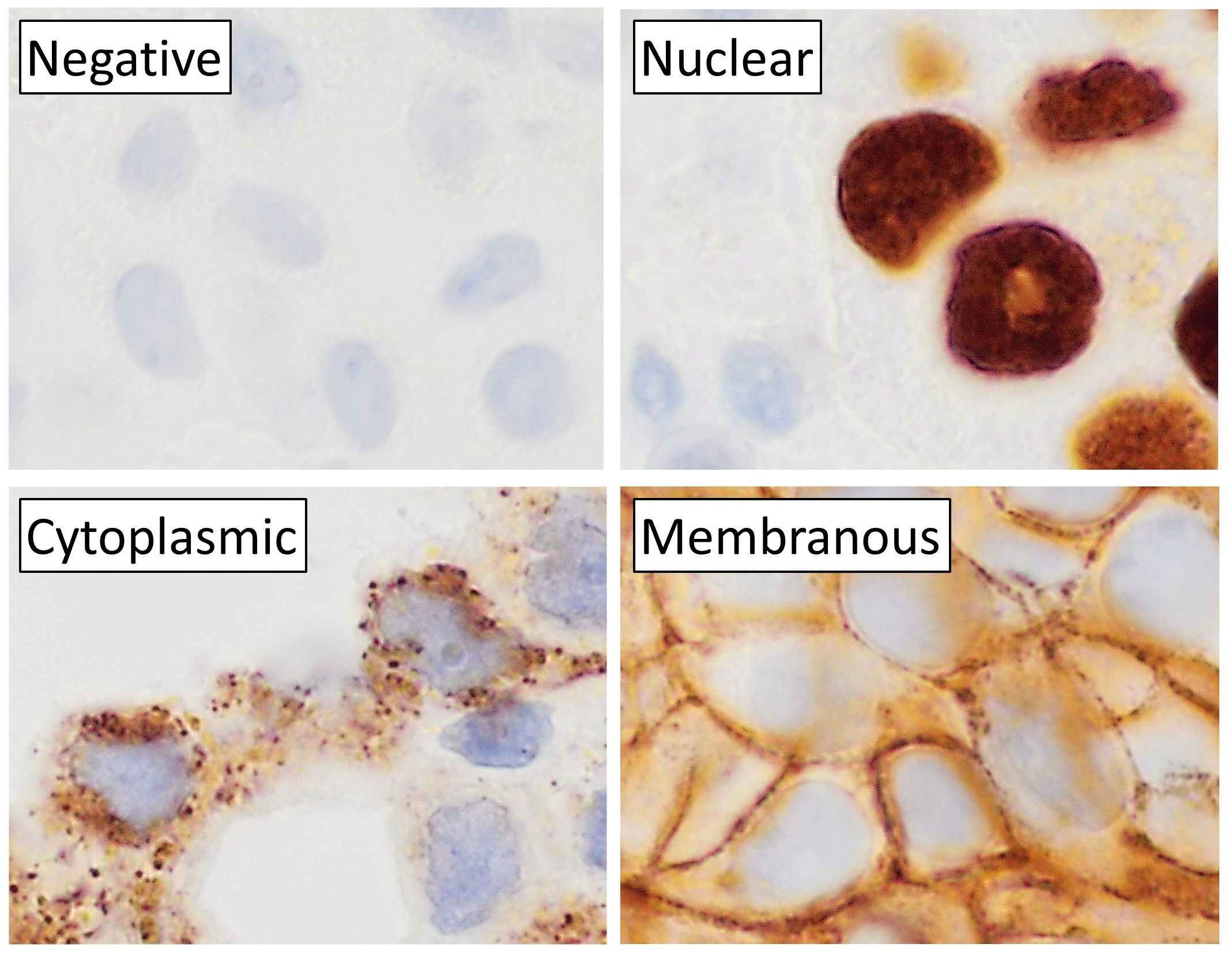
Immunohistochemistry (IHC) is the most common application of immunostaining. It involves the process of selectively identifying antigens (proteins) in cells of a tissue section by exploiting the principle of antibodies binding specifically to antigens in biological tissues. IHC takes its name from the roots "immuno", in reference to antibodies used in the procedure, and "histo", meaning tissue (compare to immunocytochemistry). Albert Coons conceptualized and first implemented the procedure in 1941. Visualising an antibody-antigen interaction can be accomplished in a number of ways, mainly either of the following: * ''Chromogenic immunohistochemistry'' (CIH), wherein an antibody is conjugated to an enzyme, such as peroxidase (the combination being termed immunoperoxidase), that can catalyse a colour-producing reaction. * ''Immunofluorescence'', where the antibody is tagged to a fluorophore, such as fluorescein or rhodamine. Immunohistochemical staining is widely used in the ... [...More Info...] [...Related Items...] OR: [Wikipedia] [Google] [Baidu] |
Patching And Capping
The aggregation of fluorescently tagged antibodies that are associated with proteins on membranes of living cells. The aggregation appears as a cap or a patch in the fluorescence microscope and is due to the bivalent nature of antibodies. Patching and capping were critical in demonstrating the fluid nature of plasma membranes. Variations in density within the specimen are amplified to enhance contrast in unstained cells which is especially useful for examining living unpigmented cells. In other words, phase contrast is a contrast-enhancing optical technique that can be used to produce high contrast images such as living cells and subcellular including nuclei and other organelles. One of the major advantages of using phase contrast microscopy is that living cells can be examined in their natural state without being killed, fixed, or especially stained. As a result, biological processes in the cell can be observed and recorded in high contrast with sharp clarity of minute specimen ... [...More Info...] [...Related Items...] OR: [Wikipedia] [Google] [Baidu] |
Cutaneous Conditions With Immunofluorescence Findings
Several cutaneous conditions can be diagnosed with the aid of immunofluorescence studies. Cutaneous conditions with positive direct or indirect immunofluorescence when using salt-split skin include: For several subtypes of pemphigus a variety of substrates are used for indirect immunofluorescence: See also * List of cutaneous conditions * List of genes mutated in cutaneous conditions * List of cutaneous conditions caused by mutations in keratins There are many different keratin proteins normally expressed in the human integumentary system. Mutations in keratin proteins in the skin can cause disease. Of note, other structural proteins in the epidermis of the skin that are closely rel ... References * * {{DEFAULTSORT:Immunofluorescence findings for autoimmune bullous conditions Cutaneous conditions Dermatology-related lists ... [...More Info...] [...Related Items...] OR: [Wikipedia] [Google] [Baidu] |
Antibodies
An antibody (Ab), also known as an immunoglobulin (Ig), is a large, Y-shaped protein used by the immune system to identify and neutralize foreign objects such as pathogenic bacteria and viruses. The antibody recognizes a unique molecule of the pathogen, called an antigen. Each tip of the "Y" of an antibody contains a paratope (analogous to a lock) that is specific for one particular epitope (analogous to a key) on an antigen, allowing these two structures to bind together with precision. Using this binding mechanism, an antibody can ''tag'' a microbe or an infected cell for attack by other parts of the immune system, or can neutralize it directly (for example, by blocking a part of a virus that is essential for its invasion). To allow the immune system to recognize millions of different antigens, the antigen-binding sites at both tips of the antibody come in an equally wide variety. In contrast, the remainder of the antibody is relatively constant. It only occurs in a few va ... [...More Info...] [...Related Items...] OR: [Wikipedia] [Google] [Baidu] |
Lupus Band Test
Lupus band test is done upon skin biopsy, with direct immunofluorescence staining, in which, if positive, IgG and complement depositions are found at the dermoepidermal junction.Marks, James G; Miller, Jeffery (2006). ''Lookingbill and Marks' Principles of Dermatology'' (4th ed.). Elsevier Inc. . This test can be helpful in distinguishing systemic lupus erythematosus (SLE) from cutaneous lupus, because in SLE the lupus band test will be positive in both involved and uninvolved skin, whereas with cutaneous lupus only the involved skin will be positive. The minimum criteria for positivity are:Ther Clin Risk Manag. 2011; 7: 27–32. The lupus band test in systemic lupus erythematosus patients. Adam Reich, Katarzyna Marcinow, and Rafal Bialynicki-Birula * In sun-exposed skin: presence of a band of deposits of IgM along the epidermal basement membrane in 50% of the biopsy, intermediate (2+) intensity or more. * In sun-protected skin : presence of interrupted (i.e. less than 50%) de ... [...More Info...] [...Related Items...] OR: [Wikipedia] [Google] [Baidu] |
Fluorophore
A fluorophore (or fluorochrome, similarly to a chromophore) is a fluorescent chemical compound that can re-emit light upon light excitation. Fluorophores typically contain several combined aromatic groups, or planar or cyclic molecules with several π bonds. Fluorophores are sometimes used alone, as a tracer in fluids, as a dye for staining of certain structures, as a substrate of enzymes, or as a probe or indicator (when its fluorescence is affected by environmental aspects such as polarity or ions). More generally they are covalently bonded to a macromolecule, serving as a marker (or dye, or tag, or reporter) for affine or bioactive reagents ( antibodies, peptides, nucleic acids). Fluorophores are notably used to stain tissues, cells, or materials in a variety of analytical methods, i.e., fluorescent imaging and spectroscopy. Fluorescein, via its amine-reactive isothiocyanate derivative fluorescein isothiocyanate (FITC), has been one of the most popular fluorophores. F ... [...More Info...] [...Related Items...] OR: [Wikipedia] [Google] [Baidu] |
Blood Vessels In Porcine Skin - SMA A488 - 20x
Blood is a body fluid in the circulatory system of humans and other vertebrates that delivers necessary substances such as nutrients and oxygen to the cells, and transports metabolic waste products away from those same cells. Blood in the circulatory system is also known as ''peripheral blood'', and the blood cells it carries, ''peripheral blood cells''. Blood is composed of blood cells suspended in blood plasma. Plasma, which constitutes 55% of blood fluid, is mostly water (92% by volume), and contains proteins, glucose, mineral ions, hormones, carbon dioxide (plasma being the main medium for excretory product transportation), and blood cells themselves. Albumin is the main protein in plasma, and it functions to regulate the colloidal osmotic pressure of blood. The blood cells are mainly red blood cells (also called RBCs or erythrocytes), white blood cells (also called WBCs or leukocytes) and platelets (also called thrombocytes). The most abundant cells in vertebrate blo ... [...More Info...] [...Related Items...] OR: [Wikipedia] [Google] [Baidu] |
HSP IF IgA
HSP may refer to: Biology, chemistry, and medicine * Hansen solubility parameters *Heat shock protein *Henoch–Schönlein purpura *Hereditary spastic paraplegia *Highly sensitive person, with high sensory processing sensitivity Mathematics, software, and technology * Hidden subgroup problem, in mathematics *High Speed Photometer, Hubble Space Telescope instrument * Host signal processing, software emulating hardware * Hot Soup Processor, a programming language *High-Scoring Segment Pair, in the BLAST algorithm * List of Bluetooth profiles#Headset Profile (HSP) Education * Harvard Sussex Program, an inter-university collaboration *Holy Spirit Preparatory School, in Atlanta, Georgia, United States Political parties * Croatian Party of Rights (Croatian: ') * People's Voice Party (Turkish: '), Turkey Other uses *Halal snack pack A halal snack pack (HSP) is a fast food dish, popular in Australia, which consists of halal-Halal certification in Australia, certified do ... [...More Info...] [...Related Items...] OR: [Wikipedia] [Google] [Baidu] |
Photobleaching
In optics, photobleaching (sometimes termed fading) is the photochemical alteration of a dye or a fluorophore molecule such that it is permanently unable to fluoresce. This is caused by cleaving of covalent bonds or non-specific reactions between the fluorophore and surrounding molecules. Such irreversible modifications in covalent bonds are caused by transition from a singlet state to the triplet state of the fluorophores. The number of excitation cycles to achieve full bleaching varies. In microscopy, photobleaching may complicate the observation of fluorescent molecules, since they will eventually be destroyed by the light exposure necessary to stimulate them into fluorescing. This is especially problematic in time-lapse microscopy. However, photobleaching may also be used prior to applying the (primarily antibody-linked) fluorescent molecules, in an attempt to quench autofluorescence. This can help improve the signal-to-noise ratio. Photobleaching may also be exploited to ... [...More Info...] [...Related Items...] OR: [Wikipedia] [Google] [Baidu] |


