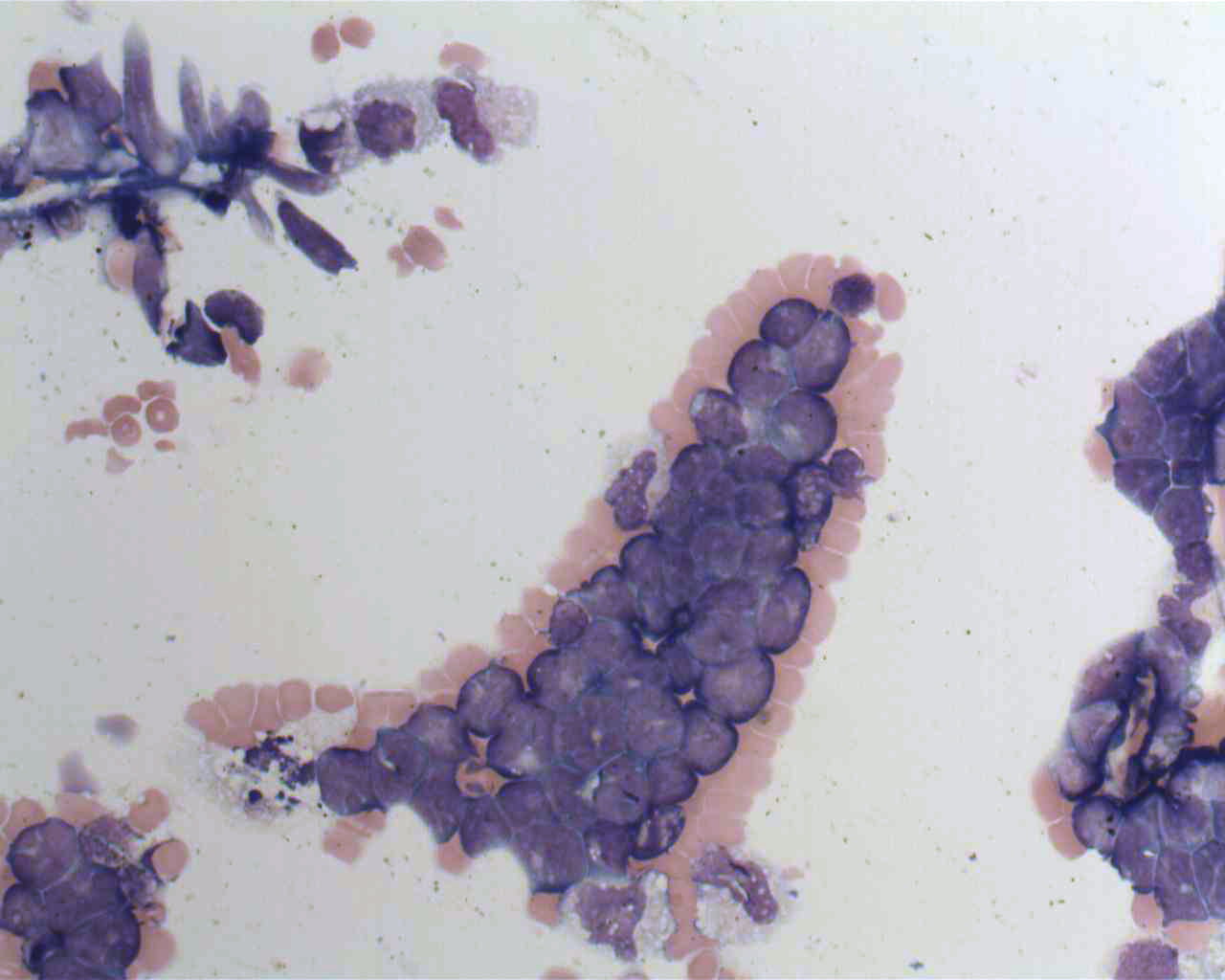
cytocentrifugation on:
[Wikipedia]
[Google]
[Amazon]
 A cytocentrifuge, sometimes referred to as a cytospin, is a specialized
 Some applications of cytocentrifuges include:
* Performing differential cell counts on body fluids, such as
 A cytocentrifuge, sometimes referred to as a cytospin, is a specialized
 centrifuge
A centrifuge is a device that uses centrifugal force to separate various components of a fluid. This is achieved by spinning the fluid at high speed within a container, thereby separating fluids of different densities (e.g. cream from milk) or l ...
used to concentrate cells in fluid specimens onto a microscope slide
A microscope slide is a thin flat piece of glass, typically 75 by 26 mm (3 by 1 inches) and about 1 mm thick, used to hold objects for examination under a microscope. Typically the object is mounted (secured) on the slide, and then ...
so that they can be stain
A stain is a discoloration that can be clearly distinguished from the surface, material, or medium it is found upon. They are caused by the chemical or physical interaction of two dissimilar materials. Accidental staining may make materials app ...
ed and examined. Cytocentrifuges are used in various areas of the clinical laboratory, such as cytopathology, hematology
Hematology ( always spelled haematology in British English) is the branch of medicine concerned with the study of the cause, prognosis, treatment, and prevention of diseases related to blood. It involves treating diseases that affect the produc ...
and microbiology
Microbiology () is the scientific study of microorganisms, those being unicellular (single cell), multicellular (cell colony), or acellular (lacking cells). Microbiology encompasses numerous sub-disciplines including virology, bacteriology, p ...
, as well as in biological research. The method can be used on many different types of specimens, including fine needle aspirates, cerebrospinal fluid
Cerebrospinal fluid (CSF) is a clear, colorless body fluid found within the tissue that surrounds the brain and spinal cord of all vertebrates.
CSF is produced by specialised ependymal cells in the choroid plexus of the ventricles of the b ...
, serous
In physiology, serous fluid or serosal fluid (originating from the Medieval Latin word ''serosus'', from Latin ''serum'') is any of various body fluids resembling serum, that are typically pale yellow or transparent and of a benign nature. The ...
and synovial fluid
Synovial fluid, also called synovia, elp 1/sup> is a viscous, non-Newtonian fluid found in the cavities of synovial joints. With its egg white–like consistency, the principal role of synovial fluid is to reduce friction between the articular ...
, and urine
Urine is a liquid by-product of metabolism in humans and in many other animals. Urine flows from the kidneys through the ureters to the urinary bladder. Urination results in urine being excreted from the body through the urethra.
Cellular m ...
.
Procedure
To prepare cytocentrifuge smears, a funnel assembly is attached to the front of a microscope slide. The surface of the funnel assembly that is in contact with the slide is lined with filter paper to absorb excess fluid. A few drops of fluid are placed in the funnel. The assembly is placed in the cytocentrifuge, which operates at a lowforce
In physics, a force is an influence that can change the motion of an object. A force can cause an object with mass to change its velocity (e.g. moving from a state of rest), i.e., to accelerate. Force can also be described intuitively as a ...
(600–800 x g) to preserve cellular structure. Centrifugal force
In Newtonian mechanics, the centrifugal force is an inertial force (also called a "fictitious" or "pseudo" force) that appears to act on all objects when viewed in a rotating frame of reference. It is directed away from an axis which is parallel ...
pushes the fluid through the funnel's opening and concentrates the cells in a small area of the slide. The centrifugation process concentrates cells by about twenty-fold and creates a one-cell-thick monolayer, allowing for assessment of cellular morphology. The slide can then be fixed and stained.
Applications
 Some applications of cytocentrifuges include:
* Performing differential cell counts on body fluids, such as
 serous
In physiology, serous fluid or serosal fluid (originating from the Medieval Latin word ''serosus'', from Latin ''serum'') is any of various body fluids resembling serum, that are typically pale yellow or transparent and of a benign nature. The ...
, synovial and cerebrospinal fluid
Cerebrospinal fluid (CSF) is a clear, colorless body fluid found within the tissue that surrounds the brain and spinal cord of all vertebrates.
CSF is produced by specialised ependymal cells in the choroid plexus of the ventricles of the b ...
* Cytopathology examination of liquid specimens such as body fluids and fine needle aspirates
* Gram stain
In microbiology and bacteriology, Gram stain (Gram staining or Gram's method), is a method of staining used to classify bacterial species into two large groups: gram-positive bacteria and gram-negative bacteria. The name comes from the Danish ...
ing of fluid specimens for identification of microorganism
A microorganism, or microbe,, ''mikros'', "small") and ''organism'' from the el, ὀργανισμός, ''organismós'', "organism"). It is usually written as a single word but is sometimes hyphenated (''micro-organism''), especially in olde ...
s
Limitations
The cytocentrifugation process can cause cells to appear distorted. Cells located at the centre of the smear may look compressed compared to cells at the periphery.Cell nuclei
The cell nucleus (pl. nuclei; from Latin or , meaning ''kernel'' or ''seed'') is a membrane-bound organelle found in eukaryotic cells. Eukaryotic cells usually have a single nucleus, but a few cell types, such as mammalian red blood cells, ha ...
may develop artifactual clefts, lobes, or holes, and the cytoplasm
In cell biology, the cytoplasm is all of the material within a eukaryotic cell, enclosed by the cell membrane, except for the cell nucleus. The material inside the nucleus and contained within the nuclear membrane is termed the nucleoplasm. Th ...
may appear vacuolated or develop irregular projections. Cytoplasmic granules may be pushed to the periphery of the cell. If the cell count is high, cells may be distorted due to crowding; therefore, samples with high cell counts are diluted prior to smear preparation.
History
Examination of cells in body fluids was historically performed using ahemocytometer
The hemocytometer (or haemocytometer) is a counting-chamber device originally designed and usually used for counting blood cells.
The hemocytometer was invented by Louis-Charles Malassez and consists of a thick glass microscope slide with a ...
, a chamber designed for counting cells microscopically. This technique was limited by poor discrimination between cell types (cells could only be classified as mononuclear or polymorphonuclear) and the low number of cells present in unconcentrated body fluids. Moreover, this technique did not produce a permanent record of the specimen. In a 1966 paper, Watson P. described the first cytocentrifuge, calling it "an apparatus for concentrating cells in suspension onto a microscope slide". The device was sold commercially in the 1970s and in 1983 it was patented by Shandon (now Thermo Scientific
Thermo Fisher Scientific Inc. is an American supplier of scientific instrumentation, reagents and consumables, and software services. Based in Waltham, Massachusetts, Thermo Fisher was formed through the merger of Thermo Electron and Fisher Sci ...
). As of 2012, numerous brands of cytocentrifuge exist on the market.
References
{{reflist, refs= {{cite book, author=Behdad Shambayati, title=Cytopathology, url=https://books.google.com/books?id=rVucAQAAQBAJ&pg=PA24, date=17 February 2011, publisher=OUP Oxford, isbn=978-0-19-953392-3, pages=24 {{cite book, author=Mary Louise Turgeon, title=Linné & Ringsrud's Clinical Laboratory Science: Concepts, Procedures, and Clinical Applications, url=https://books.google.com/books?id=QyvRoQEACAAJ, edition=7th, date=23 March 2015, publisher=Elsevier Mosby, isbn=978-0-323-22545-8, page=146 {{cite journal, last1=Stokes, first1=Barry O., title=Principles of Cytocentrifugation, journal=Laboratory Medicine, volume=35, issue=7, year=2004, pages=434–437, issn=0007-5027, doi=10.1309/FTT59GWKDWH69FB0, doi-access=free {{cite book, author1=Linda McManus , author2=Richard Mitchell , title=Pathobiology of Human Disease: A Dynamic Encyclopedia of Disease Mechanisms, url=https://books.google.com/books?id=uQB0AwAAQBAJ&pg=PA3365, date=1 August 2014, publisher=Elsevier Science, isbn=978-0-12-386457-4, pages=3365 {{cite book, author1=Elaine M. Keohane, author2=Larry Smith, author3=Jeanine M. Walenga, title=Rodak's Hematology: Clinical Principles and Applications, url=https://books.google.com/books?id=jjBTBwAAQBAJ, date=19 February 2015, publisher=Elsevier Health Sciences, isbn=978-0-323-32716-9, pages=270–1 {{cite book, author=Gary Gill, title=Cytopreparation: Principles & Practice, chapter-url=https://books.google.com/books?id=mvJPkbeXxUoC&pg=PA71, date=19 October 2012, publisher=Springer Science & Business Media, isbn=978-1-4614-4932-4, pages=73–84, chapter=Chapter 6: Cytocentrifugation {{cite journal, author=Watson P, title=A slide centrifuge: an apparatus for concentrating cells in suspension onto a microscope slide. , journal=J Lab Clin Med , year= 1966 , volume= 68 , issue= 3 , pages= 494–501 , pmid=5922759 , url=https://www.ncbi.nlm.nih.gov/entrez/eutils/elink.fcgi?dbfrom=pubmed&tool=sumsearch.org/cite&retmode=ref&cmd=prlinks&id=5922759 {{cite book, author=Nancy A. Brunzel, title=Fundamentals of Urine and Body Fluid Analysis, chapter-url=https://books.google.com/books?id=_D5yDQAAQBAJ&pg=PA356, date=5 November 2016, publisher=Elsevier Health Sciences, isbn=978-0-323-39636-3, pages=356–58, chapter=Chapter 17: Body fluid analysis {{cite book, author1=Connie R. Mahon, author2=Donald C. Lehman, author3=George Manuselis, title=Textbook of Diagnostic Microbiology - E-Book, url=https://books.google.com/books?id=VloMBAAAQBAJ&pg=PA129, date=25 March 2014, publisher=Elsevier Health Sciences, isbn=978-0-323-29262-7, pages=129 {{cite book, author=Denise Harmening, title=Clinical Hematology and Fundamentals of Hemostasis, url=https://books.google.com/books?id=W_NGPgAACAAJ, edition=5th, year=2009, publisher=F. A. Davis Company, isbn=978-0-8036-1732-2, chapter=Chapter 30: Body fluid examination: qualitative, quantitative and morphologic analysis, pages=720–757 {{cite book, author=Katherine A. Galagan, title=Color Atlas of Body Fluids: An Illustrated Field Guide Based on Proficiency Testing, url=https://books.google.com/books?id=_0E0AAAACAAJ, year=2006, publisher=College of American Pathologists
The College of American Pathologists (CAP) is a member-based physician organization founded in 1946 comprising approximately 18,000 board-certified pathologists. It serves patients, pathologists, and the public by fostering and advocating ...
, isbn=978-0-930304-91-1, pages=13–14
Centrifuges
Medical devices
Cytopathology