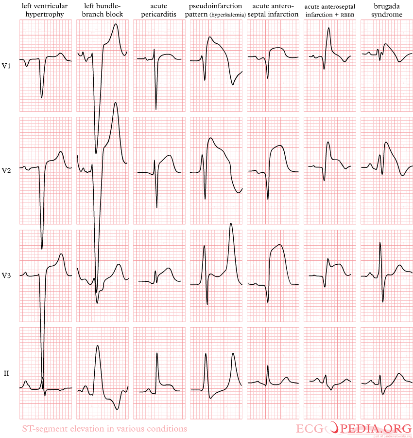
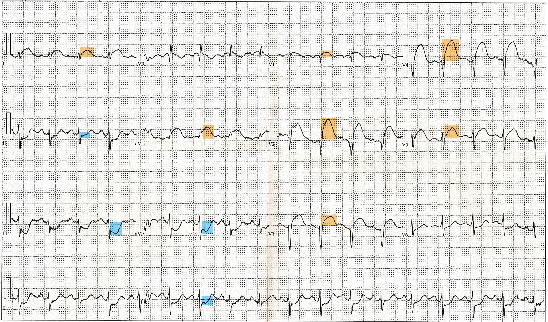
ST Elevations on:
[Wikipedia]
[Google]
[Amazon]
 ST elevation refers to a finding on an electrocardiogram wherein the trace in the

 An ST elevation is considered significant if the vertical distance inside the ECG trace and the baseline at a point 0.04 seconds after the
 When there is a blockage of the
 ST elevation refers to a finding on an electrocardiogram wherein the trace in the
 ST segment
In electrocardiography, the ST segment connects the QRS complex and the T wave and has a duration of 0.005 to 0.150 sec (5 to 150 ms).
It starts at the J point (junction between the QRS complex and ST segment) and ends at the beginning of the ...
is abnormally high above the baseline.
Electrophysiology
The ST segment starts from the J point (termination ofQRS complex
The QRS complex is the combination of three of the graphical deflections seen on a typical electrocardiogram (ECG or EKG). It is usually the central and most visually obvious part of the tracing. It corresponds to the depolarization of the r ...
and the beginning of ST segment) and ends with the T wave
In electrocardiography, the T wave represents the repolarization of the ventricles. The interval from the beginning of the QRS complex to the apex of the T wave is referred to as the ''absolute refractory period''. The last half of the T wave ...
. The ST segment is the plateau phase, in which the majority of the myocardial cells had gone through depolarization
In biology, depolarization or hypopolarization is a change within a cell, during which the cell undergoes a shift in electric charge distribution, resulting in less negative charge inside the cell compared to the outside. Depolarization is ess ...
but not repolarization
In neuroscience, repolarization refers to the change in membrane potential that returns it to a negative value just after the depolarization phase of an action potential which has changed the membrane potential to a positive value. The repolarizati ...
. The ST segment is the isoelectric line because there is no voltage difference across cardiac muscle
Cardiac muscle (also called heart muscle, myocardium, cardiomyocytes and cardiac myocytes) is one of three types of vertebrate muscle tissues, with the other two being skeletal muscle and smooth muscle. It is an involuntary, striated muscle tha ...
cell membrane during this state. Any distortion in the shape, duration, or height of the cardiac action potential
The cardiac action potential is a brief change in voltage (membrane potential) across the cell membrane of heart cells. This is caused by the movement of charged atoms (called ions) between the inside and outside of the cell, through proteins cal ...
can distort the ST segment.
Abnormalities

 An ST elevation is considered significant if the vertical distance inside the ECG trace and the baseline at a point 0.04 seconds after the
 J-point
The QRS complex is the combination of three of the graphical deflections seen on a typical electrocardiogram (ECG or EKG). It is usually the central and most visually obvious part of the tracing. It corresponds to the depolarization of the ri ...
is at least 0.1 mV (usually representing 1 mm or 1 small square) in a limb lead or 0.2 mV (2 mm or 2 small squares) in a precordial lead
Electrocardiography is the process of producing an electrocardiogram (ECG or EKG), a recording of the heart's electrical activity. It is an electrogram of the heart which is a graph of voltage versus time of the electrical activity of the hea ...
. The baseline is either the PR interval or the TP interval. This measure has a false positive
A false positive is an error in binary classification in which a test result incorrectly indicates the presence of a condition (such as a disease when the disease is not present), while a false negative is the opposite error, where the test resul ...
rate of 15–20% (which is slightly higher in women than men) and a false negative
A false positive is an error in binary classification in which a test result incorrectly indicates the presence of a condition (such as a disease when the disease is not present), while a false negative is the opposite error, where the test result ...
rate of 20–30%.
Myocardial infarction
 When there is a blockage of the
 coronary artery
The coronary arteries are the arterial blood vessels of coronary circulation, which transport oxygenated blood to the heart muscle. The heart requires a continuous supply of oxygen to function and survive, much like any other tissue or organ o ...
, there will be lack of oxygen supply to all three layers of cardiac muscle
Cardiac muscle (also called heart muscle, myocardium, cardiomyocytes and cardiac myocytes) is one of three types of vertebrate muscle tissues, with the other two being skeletal muscle and smooth muscle. It is an involuntary, striated muscle tha ...
(transmural ischemia). The leads facing the injured cardiac muscle cells will record the action potential as ST elevation during systole
Systole ( ) is the part of the cardiac cycle during which some chambers of the heart contract after refilling with blood. The term originates, via New Latin, from Ancient Greek (''sustolē''), from (''sustéllein'' 'to contract'; from ' ...
while during diastole
Diastole ( ) is the relaxed phase of the cardiac cycle when the chambers of the heart are re-filling with blood. The contrasting phase is systole when the heart chambers are contracting. Atrial diastole is the relaxing of the atria, and ventri ...
, there will be depression of the PR segment and the PT segment. Since PR and PT interval are regarded as baseline, ST segment elevation is regarded as a sign of myocardial ischemia. The opposing leads (such as V3 and V4 versus posterior leads V7–V9) always show reciprocal ST segment changes (ST elevation in one lead is followed by ST depression in the opposing lead). This is highly specific for myocardial infarction. An upsloping, convex ST segment is highly predictive of a myocardial infarction ( Pardee sign) while a concave ST elevation is less suggestive and can be found in other non-ischaemic causes. Following infarction, ventricular aneurysm
Ventricular aneurysms are one of the many complications that may occur after a heart attack. The word aneurysm refers to a bulge or 'pocketing' of the wall or lining of a vessel commonly occurring in the blood vessels at the base of the septum, o ...
can develop, which leads to persistent ST elevation, loss of S wave, and T wave inversion.
Weakening of the electrical activity of the cardiac muscles causes the decrease in height of the R wave
The QRS complex is the combination of three of the graphical deflections seen on a typical electrocardiogram (ECG or EKG). It is usually the central and most visually obvious part of the tracing. It corresponds to the depolarization of the ri ...
in those leads facing it. In opposing leads, it manifests as Q wave. However, Q waves may be found in healthy individuals at lead I, aVL, V5 and V6 due to left to right depolarisation.
Myocarditis/pericarditis
In these conditions, there will mostly be concave ST elevations in almost all the leads except for aVR and V1. These two leads, ST depression will be seen because they are the opposing leads of the cardiac axis. PR segment depression is highly suggestive of pericarditis. R wave in most cases will be unaltered. In two weeks after pericarditis, there will be upward concave ST elevation, positive T wave, and PR depression. After several more weeks, PR and ST segments normalised with flattened T wave. At last, there will be T wave inversion which will take weeks or months to vanish.Associated conditions
The topology and distribution of the affected areas depend on the underlying condition. Thus, ST elevation may be present on all or some leads of ECG. It can be associated with: *Myocardial infarction
A myocardial infarction (MI), commonly known as a heart attack, occurs when blood flow decreases or stops to the coronary artery of the heart, causing damage to the heart muscle. The most common symptom is chest pain or discomfort which ...
(see also ECG in myocardial infarction). ST elevation in select leads is more common with myocardial infarction. ST elevation only occurs in full thickness infarction
* Prinzmetal's angina
Variant angina, also known as Prinzmetal angina, vasospastic angina, angina inversa, coronary vessel spasm, or coronary artery vasospasm, is a syndrome typically consisting of angina (cardiac chest pain). Variant angina differs from stable angina ...
* Acute pericarditis
Acute pericarditis is a type of pericarditis (inflammation of the sac surrounding the heart, the pericardium) usually lasting less than 6 weeks. It is the most common condition affecting the pericardium.
Signs and symptoms
Chest pain is one of th ...
ST elevation in all leads (diffuse ST elevation) is more common with acute pericarditis.
* Left ventricular aneurysm
Ventricular aneurysms are one of the many complications that may occur after a heart attack. The word aneurysm refers to a bulge or 'pocketing' of the wall or lining of a vessel commonly occurring in the blood vessels at the base of the septum, o ...
* Blunt trauma
Blunt trauma, also known as blunt force trauma or non-penetrating trauma, is physical traumas, and particularly in the elderly who fall. It is contrasted with penetrating trauma which occurs when an object pierces the skin and enters a tissu ...
to the chest resulting in a cardiac contusion
A bruise, also known as a contusion, is a type of hematoma of tissue, the most common cause being capillaries damaged by trauma, causing localized bleeding that extravasates into the surrounding interstitial tissues. Most bruises occur clos ...
* Hyperkalemia
Hyperkalemia is an elevated level of potassium (K+) in the blood. Normal potassium levels are between 3.5 and 5.0 mmol/L (3.5 and 5.0 mEq/L) with levels above 5.5mmol/L defined as hyperkalemia. Typically hyperkalemia does not cause symptoms. Occa ...
* Acute myocarditis
Myocarditis, also known as inflammatory cardiomyopathy, is an acquired cardiomyopathy due to inflammation of the heart muscle. Symptoms can include shortness of breath, chest pain, decreased ability to exercise, and an irregular heartbeat. The ...
* Pulmonary embolism
Pulmonary embolism (PE) is a blockage of an artery in the lungs by a substance that has moved from elsewhere in the body through the bloodstream (embolism). Symptoms of a PE may include shortness of breath, chest pain particularly upon breathing ...
* Brugada syndrome
Brugada syndrome (BrS) is a genetic disorder in which the electrical activity of the heart is abnormal due to channelopathy. It increases the risk of abnormal heart rhythms and sudden cardiac death. Those affected may have episodes of syn ...
* Hypothermia
Hypothermia is defined as a body core temperature below in humans. Symptoms depend on the temperature. In mild hypothermia, there is shivering and mental confusion. In moderate hypothermia, shivering stops and confusion increases. In severe h ...
* J-point
The QRS complex is the combination of three of the graphical deflections seen on a typical electrocardiogram (ECG or EKG). It is usually the central and most visually obvious part of the tracing. It corresponds to the depolarization of the ri ...
elevation
* Early repolarization
* Subarachnoid hemorrhage
See also
*ST segment
In electrocardiography, the ST segment connects the QRS complex and the T wave and has a duration of 0.005 to 0.150 sec (5 to 150 ms).
It starts at the J point (junction between the QRS complex and ST segment) and ends at the beginning of the ...
* ST depression
ST depression refers to a finding on an electrocardiogram, wherein the trace in the ST segment is abnormally low below the baseline.
Causes
It is often a sign of myocardial ischemia, of which coronary insufficiency is a major cause. Other isch ...
References
{{Heart diseases Cardiac arrhythmia