Penfield Homunculus on:
[Wikipedia]
[Google]
[Amazon]
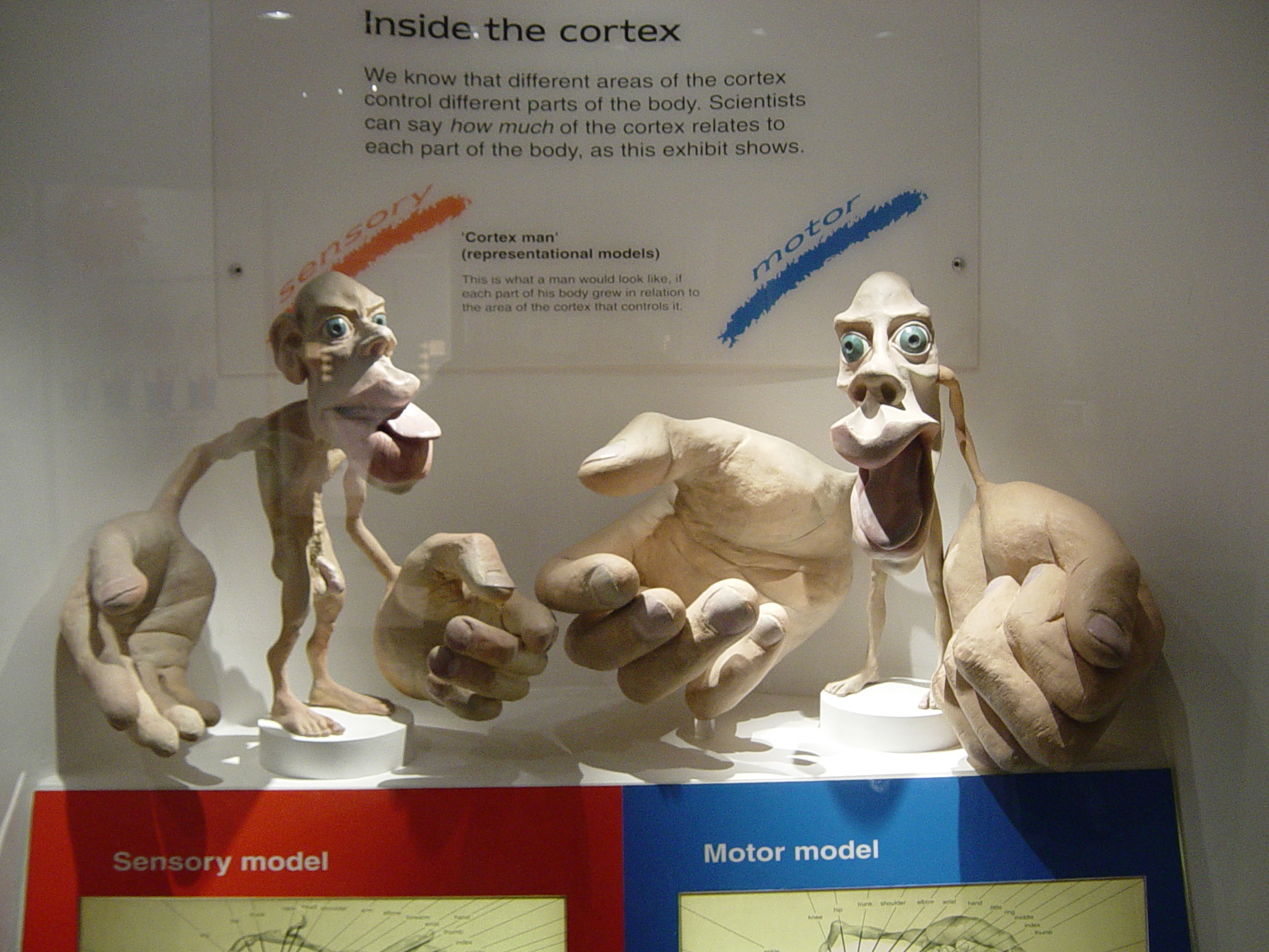
 A cortical homunculus () is a distorted representation of the
 A motor homunculus represents a map of brain areas dedicated to ''motor'' processing for different anatomical divisions of the body. The
 Dr.
Dr.
File:Side-black.gif, alt=Sharon Price-James' sensory homunculus from the side
File:Front of Sensory Homunculus.gif, alt=Sharon Price-James' sensory homunculus from the front
File:Rear of Sensory Homunculus.jpg, alt=Sharon Price-James' sensory homunculus from the back
In a 2021 article by Haven Wright and Preston Foerder published in the peer-reviewed journal ''Leonardo'' entitled "The Missing Female Homunculus", the authors revisit the history of the homunculus, shed light on current research in neuroscience on the female brain, and reveal what they believe to be the first sculpture of the female homunculus, done by the artist and first author Haven Wright, based on the current research available.
Mole-ratunculus
— an analog of a sensory homunculus for a
human body
The human body is the structure of a Human, human being. It is composed of many different types of Cell (biology), cells that together create Tissue (biology), tissues and subsequently organ systems. They ensure homeostasis and the life, viabi ...
, based on a neurological "map" of the areas and proportions of the human brain
The human brain is the central organ of the human nervous system, and with the spinal cord makes up the central nervous system. The brain consists of the cerebrum, the brainstem and the cerebellum. It controls most of the activities of the ...
dedicated to processing motor function
Motor control is the regulation of movement in organisms that possess a nervous system. Motor control includes reflexes as well as directed movement.
To control movement, the nervous system must integrate multimodal sensory information (both f ...
s, or sensory functions, for different parts of the body. Nerve fibres
An axon (from Greek ἄξων ''áxōn'', axis), or nerve fiber (or nerve fibre: see spelling differences), is a long, slender projection of a nerve cell, or neuron, in vertebrates, that typically conducts electrical impulses known as action po ...
conducting somatosensory information from all over the bodyterminate in various areas of the parietal lobe
The parietal lobe is one of the four major lobes of the cerebral cortex in the brain of mammals. The parietal lobe is positioned above the temporal lobe and behind the frontal lobe and central sulcus.
The parietal lobe integrates sensory informa ...
in the cerebral cortex
The cerebral cortex, also known as the cerebral mantle, is the outer layer of neural tissue of the cerebrum of the brain in humans and other mammals. The cerebral cortex mostly consists of the six-layered neocortex, with just 10% consisting of ...
, forming a representational map of the body.
Types
primary motor cortex
The primary motor cortex (Brodmann area 4) is a brain region that in humans is located in the dorsal portion of the frontal lobe. It is the primary region of the motor system and works in association with other motor areas including premotor co ...
is located in the precentral gyrus
The precentral gyrus is a prominent gyrus on the surface of the posterior frontal lobe of the brain. It is the site of the primary motor cortex that in humans is cytoarchitecturally defined as Brodmann area 4.
Structure
The precentral gyrus l ...
, and handles signals coming from the premotor area of the frontal lobe
The frontal lobe is the largest of the four major lobes of the brain in mammals, and is located at the front of each cerebral hemisphere (in front of the parietal lobe and the temporal lobe). It is parted from the parietal lobe by a groove betwe ...
s.
A sensory homunculus represents a map of brain areas dedicated to ''sensory'' processing for different anatomical divisions of the body. The primary sensory cortex
In neuroanatomy, the postcentral gyrus is a prominent gyrus in the lateral parietal lobe of the human brain. It is the location of the primary somatosensory cortex, the main sensory receptive area for the sense of touch. Like other sensory areas, ...
is located in the postcentral gyrus
In neuroanatomy, the postcentral gyrus is a prominent gyrus in the lateral parietal lobe of the human brain. It is the location of the primary somatosensory cortex, the main sensory receptive area for the somatosensory system, sense of touch. Lik ...
, and handles signals coming from the thalamus
The thalamus (from Greek θάλαμος, "chamber") is a large mass of gray matter located in the dorsal part of the diencephalon (a division of the forebrain). Nerve fibers project out of the thalamus to the cerebral cortex in all directions, ...
.
The thalamus itself receives corresponding signals from the brain stem
The brainstem (or brain stem) is the posterior stalk-like part of the brain that connects the cerebrum with the spinal cord. In the human brain the brainstem is composed of the midbrain, the pons, and the medulla oblongata. The midbrain is conti ...
and spinal cord
The spinal cord is a long, thin, tubular structure made up of nervous tissue, which extends from the medulla oblongata in the brainstem to the lumbar region of the vertebral column (backbone). The backbone encloses the central canal of the spi ...
.
Arrangement
Along the length of the primary motor and sensory cortices, the areas specializing in different parts of the body are arranged in an orderly manner, although ordered differently than one might expect. The toes are represented at the top of the cerebral hemisphere (or more accurately, "the upper end," since the cortex curls inwards and down at the top), and then as one moves down the hemisphere, progressively higher parts of the body are represented, assuming a body that is faceless and has arms raised. Going further down the cortex, the different areas of the face are represented, in approximately top-to-bottom order, rather than bottom-to-top as before. The homunculus is split in half, with motor and sensory representations for the left side of the body on the right side of the brain, and vice versa. The amount of cortex devoted to any given body region is not proportional to that body region's surface area or volume, but rather to how richly innervated that region is. Areas of the body with more complex and/or more numerous sensory or motor connections are represented as larger in the homunculus, while those with less complex and/or less numerous connections are represented as smaller. The resulting image is that of a distorted human body, with disproportionately huge hands, lips, and face. In the sensory homunculus, below the areas handling sensation for the teeth, gums, jaw, tongue, and pharynx lies an area for intra-abdominal sensation. At the very top end of the primary sensory cortex, beyond the area for the toes, it has traditionally been believed that the sensory neural networks for the genitals occur. However, more recent research has suggested that there may be two different cortical areas for the genitals, possibly differentiated by one dealing with erogenous stimulation and the other dealing with non-erogenous stimulation.Discovery
 Dr.
Dr. Wilder Penfield
Wilder Graves Penfield (January 26, 1891April 5, 1976) was an American Canadians, American-Physicians in Canada, Canadian neurosurgeon. He expanded brain surgery's methods and techniques, including mapping the functions of various regions of th ...
and his co-investigators Edwin Boldrey and Theodore Rasmussen are considered to be the originators of the sensory and motor homunculi. They were not the first scientists to attempt to objectify human brain function by means of a homunculus. However, they were the first to differentiate between sensory and motor function and to map the two across the brain separately, resulting in two different homunculi. In addition, their drawings and later drawings derived from theirs became perhaps the most famous conceptual maps in modern neuroscience because they compellingly illustrated the data at a single glance.
Penfield first conceived of his homunculi as a thought experiment, and went so far as to envision an imaginary world in which the homunculi lived, which he referred to as "if". He and his colleagues went on to experiment with electrical stimulation of different brain areas of patients undergoing open brain surgery to control epilepsy, and were thus able to produce the topographical brain maps and their corresponding homunculi.
More recent studies have improved this understanding of somatotopic arrangement
Somatotopy is the point-for-point correspondence of an area of the body to a specific point on the central nervous system. Typically, the area of the body corresponds to a point on the primary somatosensory cortex (postcentral gyrus). This cortex i ...
using techniques such as functional magnetic resonance imaging
Functional magnetic resonance imaging or functional MRI (fMRI) measures brain activity by detecting changes associated with blood flow. This technique relies on the fact that cerebral blood flow and neuronal activation are coupled. When an area o ...
(fMRI).
Representation
Penfield referred to his creations as "grotesque creatures" due to their strange-looking proportions. For example, the sensory nerves arriving from the hands terminate over large areas of the brain, resulting in the hands of the homunculus being correspondingly large. In contrast, the nerves emanating from the torso or arms cover a much smaller area, thus the torso and arms of the homunculus look comparatively small and weak. Penfield's homunculi are usually shown as 2-D diagrams. This is an oversimplification, as it cannot fully show the data set Penfield collected from his brain surgery patients. Rather than the sharp delineation between different body areas shown in the drawings, there is actually significant overlap between neighboring regions. The simplification suggests that lesions of the motor cortex will give rise to specific deficits in specific muscles. However, this is a misconception, as lesions produce deficits in groups of synergistic muscles. This finding suggests that themotor cortex
The motor cortex is the region of the cerebral cortex believed to be involved in the planning, control, and execution of voluntary movements.
The motor cortex is an area of the frontal lobe located in the posterior precentral gyrus immediately a ...
functions in terms of overall movements as coordinated groups of individual motions.
The sensorimotor homunculi can also be represented as 3-D figures (such as the sensory homunculus sculpted by Sharon Price-James shown from different angles below), which can make it easier for laymen to understand the ratios between the different body regions' levels of motor or sensory innervation. However, these 3-D models do not illustrate which areas of the brain are associated with which parts of the body.
References
External links
Mole-ratunculus
— an analog of a sensory homunculus for a
mole-rat Mole-rat or mole rat can refer to several groups of burrowing Old World rodents:
* Bathyergidae, a family of about 20 hystricognath species in six genera from Africa also called blesmols.
*'' Heterocephalus glaber'', the naked mole-rat.
* Spalaci ...
, from the paper:
*{{cite journal , title=Somatosensory cortex dominated by the representation of teeth in the naked mole-rat brain , volume=99 , issue=8 , bibcode=2002PNAS...99.5692C , last1=Catania , first1=Kenneth C. , last2=Remple , first2=Michael S. , journal=Proceedings of the National Academy of Sciences , year=2002 , page=5692 , doi=10.1073/pnas.072097999 , pmid=11943853 , s2cid=8869228 , quote=Fig. 3: ...This “mole-ratunculus” provides a graphic illustration of the cortical magnification of the incisors and head , doi-access=free
Sensory systems
Cerebrum
Thought experiments