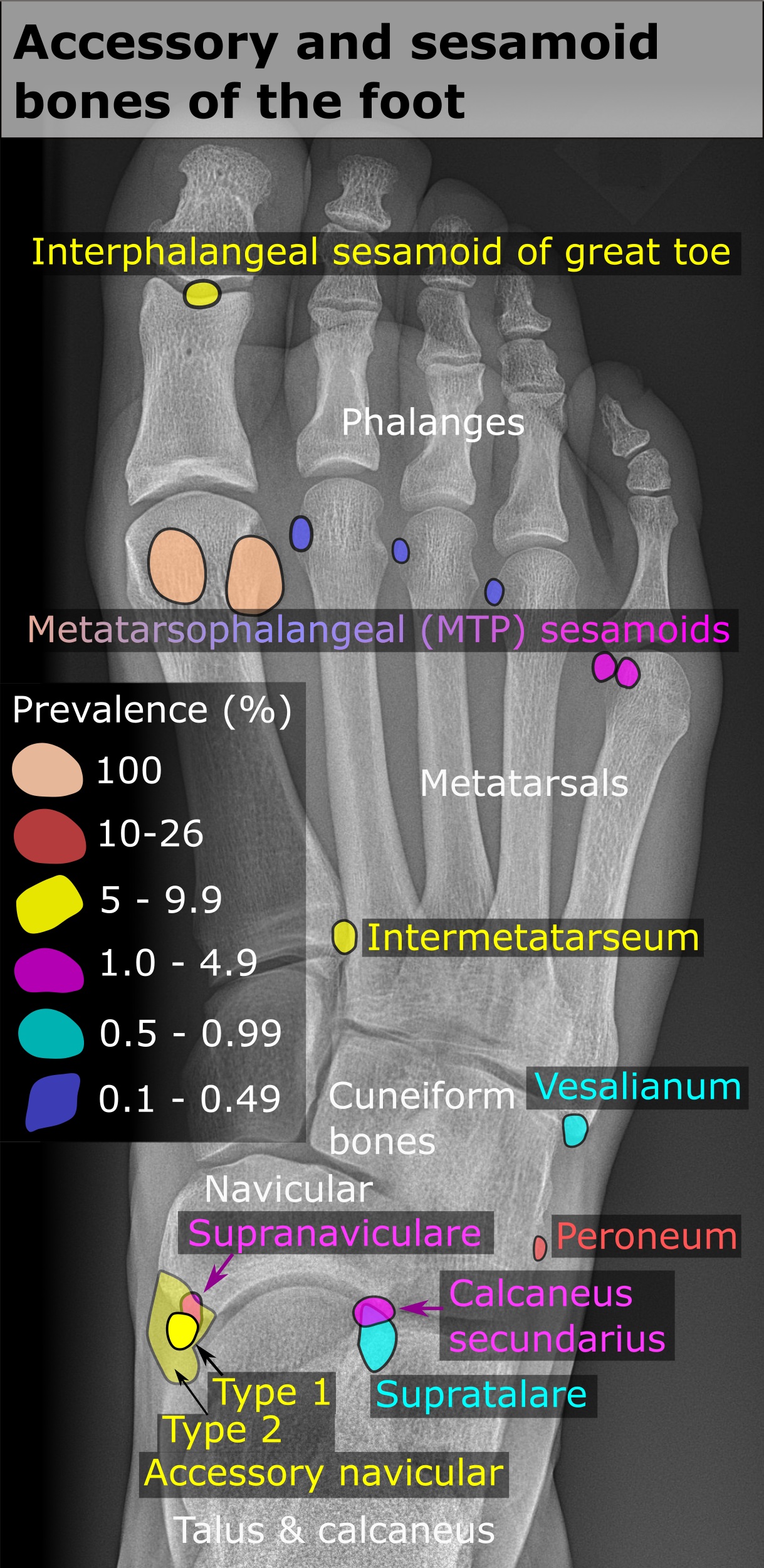
Os Peroneum on:
[Wikipedia]
[Google]
[Amazon]
 An accessory bone or supernumerary bone is a

 Accessory bones at the ankle mainly include:
*''Os subtibiale'', with a prevalence of approximately 1%. It is a secondary

 An
An
File:Os trigonum - Os talonaviculare.jpg
File:Os trigonum 1.jpg
File:Os trigonum2.jpg
File:Os trigonum 3.jpg

 An accessory bone or supernumerary bone is a
 bone
A bone is a rigid organ that constitutes part of the skeleton in most vertebrate animals. Bones protect the various other organs of the body, produce red and white blood cells, store minerals, provide structure and support for the body, an ...
that is not normally present in the body, but can be found as a variant in a significant number of people. It poses a risk of being misdiagnosed as bone fracture
A bone fracture (abbreviated FRX or Fx, Fx, or #) is a medical condition in which there is a partial or complete break in the continuity of any bone in the body. In more severe cases, the bone may be broken into several fragments, known as a '' ...
s on radiography
Radiography is an imaging technique using X-rays, gamma rays, or similar ionizing radiation and non-ionizing radiation to view the internal form of an object. Applications of radiography include medical radiography ("diagnostic" and "therapeuti ...
.
Wrist and hand

Os ulnostyloideum
The ''os ulnostyloideum'' is anulnar styloid process
The styloid process of the ulna is a bony prominence found at distal end of the ulna in the forearm.
Structure
The styloid process of the ulna projects from the medial and back part of the ulna. It descends a little lower than the head. The head ...
that is not fused to the rest of the ulna
The ulna (''pl''. ulnae or ulnas) is a long bone found in the forearm that stretches from the elbow to the smallest finger, and when in anatomical position, is found on the medial side of the forearm. That is, the ulna is on the same side of t ...
bone.R. O'Rahilly. ''A survey of carpal and tarsal anomalies.'' J Bone Joint Surg Am. 1953; 35: 626–642 On X-ray
X-rays (or rarely, ''X-radiation'') are a form of high-energy electromagnetic radiation. In many languages, it is referred to as Röntgen radiation, after the German scientist Wilhelm Conrad Röntgen, who discovered it in 1895 and named it ' ...
s, an ''os ulnostyloideum'' is sometimes mistaken for an avulsion fracture
An avulsion fracture is a bone fracture which occurs when a fragment of bone tears away from the main mass of bone as a result of physical trauma. This can occur at the ligament by the application of forces external to the body (such as a fal ...
of the styloid process. However, the distinction between these is extremely difficult.T.E. Keats, M.W. Anderson. ''Atlas of normal roentgen variants that may simulate disease''. 7th edition, Mosby Inc. 2001 It is alleged that the os ulnostyloideum has a close relationship with or is synonymous with the os triquetrum secundarium.
Os centrale
The ''os carpi centrale'' (also briefly ''os centrale'') is, where present, located on the dorsal side of the wrist between thescaphoid
The scaphoid bone is one of the carpal bones of the wrist. It is situated between the hand and forearm on the thumb side of the wrist (also called the lateral or radial side). It forms the radial border of the carpal tunnel. The scaphoid bone ...
, the trapezoid
A quadrilateral with at least one pair of parallel sides is called a trapezoid () in American and Canadian English. In British and other forms of English, it is called a trapezium ().
A trapezoid is necessarily a convex quadrilateral in Eucli ...
and capitate
The capitate bone is a bone in the human wrist found in the center of the carpal bone region, located at the distal end of the radius and ulna bones. It articulates with the third metacarpal bone (the middle finger) and forms the third carpomet ...
, radially to the deep fossa of the capitate. The bone is present in almost every human embryo of 17–49 mm length, but then usually fuses with the ulnar side of the scaphoid. Sometimes it fuses with the capitate or the trapezoid. The literature also refers to an ''os centrale'' at the palm of the carpus, but this existence is questioned.
In most primates, including orangutan
Orangutans are great apes native to the rainforests of Indonesia and Malaysia. They are now found only in parts of Borneo and Sumatra, but during the Pleistocene they ranged throughout Southeast Asia and South China. Classified in the gen ...
s and gibbon
Gibbons () are apes in the family Hylobatidae (). The family historically contained one genus, but now is split into four extant genera and 20 species. Gibbons live in subtropical and tropical rainforest from eastern Bangladesh to Northeast Indi ...
s, the os centrale is an independent bone that is attached to the scaphoid by strong ligaments. Conversely, in African apes
Homininae (), also called "African hominids" or "African apes", is a subfamily of Hominidae. It includes two tribes, with their extant as well as extinct species: 1) the tribe Hominini (with the genus ''Homo'' including modern humans and numerou ...
and humans, the os centrale normally fuses to the scaphoid early in development. In chimpanzees, the bone fuses with the scaphoid first after birth, while in gibbons and orangutans this occurs first at older age. A good number of scholars have construed the scaphoid-centrale fusion as a functional adaptation to knuckle-walking
Knuckle-walking is a form of quadrupedal walking in which the forelimbs hold the fingers in a partially flexed posture that allows body weight to press down on the ground through the knuckles. Gorillas, bonobos, and chimpanzees use this style of ...
, since a fused morphology would better cope with the increased shear stress on this joint during this kind of quadrupedal locomotion. The results from a simulation study have shown that fused scaphoid-centrales show lower stress values as compared to non fused morphologies, thus supporting a biomechanical explanation for the scaphoid-centrale fusion as a functional adaptation for knuckle-walking.
Ankle
 Accessory bones at the ankle mainly include:
*''Os subtibiale'', with a prevalence of approximately 1%. It is a secondary
 ossification center
An ossification center is a point where ossification of the cartilage begins. The first step in ossification is that the cartilage cells at this point enlarge and arrange themselves in rows.Gray and Spitzka (1910), page 44.
The matrix in which t ...
of the distal tibia that appears during the first year of life, and which in most people fuses with the shaft at approximately 15 years in females and approximately 17 years in males.
*''Os subfibulare'', with a prevalence of approximately 0.2%.
''Os trigonum'' (further described below) may also be seen on an ankle X-ray.
Foot

Accessory navicular
 An
An accessory navicular bone
An accessory navicular bone is an accessory bone of the foot that occasionally develops abnormally in front of the ankle towards the inside of the foot. This bone may be present in approximately 2-21% of the general population and is usually asym ...
, also called ''os tibiale externum'', occasionally develops in front of the ankle
The ankle, or the talocrural region, or the jumping bone (informal) is the area where the foot and the leg meet. The ankle includes three joints: the ankle joint proper or talocrural joint, the subtalar joint, and the inferior tibiofibular ...
towards the inside of the foot. This bone may be present in approximately 2–21% of the general population and is usually asymptomatic. When it is symptomatic, surgery may be necessary.
The Geist classification divides the accessory navicular bones into three types.
* Type 1: An os tibiale externum is a 2–3 mm sesamoid bone in the distal posterior tibialis tendon. Usually asymptomatic.
* Type 2: Triangular or heart-shaped ossicle measuring up to 12 mm, which represents a secondary ossification center connected to the navicular tuberosity by a 1–2 mm layer of fibrocartilage or hyaline cartilage. Portions of the posterior tibialis tendon sometimes insert onto the accessory ossicle, which can cause dysfunction, and therefore, symptoms.
* Type 3: A cornuate navicular bone represents an enlarged navicular tuberosity, which may represent a fused Type 2 accessory bone. Occasionally symptomatic due to bunion formation.
Os trigonum
The ''os trigonum'' or ''accessory talus'' represents a failure of fusion of the lateral tubercle of the posterior process of thetalus bone
The talus (; Latin for ankle or ankle bone), talus bone, astragalus (), or ankle bone is one of the group of foot bones known as the tarsus. The tarsus forms the lower part of the ankle joint. It transmits the entire weight of the body from the ...
. Is estimated to be present in 7–25% of adults. It can be mistaken for an avulsion fracture
An avulsion fracture is a bone fracture which occurs when a fragment of bone tears away from the main mass of bone as a result of physical trauma. This can occur at the ligament by the application of forces external to the body (such as a fal ...
of lateral tubercle of talus (Shepherd fracture) or a fracture of the Stieda process. In most cases, Os Trigonum will go unnoticed, but with some ankle injuries it can get trapped between the heel and ankle bones which irritates the surrounding structures, leading to Os Trigonum Syndrome.
Less common accessory bones

Other locations
Neck
*Nodules in the posterior margin of thenuchal ligament
The nuchal ligament is a ligament at the back of the neck that is continuous with the supraspinous ligament.
Structure
The nuchal ligament extends from the external occipital protuberance on the skull and median nuchal line to the spinous pro ...
form bone tissue
A bone is a rigid organ that constitutes part of the skeleton in most vertebrate animals. Bones protect the various other organs of the body, produce red and white blood cells, store minerals, provide structure and support for the body, and ...
in approximately 11% of males and 3–5% in females after the third decade of life, and may then be regarded to be sesamoid bone
In anatomy, a sesamoid bone () is a bone embedded within a tendon or a muscle. Its name is derived from the Arabic word for 'sesame seed', indicating the small size of most sesamoids. Often, these bones form in response to strain, or can be prese ...
s.
Shoulder
*An os acromiale forms when any of its fourossification center
An ossification center is a point where ossification of the cartilage begins. The first step in ossification is that the cartilage cells at this point enlarge and arrange themselves in rows.Gray and Spitzka (1910), page 44.
The matrix in which t ...
s fail to fuse. These four ossification centers are called (from tip to base) pre-acromion, meso-acromion, meta-acromion, and basi-acromion. In most cases, the first three fuse at 15–18 years, whereas the base part fuses to the scapular spine at 12 years. Such failure to fuse occurs in between 1% and 15% of cases. It rarely causes pain.
Vertebral column
*An ''Oppenheimer ossicle'' is found in approximately 4% (range 1–7%) of individuals. It is associated with the facet joints of lumbar spines. It usually occurs as a single, unilateral ossicle at the inferiorarticular processes
The articular processes or zygapophyses (Greek ζυγον = "yoke" (because it links two vertebrae) + απο = "away" + φυσις = " process") of a vertebra are projections of the vertebra that serve the purpose of fitting with an adjacent verteb ...
, but can also occur at the superior articular processes.
Knee
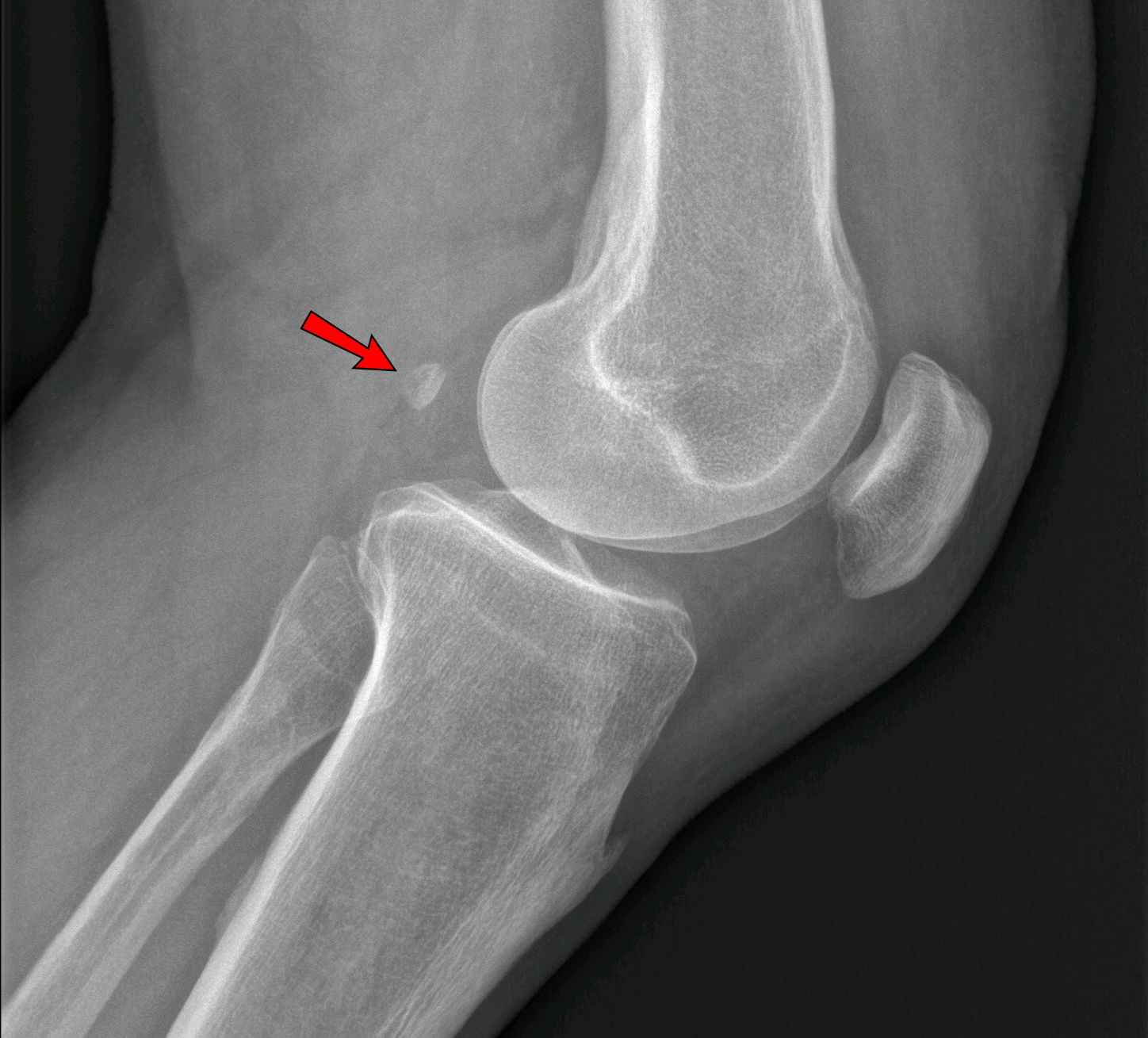
*Thefabella
The fabella is a small sesamoid bone found in some mammals embedded in the tendon of the lateral head of the gastrocnemius muscle behind the lateral condyle of the femur. It is an accessory bone, an anatomical variation present in 39% of human ...
is present in 10% to 30% of individuals.
See also
*Accessory muscle
An accessory muscle is a relatively rare anatomical variation where duplication of a muscle may appear anywhere in the muscular system. Treatment is not indicated unless the accessory muscle interferes with normal function. Examples are the stern ...
References
{{Bones of skeleton Skeletal system