microvascular dysfunction on:
[Wikipedia]
[Google]
[Amazon]
Microangiopathy (also known as microvascular disease, small vessel disease (SVD) or microvascular dysfunction) is a disease of the

 Optical coherence tomography angiography (OCTA) is another imaging modality that offers high-resolution visualization of the retinal capillary network and can be used to evaluate microcirculation in conditions such as diabetic retinopathy.
Many studies have demonstrated that evaluation of the retinal microvascular changes using OCTA or other methods such as fluorescein angiography may reflect the systemic microvascular functions as in patients with coronary microvascular disease, cerebral small vessel diseases or systemic sclerosis (The potential of retinal microvascularopathy as a biomarker for assessing microvascular status of other circulations).
Unlike the retinal microcirculation, the coronary microvasculature cannot be directly imaged. Instead, a number of different tests can be used to measure how much blood is flowing through the coronary microvasculature. These tests can be used to assess how well the coronary microvasculature is functioning and to diagnose coronary microvascular disease. They include non-invasive measures such as cardiac MRI and invasive measures such as intracoronary Doppler wire.
Similarly, CSVD is typically recognized on both brain magnetic resonance imaging (MRI) and computed tomography (CT) scans, but MRI has greater sensitivity and specificity. Neuroimaging of CSVD primarily involves visualizing radiological phenotypes of CSVD such as recent subcortical infarcts or cerebral microbleeds (CMBs).
microvessel
The microcirculation is the circulatory system, circulation of the blood in the smallest blood vessels, the microvessels of the microvasculature present within organ (anatomy), organ Tissue (biology), tissues. The microvessels include terminal ar ...
s, small blood vessel
Blood vessels are the tubular structures of a circulatory system that transport blood throughout many Animal, animals’ bodies. Blood vessels transport blood cells, nutrients, and oxygen to most of the Tissue (biology), tissues of a Body (bi ...
s in the microcirculation
The microcirculation is the circulation of the blood in the smallest blood vessels, the microvessels of the microvasculature present within organ tissues. The microvessels include terminal arterioles, metarterioles, capillaries, and venules. ...
. It can be contrasted to macroangiopathies such as atherosclerosis
Atherosclerosis is a pattern of the disease arteriosclerosis, characterized by development of abnormalities called lesions in walls of arteries. This is a chronic inflammatory disease involving many different cell types and is driven by eleva ...
, where large and medium-sized arteries (e.g., aorta
The aorta ( ; : aortas or aortae) is the main and largest artery in the human body, originating from the Ventricle (heart), left ventricle of the heart, branching upwards immediately after, and extending down to the abdomen, where it splits at ...
, carotid
In anatomy, the left and right common carotid arteries (carotids) () are arteries that supply the head and neck with oxygenated blood; they divide in the neck to form the external and internal carotid arteries.
Structure
The common carotid a ...
and coronary arteries
The coronary arteries are the arteries, arterial blood vessels of coronary circulation, which transport oxygenated blood to the Cardiac muscle, heart muscle. The heart requires a continuous supply of oxygen to function and survive, much like any ...
) are primarily affected.
Small vessel diseases (SVDs) affect primarily organs that receive significant portions of cardiac output such as the brain
The brain is an organ (biology), organ that serves as the center of the nervous system in all vertebrate and most invertebrate animals. It consists of nervous tissue and is typically located in the head (cephalization), usually near organs for ...
, the kidney
In humans, the kidneys are two reddish-brown bean-shaped blood-filtering organ (anatomy), organs that are a multilobar, multipapillary form of mammalian kidneys, usually without signs of external lobulation. They are located on the left and rig ...
, and the retina
The retina (; or retinas) is the innermost, photosensitivity, light-sensitive layer of tissue (biology), tissue of the eye of most vertebrates and some Mollusca, molluscs. The optics of the eye create a focus (optics), focused two-dimensional ...
. Thus, SVDs are a major etiologic cause in debilitating conditions such as renal failure
Kidney failure, also known as renal failure or end-stage renal disease (ESRD), is a medical condition in which the kidneys can no longer adequately filter waste products from the blood, functioning at less than 15% of normal levels. Kidney fa ...
, blindness
Visual or vision impairment (VI or VIP) is the partial or total inability of visual perception. In the absence of treatment such as corrective eyewear, assistive devices, and medical treatment, visual impairment may cause the individual difficul ...
, lacunar infarcts, and dementia
Dementia is a syndrome associated with many neurodegenerative diseases, characterized by a general decline in cognitive abilities that affects a person's ability to perform activities of daily living, everyday activities. This typically invo ...
.
Types
Microangiopathies are involved in a variety of different diseases including:Pathophysiology
The main target of small vessel diseases is theendothelium
The endothelium (: endothelia) is a single layer of squamous endothelial cells that line the interior surface of blood vessels and lymphatic vessels. The endothelium forms an interface between circulating blood or lymph in the lumen and the r ...
, which plays a key role in vascular homeostasis. The pathogenesis of SVDs in various organs is characterized by endothelial dysfunction, capillary rarefaction, microthrombi and microvascular remodeling.
Diabetic microangiopathy, which is the most common cause of microangiopathy, is more prevalent in the kidney
In humans, the kidneys are two reddish-brown bean-shaped blood-filtering organ (anatomy), organs that are a multilobar, multipapillary form of mammalian kidneys, usually without signs of external lobulation. They are located on the left and rig ...
, retina
The retina (; or retinas) is the innermost, photosensitivity, light-sensitive layer of tissue (biology), tissue of the eye of most vertebrates and some Mollusca, molluscs. The optics of the eye create a focus (optics), focused two-dimensional ...
and vascular endothelium since glucose transport in these sites isn’t regulated by insulin and these tissues cannot stop glucose from entering cells when blood sugar levels are high. Among all biochemical mechanisms involved in diabetic vascular damage such as the polyol pathway and the renin–angiotensin system
The renin–angiotensin system (RAS), or renin–angiotensin–aldosterone system (RAAS), is a hormone system that regulates blood pressure, fluid, and electrolyte balance, and systemic vascular resistance.
When renal blood flow is reduced, ...
(RAS), the advanced glycation end products (AGEs) pathway appears to be the most important in the pathogenesis and progression of microvascular complications.
Chronic high blood sugar levels lead to the attachment of sugar molecules to various proteins, including collagen
Collagen () is the main structural protein in the extracellular matrix of the connective tissues of many animals. It is the most abundant protein in mammals, making up 25% to 35% of protein content. Amino acids are bound together to form a trip ...
, laminin
Laminins are a family of glycoproteins of the extracellular matrix of all animals. They are major constituents of the basement membrane, namely the basal lamina (the protein network foundation for most cells and organs). Laminins are vital to bi ...
, and peripheral nerve proteins. This process, called glycosylation
Glycosylation is the reaction in which a carbohydrate (or ' glycan'), i.e. a glycosyl donor, is attached to a hydroxyl or other functional group of another molecule (a glycosyl acceptor) in order to form a glycoconjugate. In biology (but not ...
, creates advanced glycation end products (AGEs). AGEs formation cross-links these proteins, making them resistant to degradation. This leads to accumulation of AGEs, thickening of the basement membrane
The basement membrane, also known as base membrane, is a thin, pliable sheet-like type of extracellular matrix that provides cell and tissue support and acts as a platform for complex signalling. The basement membrane sits between epithelial tis ...
, narrowing the blood vessels, reducing blood flow to the tissues and causing ischemic
Ischemia or ischaemia is a restriction in blood supply to any tissue, muscle group, or organ of the body, causing a shortage of oxygen that is needed for cellular metabolism (to keep tissue alive). Ischemia is generally caused by problems ...
injury.
In addition, oxidative stress
Oxidative stress reflects an imbalance between the systemic manifestation of reactive oxygen species and a biological system's ability to readily detoxify the reactive intermediates or to repair the resulting damage. Disturbances in the normal ...
, caused by AGEs and the other pathways, causes apoptosis
Apoptosis (from ) is a form of programmed cell death that occurs in multicellular organisms and in some eukaryotic, single-celled microorganisms such as yeast. Biochemistry, Biochemical events lead to characteristic cell changes (Morphology (biol ...
of pericytes
Pericytes (formerly called Rouget cells) are multi-functional mural cells of the microcirculation that wrap around the endothelial cells that line the capillaries throughout the body. Pericytes are embedded in the basement membrane of blood capil ...
and podocytes
Podocytes are cells in Bowman's capsule in the kidneys that wrap around capillaries of the glomerulus. Podocytes make up the epithelial lining of Bowman's capsule, the third layer through which filtration of blood takes place. Bowman's capsule ...
in the retina and the kidneys respectively leading to capillary wall fragility and increased vascular leakage. This results in local swelling (e.g. macular edema
Macular edema occurs when fluid and protein deposits collect on or under the macula of the eye (a yellow central area of the retina) and causes it to thicken and swell ( edema). The swelling may distort a person's central vision, because the ma ...
) and impaired tissue function.
Microvascular diseases as a multisystem disorder
Some researchers have suggested that SVD may be a multisystem disorder, meaning that it can affect multiple organs in the body, including the heart and brain. This is supported by multiple studies stating that cardiac pathologies are more prevalent in patients with pathological evidence of cerebrovascular SVD and vice versa. Coronary microvascular diseases (CMDs) can be caused by: On the other hand, Cerebral SVD encompasses a range of vascular pathologies including arteriosclerosis-related CSVD, where lipohyalinosis causes stenosis of the lumen of the arterioles and amyloid-related CSVD, characterized by the build-up of β-amyloid deposits in small- and medium-caliber cerebral vessels. The vascular anatomy of the heart and brain is similar in that conduit arteries are distributed on the surface of these organs with tissue perfusion achieved through deep penetrating arteries. Both coronary and cerebral microvascular diseases do share some common risk factors such as hypertension. Why some patients with microvascular angina subsequently develop vascular cognitive impairment and others do not is an unanswered question. Potential underpinning mechanisms include premature vascular aging and clustering of vascular risk factors leading to an accelerated cardiovascular risk.Diagnosis
The diagnosis of microangiopathies can be based on direct visualization of the microcirculation, imaging modalities (e.g.MRI
Magnetic resonance imaging (MRI) is a medical imaging technique used in radiology to generate pictures of the anatomy and the physiological processes inside the body. MRI scanners use strong magnetic fields, magnetic field gradients, and rad ...
), conventional testings (e.g. ophthalmoscopy
Ophthalmoscopy, also called funduscopy, is a test that allows a health professional to see inside the fundus of the eye and other structures using an ophthalmoscope (or funduscope). It is done as part of an eye examination and may be done as part ...
for diabetic retinopathy) or other diagnostic measures (e.g. blood smear
A blood smear, peripheral blood smear or blood film is a thin layer of blood smeared on a glass microscope slide and then stained in such a way as to allow the various blood cells to be examined microscopically. Blood smears are examined in the i ...
for schistocytes in thrombotic microangiopathies).
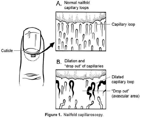
For assessment of the morphological and functional aspects of microcirculation, nailfold videocapillaroscopy (NVC) can be used, in which videocapillaroscopy is performed at the nailfold, where capillaries are arranged with the longitudinal axis parallel to the skin surface, so that they can be examined along their entire length.
NVC has been largely used not only for investigating peripheral microangiopathy, but also as a sort of "window" to systemic microvascular dysfunction. Although its main application is within the connective tissue diseases such as systemic scleroderma and dermatomyositis
Dermatomyositis (DM) is a Chronic condition, long-term inflammatory disorder, inflammatory Autoimmune disease, autoimmune disorder which affects the skin and the muscles. Its symptoms are generally a skin rash and worsening muscle weakness over ...
, it has been employed in non-rheumatic diseases with microvascular involvement such as diabetes mellitus, essential hypertension and COVID-19 infection.

 Optical coherence tomography angiography (OCTA) is another imaging modality that offers high-resolution visualization of the retinal capillary network and can be used to evaluate microcirculation in conditions such as diabetic retinopathy.
Many studies have demonstrated that evaluation of the retinal microvascular changes using OCTA or other methods such as fluorescein angiography may reflect the systemic microvascular functions as in patients with coronary microvascular disease, cerebral small vessel diseases or systemic sclerosis (The potential of retinal microvascularopathy as a biomarker for assessing microvascular status of other circulations).
Unlike the retinal microcirculation, the coronary microvasculature cannot be directly imaged. Instead, a number of different tests can be used to measure how much blood is flowing through the coronary microvasculature. These tests can be used to assess how well the coronary microvasculature is functioning and to diagnose coronary microvascular disease. They include non-invasive measures such as cardiac MRI and invasive measures such as intracoronary Doppler wire.
Similarly, CSVD is typically recognized on both brain magnetic resonance imaging (MRI) and computed tomography (CT) scans, but MRI has greater sensitivity and specificity. Neuroimaging of CSVD primarily involves visualizing radiological phenotypes of CSVD such as recent subcortical infarcts or cerebral microbleeds (CMBs).
Treatment
Treatment options of microangiopathies can be directed at: A better understanding of the mechanisms leading to damage of small blood vessels may be associated with novel therapeutic approaches, the safety and efficacy of some of which will need to be further investigated. Examples include calcium dobesilate and aldose reductase inhibitors in diabetic microangiopathies and endothelin receptor antagonists forpulmonary hypertension
Pulmonary hypertension (PH or PHTN) is a condition of increased blood pressure in the pulmonary artery, arteries of the lungs. Symptoms include dypsnea, shortness of breath, Syncope (medicine), fainting, tiredness, chest pain, pedal edema, swell ...
.
References
External links
{{Vascular diseases Histopathology