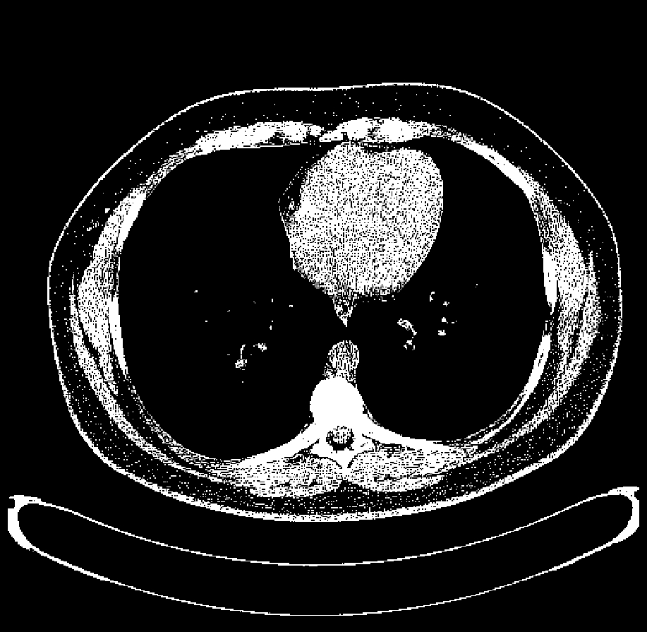
Hounsfield units on:
[Wikipedia]
[Google]
[Amazon]
The Hounsfield scale , named after Sir


 HU-based differentiation of material applies to medical-grade dual-energy CT scans but not to cone beam computed tomography (CBCT) scans, as CBCT scans provide unreliable HU readings.
Values reported here are approximations. Different dynamics are reported from one study to another.
Exact HU dynamics can vary from one CT acquisition to another due to CT acquisition and reconstruction parameters (kV, filters, reconstruction algorithms, etc.). The use of contrast agents modifies HU as well in some body parts (mainly blood).
A practical application of this is in evaluation of tumors, where, for example, an
 medscape >Adrenal Adenoma Imaging
Author: Perry J Horwich. Chief Editor: Eugene C Lin. Updated: Apr 21, 2011
Hounsfield Unit
- fpnotebook.com *{{cite web, url=http://www.intl.elsevierhealth.com/e-books/pdf/940.pdf, title=Introduction to CT physics, publisher=elsevierhealth.com, archive-url=https://web.archive.org/web/20070926231241/http://www.intl.elsevierhealth.com/e-books/pdf/940.pdf, archive-date=2007-09-26
Imaging of deep brain stimulation leads using extended Hounsfield unit CT. Stereotact Funct Neurosurg. 2009;87(3):155-60. doi: 10.1159/000209296
Radiology
Godfrey Hounsfield
Sir Godfrey Newbold Hounsfield (28 August 1919 – 12 August 2004) was an English electrical engineer who shared the 1979 Nobel Prize for Physiology or Medicine with Allan MacLeod Cormack for his part in developing the diagnostic technique of ...
, is a quantitative scale for describing radiodensity. It is frequently used in CT scans, where its value is also termed CT number.
Definition
The Hounsfield unit (HU) scale is a linear transformation of the original linear attenuation coefficient measurement into one in which the radiodensity of distilled water at standard pressure and temperature ( STP) is defined as 0 Hounsfield units (HU), while the radiodensity ofair
The atmosphere of Earth is the layer of gases, known collectively as air, retained by Earth's gravity that surrounds the planet and forms its planetary atmosphere. The atmosphere of Earth protects life on Earth by creating pressure allowing for ...
at STP is defined as −1000 HU. In a voxel with average linear attenuation coefficient , the corresponding HU value is therefore given by:
where and are respectively the linear attenuation coefficients of water and air.
Thus, a change of one Hounsfield unit (HU) represents a change of 0.1% of the attenuation coefficient of water since the attenuation coefficient of air is nearly zero.
Calibration tests of HU with reference to water and other materials may be done to ensure standardised response. This is particularly important for CT scans used in radiotherapy treatment planning
In radiotherapy, radiation treatment planning (RTP) is the process in which a team consisting of radiation oncologists, radiation therapist, medical physicists and medical dosimetrists plan the appropriate external beam radiotherapy or internal br ...
, where HU is converted to electron density
In quantum chemistry, electron density or electronic density is the measure of the probability of an electron being present at an infinitesimal element of space surrounding any given point. It is a scalar quantity depending upon three spatial va ...
. Variation in the measured values of reference materials with known composition, and variation between and within slices may be used as part of test procedures.
Rationale
The above standards were chosen as they are universally available references and suited to the key application for which computed axial tomography was developed: imaging the internal anatomy of living creatures based on organized water structures and mostly living in air, ''e.g.'' humans.Values for different body tissues and material


 HU-based differentiation of material applies to medical-grade dual-energy CT scans but not to cone beam computed tomography (CBCT) scans, as CBCT scans provide unreliable HU readings.
Values reported here are approximations. Different dynamics are reported from one study to another.
Exact HU dynamics can vary from one CT acquisition to another due to CT acquisition and reconstruction parameters (kV, filters, reconstruction algorithms, etc.). The use of contrast agents modifies HU as well in some body parts (mainly blood).
A practical application of this is in evaluation of tumors, where, for example, an
 adrenal tumor
An adrenal tumor or adrenal mass is any benign or malignant neoplasms of the adrenal gland, several of which are notable for their tendency to overproduce endocrine hormones. Adrenal cancer is the presence of malignant adrenal tumors, and includes ...
with a radiodensity of less than 10 HU is rather fatty in composition and almost certainly a benign adrenal adenoma
Adrenocortical adenoma is commonly described as a benign neoplasm emerging from the cells that comprise the adrenal cortex. Like most adenomas, the adrenocortical adenoma is considered a benign tumor since the majority of them are non-functioning ...
.Author: Perry J Horwich. Chief Editor: Eugene C Lin. Updated: Apr 21, 2011
See also
* Cone beam computed tomography: Bone density and the Hounsfield scale.References
*External links
*Hounsfield Unit
- fpnotebook.com *{{cite web, url=http://www.intl.elsevierhealth.com/e-books/pdf/940.pdf, title=Introduction to CT physics, publisher=elsevierhealth.com, archive-url=https://web.archive.org/web/20070926231241/http://www.intl.elsevierhealth.com/e-books/pdf/940.pdf, archive-date=2007-09-26
Imaging of deep brain stimulation leads using extended Hounsfield unit CT. Stereotact Funct Neurosurg. 2009;87(3):155-60. doi: 10.1159/000209296
Radiology