ground glass opacity on:
[Wikipedia]
[Google]
[Amazon]
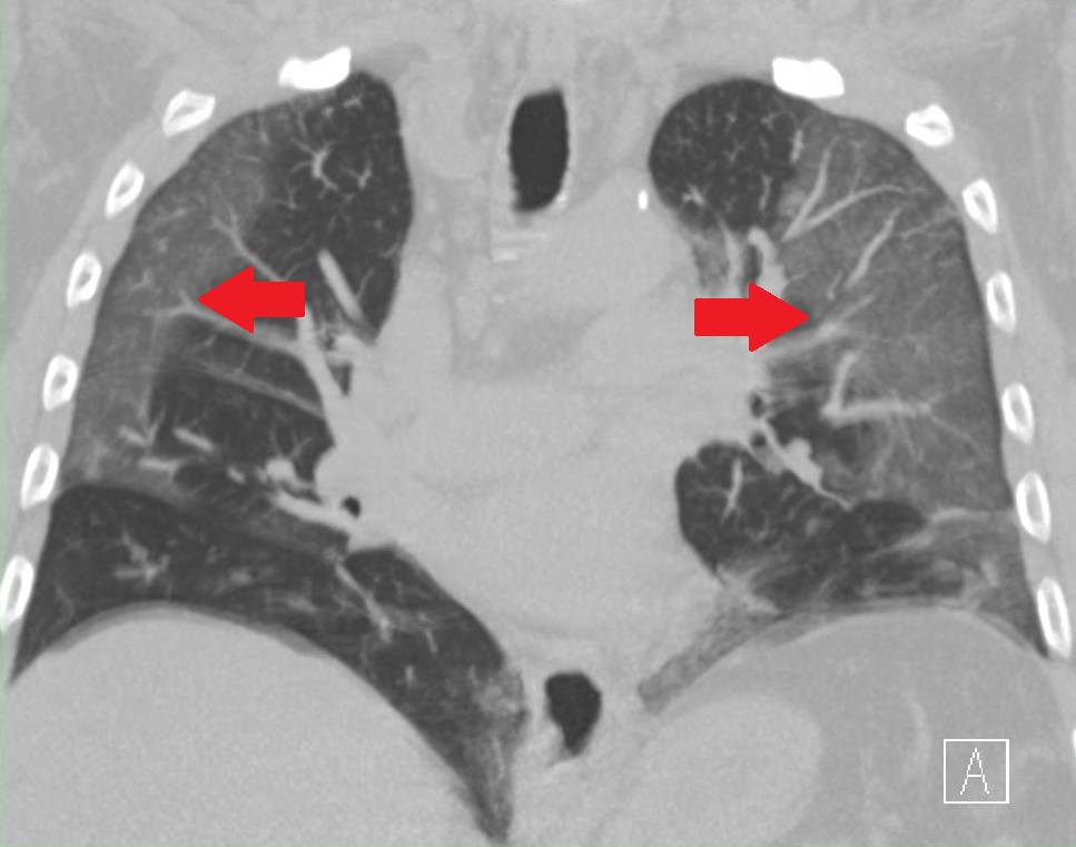 Ground-glass opacity (GGO) is a finding seen on
 *
*
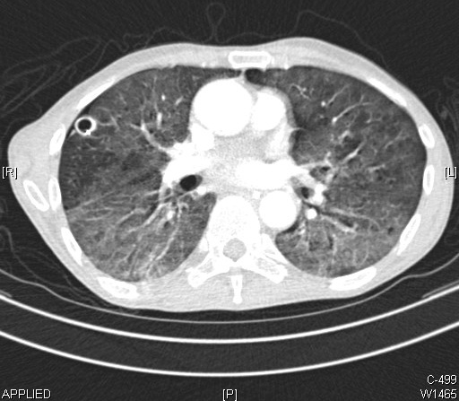
 Ground-glass opacity (GGO) is a finding seen on
 chest x-ray
A chest radiograph, called a chest X-ray (CXR), or chest film, is a projection radiograph of the chest used to diagnose conditions affecting the chest, its contents, and nearby structures. Chest radiographs are the most common film taken in med ...
(radiograph) or computed tomography (CT) imaging of the lung
The lungs are the primary organs of the respiratory system in humans and most other animals, including some snails and a small number of fish. In mammals and most other vertebrates, two lungs are located near the backbone on either side of t ...
s. It is typically defined as an area of hazy opacification (x-ray) or increased attenuation (CT) due to air displacement by fluid, airway collapse, fibrosis
Fibrosis, also known as fibrotic scarring, is a pathological wound healing in which connective tissue replaces normal parenchymal tissue to the extent that it goes unchecked, leading to considerable tissue remodelling and the formation of perma ...
, or a neoplastic process. When a substance other than air fills an area of the lung it increases that area's density. On both x-ray and CT, this appears more grey or hazy as opposed to the normally dark-appearing lungs. Although it can sometimes be seen in normal lungs, common pathologic causes include infection
An infection is the invasion of tissues by pathogens, their multiplication, and the reaction of host tissues to the infectious agent and the toxins they produce. An infectious disease, also known as a transmissible disease or communicable dise ...
s, interstitial lung disease
Interstitial lung disease (ILD), or diffuse parenchymal lung disease (DPLD), is a group of respiratory diseases affecting the interstitium (the tissue and space around the alveoli (air sacs)) of the lungs. It concerns alveolar epithelium, pulmo ...
, and pulmonary edema
Pulmonary edema, also known as pulmonary congestion, is excessive edema, liquid accumulation in the parenchyma, tissue and pulmonary alveolus, air spaces (usually alveoli) of the lungs. It leads to impaired gas exchange and may cause hypoxemia an ...
.
Definition
In both CT and chest radiographs, normal lungs appear dark due to the relative lowerdensity
Density (volumetric mass density or specific mass) is the substance's mass per unit of volume. The symbol most often used for density is ''ρ'' (the lower case Greek letter rho), although the Latin letter ''D'' can also be used. Mathematical ...
of air compared to the surrounding tissues. When air is replaced by another substance (e.g. fluid or fibrosis), the density of the area increases, causing the tissue to appear lighter or more grey.
Ground-glass opacity is most often used to describe findings in high-resolution CT
High-resolution computed tomography (HRCT) is a type of computed tomography (CT) with specific techniques to enhance image resolution. It is used in the diagnosis of various health problems, though most commonly for lung disease, by assessing t ...
imaging of the thorax
The thorax or chest is a part of the anatomy of humans, mammals, and other tetrapod animals located between the neck and the abdomen. In insects, crustaceans, and the extinct trilobites, the thorax is one of the three main divisions of the cre ...
, although it is also used when describing chest radiographs. In CT, the term refers to one or multiple areas of increased attenuation
In physics, attenuation (in some contexts, extinction) is the gradual loss of flux intensity through a medium. For instance, dark glasses attenuate sunlight, lead attenuates X-rays, and water and air attenuate both light and sound at variable att ...
(density) ''without'' concealment of the pulmonary vasculature. This appears more grey, as opposed to the normally dark-appearing (air-filled) lung on CT imaging. In chest radiographs, the term refers to one or multiple areas in which the normally darker-appearing (air-filled) lung appears more opaque, hazy, or cloudy. Ground-glass opacity is in contrast to consolidation, in which the pulmonary vascular markings are obscured. GGO can be used to describe both focal and diffuse areas of increased density. Subtypes of GGOs include diffuse, nodular, centrilobular, mosaic, crazy paving, halo sign, and reversed halo sign.
Causes
The differential diagnosis for ground-glass opacities is broad. General etiologies include infections, interstitial lung diseases, pulmonary edema,pulmonary hemorrhage
Pulmonary hemorrhage (or pulmonary haemorrhage) is an acute bleeding from the lung, from the upper respiratory tract and the trachea, and the pulmonary alveoli. When evident clinically, the condition is usually massive.
, and neoplasm. A correlation of imaging with a patient's clinical features is useful in narrowing the diagnosis. GGOs can be seen in normal lungs. Upon expiration there is less air in the lungs, leading to a relative increase in density of the tissue, and thus increased attenuation on CT. Furthermore, when a patient lays supine for a CT scan, the posterior lungs are in a dependent position, causing partial collapse of the posterior alveoli Alveolus (; pl. alveoli, adj. alveolar) is a general anatomical term for a concave cavity or pit.
Uses in anatomy and zoology
* Pulmonary alveolus, an air sac in the lungs
** Alveolar cell or pneumocyte
** Alveolar duct
** Alveolar macrophage
* ...
. This leads to an increase in density of the tissue, resulting increased attenuation and a possible ground-glass appearance on CT.
Infectious causes
In the setting ofpneumonia
Pneumonia is an inflammatory condition of the lung primarily affecting the small air sacs known as alveoli. Symptoms typically include some combination of productive or dry cough, chest pain, fever, and difficulty breathing. The severity ...
, the presence of GGO (as opposed to consolidation) is a useful diagnostic clue. Most bacterial infections lead to lobar consolidation, while atypical pneumonia
Atypical pneumonia, also known as walking pneumonia, is any type of pneumonia not caused by one of the pathogens most commonly associated with the disease. Its clinical presentation contrasts to that of "typical" pneumonia. A variety of microorgan ...
s may cause GGOs. It is important to note that while many of the pulmonary infections listed below may lead to GGOs, this does not occur in every case.

Bacterial
* Diffuse ** ''Mycoplasma pneumoniae
''Mycoplasma pneumoniae'' is a very small bacterium in the class Mollicutes. It is a human pathogen that causes the disease mycoplasma pneumonia, a form of atypical bacterial pneumonia related to cold agglutinin disease. ''M. pneumoniae'' is char ...
''
** ''Chlamydophila pneumoniae
''Chlamydia pneumoniae'' is a species of ''Chlamydia'', an obligate intracellular bacterium that infects humans and is a major cause of pneumonia. It was known as the Taiwan acute respiratory agent (TWAR) from the names of the two original isola ...
''
** '' Legionella pneumophilia''
* Focal or nodular
** ''Mycobacterium
''Mycobacterium'' is a genus of over 190 species in the phylum Actinomycetota, assigned its own family, Mycobacteriaceae. This genus includes pathogens known to cause serious diseases in mammals, including tuberculosis ('' M. tuberculosis'') and ...
''
** '' Nocardia''
** Septic emboli
Viral
*Adenovirus
Adenoviruses (members of the family ''Adenoviridae'') are medium-sized (90–100 nm), nonenveloped (without an outer lipid bilayer) viruses with an icosahedral nucleocapsid containing a double-stranded DNA genome. Their name derives from the ...
* Coronavirus
Coronaviruses are a group of related RNA viruses that cause diseases in mammals and birds. In humans and birds, they cause respiratory tract infections that can range from mild to lethal. Mild illnesses in humans include some cases of the com ...
(including MERS-CoV
''Middle East respiratory syndrome–related coronavirus'' (''MERS-CoV''), or EMC/2012 ( HCoV-EMC/2012), is the virus that causes Middle East respiratory syndrome (MERS). It is a species of coronavirus which infects humans, bats, and camels. Th ...
, SARS-CoV
Severe acute respiratory syndrome coronavirus 1 (SARS-CoV-1; or Severe acute respiratory syndrome coronavirus, SARS-CoV) is a strain of coronavirus that causes severe acute respiratory syndrome (SARS), the respiratory illness responsible for ...
, and SARS-CoV-2
Severe acute respiratory syndrome coronavirus 2 (SARS‑CoV‑2) is a strain of coronavirus that causes COVID-19 (coronavirus disease 2019), the respiratory illness responsible for the ongoing COVID-19 pandemic. The virus previously had a ...
)
* Cytomegalovirus (CMV)
* Herpes Simplex Virus (HSV)
* Human metapneumovirus (HMPV)
* Influenza
Influenza, commonly known as "the flu", is an infectious disease caused by influenza viruses. Symptoms range from mild to severe and often include fever, runny nose, sore throat, muscle pain, headache, coughing, and fatigue. These symptoms ...
 *
* Measles
Measles is a highly contagious infectious disease caused by measles virus. Symptoms usually develop 10–12 days after exposure to an infected person and last 7–10 days. Initial symptoms typically include fever, often greater than , cough, ...
* Respiratory Syncytial Virus (RSV)
* Varicella zoster
Varicella-zoster virus (VZV), also known as human herpesvirus 3 (HHV-3, HHV3) or ''Human alphaherpesvirus 3'' (taxonomically), is one of nine known herpes viruses that can infect humans. It causes chickenpox (varicella) commonly affecting chil ...
Fungal
* ''Pneumocystis jirovecii
''Pneumocystis jirovecii'' (previously ''P. carinii'') is a yeast-like fungus of the genus ''Pneumocystis''. The causative organism of ''Pneumocystis'' pneumonia, it is an important human pathogen, particularly among immunocompromised hosts. Pr ...
'' (PCP)
* Invasive aspergillosis
* Candidiasis
Candidiasis is a fungal infection due to any type of '' Candida'' (a type of yeast). When it affects the mouth, in some countries it is commonly called thrush. Signs and symptoms include white patches on the tongue or other areas of the mouth ...
* Mucormycosis
Mucormycosis, also known as black fungus, is a serious fungal infection that comes under fulminant fungal sinusitis, usually in people who are immunocompromised. It is curable only when diagnosed early. Symptoms depend on where in the body the ...
* Pulmonary cryptococcus
*Paracoccidioidomycosis
Paracoccidioidomycosis (PCM), also known as South American blastomycosis, is a fungal infection that can occur as a mouth and skin type, lymphangitic type, multi-organ involvement type (particularly lungs), or mixed type. If there are mouth ulce ...
Parasitic
* Pulmonary SchistosomiasisNon-infectious causes

Exposures
* Aspiration pneumonitis * Drug toxicity (most common includecyclophosphamide
Cyclophosphamide (CP), also known as cytophosphane among other names, is a medication used as chemotherapy and to suppress the immune system. As chemotherapy it is used to treat lymphoma, multiple myeloma, leukemia, ovarian cancer, breast cancer ...
, amiodarone
Amiodarone is an antiarrhythmic medication used to treat and prevent a number of types of cardiac dysrhythmias. This includes ventricular tachycardia (VT), ventricular fibrillation (VF), and wide complex tachycardia, as well as atrial fibrilla ...
, carmustine, methotrexate
Methotrexate (MTX), formerly known as amethopterin, is a chemotherapy agent and immune-system suppressant. It is used to treat cancer, autoimmune diseases, and ectopic pregnancies. Types of cancers it is used for include breast cancer, leuke ...
, and bleomycin
-13- (1''H''-imidazol-5-yl)methyl9-hydroxy-5- 1''R'')-1-hydroxyethyl8,10-dimethyl-4,7,12,15-tetraoxo-3,6,11,14-tetraazapentadec-1-yl}-2,4'-bi-1,3-thiazol-4-yl)carbonyl]amino}propyl)(dimethyl)sulfonium
, chemical_formula =
, C=55 , H=84 , N=1 ...
)
*Hypersensitivity pneumonitis
Hypersensitivity pneumonitis (HP) or extrinsic allergic alveolitis (EAA) is a syndrome caused by the repetitive inhalation of antigens from the environment in susceptible or sensitized people. Common antigens include molds, bacteria, bird dropping ...
* EVALI
* Radiation pneumonitis
Idiopathic interstitial pneumonia
*Acute interstitial pneumonitis
Acute interstitial pneumonitis is a rare, severe lung disease that usually affects otherwise healthy individuals. There is no known cause or cure.
Acute interstitial pneumonitis is often categorized as both an interstitial lung disease and a form ...
* Desquamative interstitial pneumonia
* Lymphocytic interstitial pneumonia
Lymphocytic interstitial pneumonia (LIP) is a syndrome secondary to autoimmune and other lymphoproliferative disorders. Symptoms include fever, cough, and shortness of breath. Lymphocytic interstitial pneumonia applies to disorders associated w ...
* Non-specific interstitial pneumonia
Non-specific interstitial pneumonia (NSIP) is a form of idiopathic interstitial pneumonia.
Symptoms
Symptoms include cough, difficulty breathing, and fatigue.
Causes
It has been suggested that idiopathic nonspecific interstitial pneumonia has a ...
* Cryptogenic organizing pneumonia
Cryptogenic organizing pneumonia (COP), formerly known as bronchiolitis obliterans organizing pneumonia (BOOP), is an inflammation of the bronchioles ( bronchiolitis) and surrounding tissue in the lungs. It is a form of idiopathic interstitial pn ...
Neoplastic processes
*
Lung adenocarcinoma
Adenocarcinoma of the lung is the most common type of lung cancer, and like other forms of lung cancer, it is characterized by distinct cellular and molecular features. It is classified as one of several non-small cell lung cancers (NSCLC), to d ...
* Adenocarcinoma in situ of the lung
Adenocarcinoma in situ (AIS) of the lung —previously included in the category of "bronchioloalveolar carcinoma" (BAC)—is a subtype of lung adenocarcinoma. It tends to arise in the distal bronchioles or alveoli and is defined by a non-invasive ...
* Atypical adenomatous hyperplasia Atypical adenomatous hyperplasia is a subtype of pneumocytic hyperplasia in the lung. It can be a precursor lesion of in situ adenocarcinoma of the lung (bronchioloalveolar carcinoma).
In prostate tissue biopsy, it can be confused for adenocarci ...
Additional causes
*Acute eosinophilic pneumonia
Acute eosinophilic pneumonia is the acute-onset form of eosinophilic pneumonia, a lung disease caused by the buildup of eosinophils, a type of white blood cell, in the lungs. It is characterized by a rapid onset of shortness of breath, cough, fat ...
*Cholesterol granulomas
*Focal interstitial fibrosis
*Granulomatosis with polyangiitis
Granulomatosis with polyangiitis (GPA), previously known as Wegener's granulomatosis (WG), is a rare long-term systemic disorder that involves the formation of granulomas and inflammation of blood vessels (vasculitis). It is a form of vasculitis ...
* Lymphatoid granulomatosis
* Pulmonary alveolar proteinosis
Pulmonary alveolar proteinosis (PAP) is a rare lung disorder characterized by an abnormal accumulation of surfactant-derived lipoprotein compounds within the alveoli of the lung. The accumulated substances interfere with the normal gas exchange and ...
*Pulmonary calcifications
*Pulmonary capillary hemangiomatosis
Pulmonary capillary hemangiomatosis (PCH) is a disease affecting the blood vessels of the lungs, where abnormal capillary proliferation and venous fibrous intimal thickening result in progressive increase in vascular resistance. It is a rare cause ...
*Pulmonary contusion
A pulmonary contusion, also known as lung contusion, is a bruise of the lung, caused by chest trauma. As a result of damage to capillaries, blood and other fluids accumulate in the lung tissue. The excess fluid interferes with gas exchange, pot ...
*Pulmonary edema
Pulmonary edema, also known as pulmonary congestion, is excessive edema, liquid accumulation in the parenchyma, tissue and pulmonary alveolus, air spaces (usually alveoli) of the lungs. It leads to impaired gas exchange and may cause hypoxemia an ...
*Pulmonary hemorrhage
Pulmonary hemorrhage (or pulmonary haemorrhage) is an acute bleeding from the lung, from the upper respiratory tract and the trachea, and the pulmonary alveoli. When evident clinically, the condition is usually massive.
* Pulmonary infarction
*Sarcoidosis
Sarcoidosis (also known as ''Besnier-Boeck-Schaumann disease'') is a disease involving abnormal collections of inflammatory cells that form lumps known as granulomata. The disease usually begins in the lungs, skin, or lymph nodes. Less commonly af ...
*Thoracic endometriosis Thoracic endometriosis is a rare form of endometriosis where endometrial-like tissue is found in the lung parenchyma and/or the pleura. It can be classified as either ''pulmonary'', or ''pleural'', respectively.Rojas, J. (2014). Endometriosis pulmon ...
Patterns
There are seven general patterns of ground-glass opacities. When combined with a patient's clinical signs and symptoms, the GGO pattern seen on imaging is useful in narrowing the differential diagnosis. It is important to note that while some disease processes present as only one pattern, many can present with a mixture of GGO patterns.Diffuse
The diffuse pattern typically refers to GGOs in multiple lobes of one or both lungs. Broadly, a diffuse pattern of GGO can be caused by displacement of air with fluid, inflammatory debris, or fibrosis. Cardiogenic pulmonary edema and ARDS are common causes of a fluid-filled lung.Diffuse alveolar hemorrhage
Pulmonary hemorrhage (or pulmonary haemorrhage) is an acute bleeding from the lung, from the upper respiratory tract and the trachea, and the pulmonary alveoli. When evident clinically, the condition is usually massive.
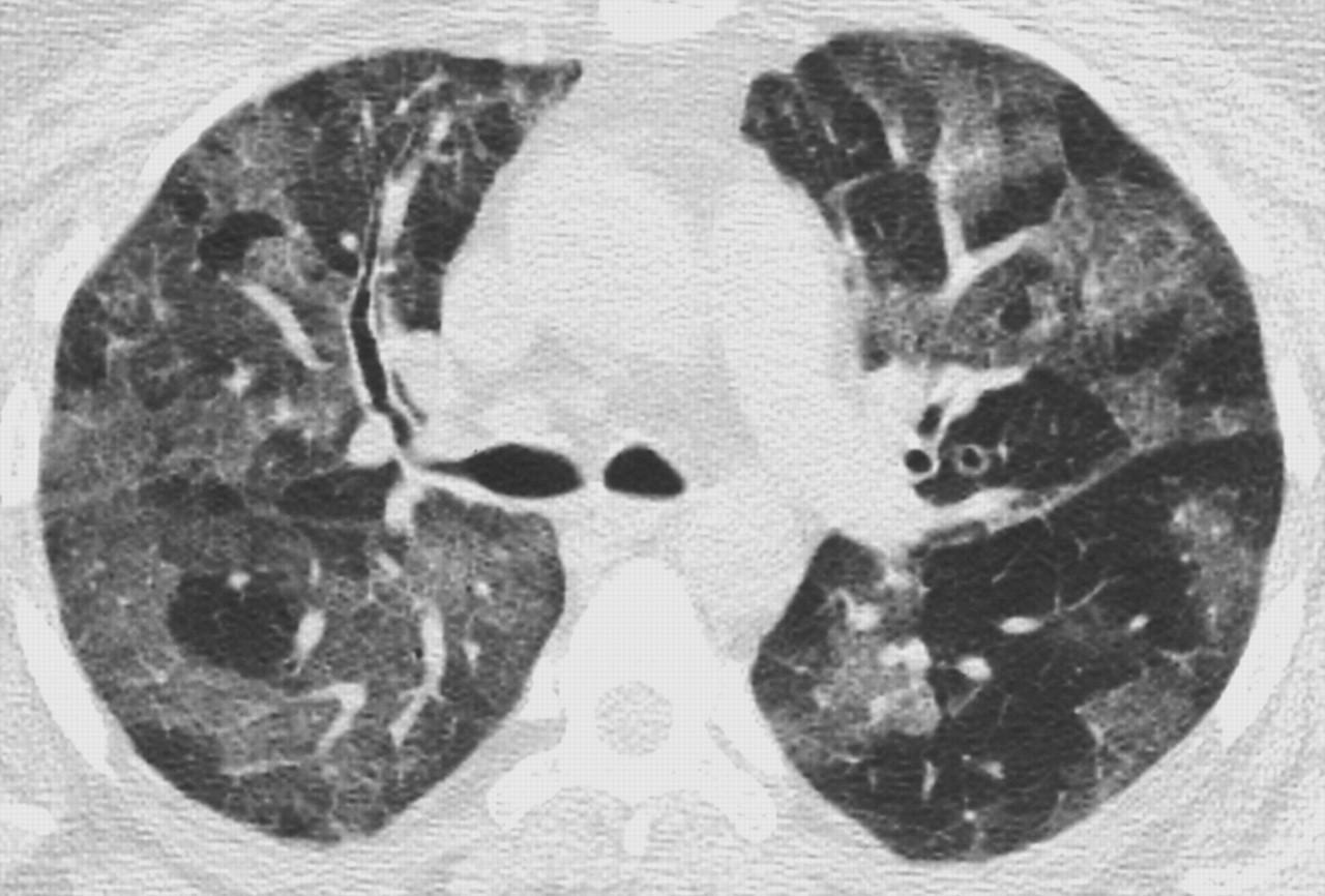
File:COVID-19 Pneumonie - 74m CTax - 003.jpg, CT showing diffuse ground-glass opacities in periphery of both lungs in patient with COVID-19.
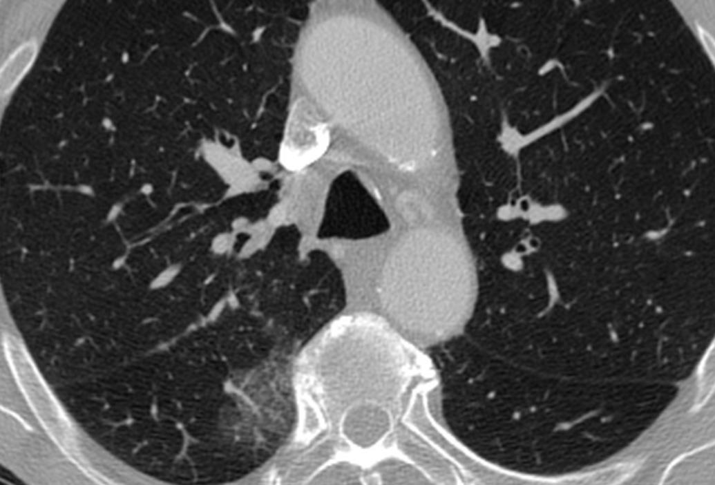
File:CT of ground glass lung nodule.png, CT image showing ground-glass nodule (circled).
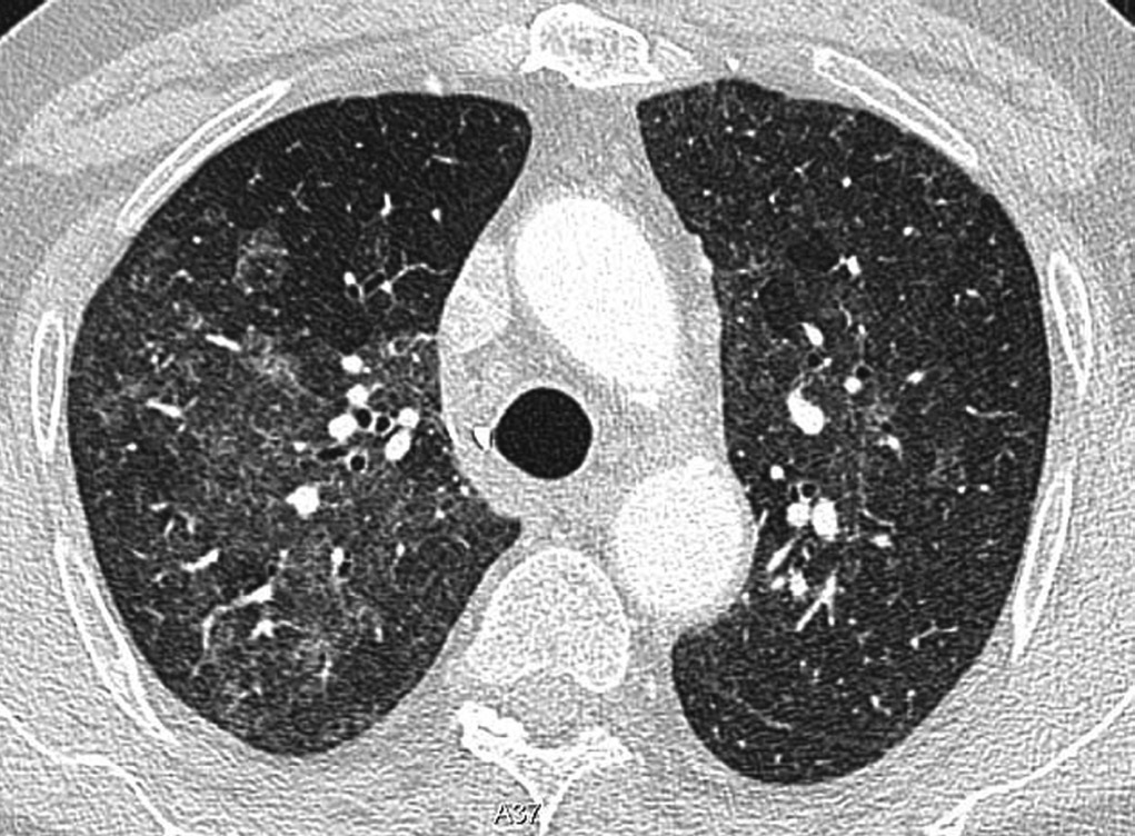
File:PulmonaryTB.png, CT image showing centrilobular pattern of GGOs in patient with pulmonary tuberculosis. Note the small, nodular areas of increased attenuation in both lungs.
File:Neumonitis por hipersensibilidad 2.jpg, CT image showing mosaic attenuation pattern in patient with hypersensitivity pneumonitis. Note the alternating, patchy areas of increased and decreased attenuation, particularly in the left lung (screen right).
File:Crazy paving pattern on chest CT scan.jpg, CT image showing crazy paving pattern of ground-glass opacities in both lungs.
File:CT of a subpleural nodule.png, CT image showing halo sign in patient with pulmonary aspergillosis. Note ground-glass opacification surrounding the area of consolidation (circled).
File:Chest CT with reversed halo sign.jpg, CT image of reversed halo sign in patient with organizing pneumonia.
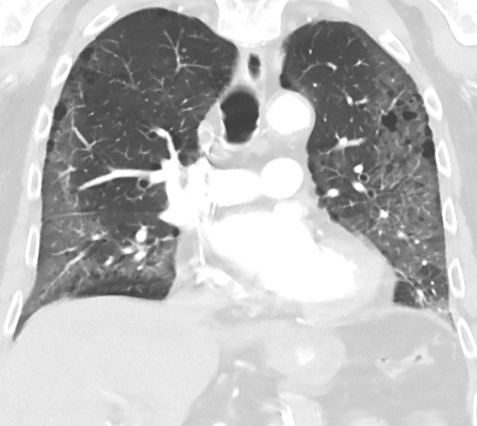 Ground-glass opacity is among the most common imaging findings in patients with confirmed
Ground-Glass Opacity of the Lung Parenchyma: A Guide to Analysis with High-Resolution CT
Radiologic signs
Inflammation
Inflammation (from la, wikt:en:inflammatio#Latin, inflammatio) is part of the complex biological response of body tissues to harmful stimuli, such as pathogens, damaged cells, or Irritation, irritants, and is a protective response involving im ...
and fibrosis can also cause diffuse GGOs. Pneumocystis
The Pneumocystidomycetes are a class of ascomycete fungi. It includes the single order Pneumocystidales, which contains the single monotypic family Pneumocystidaceae, which in turn contains the genus
Genus ( plural genera ) is a taxonomic r ...
pneumonia, an infection typically seen in immunocompromised
Immunodeficiency, also known as immunocompromisation, is a state in which the immune system's ability to fight infectious diseases and cancer is compromised or entirely absent. Most cases are acquired ("secondary") due to extrinsic factors that a ...
(e.g. patients with AIDS
Human immunodeficiency virus infection and acquired immunodeficiency syndrome (HIV/AIDS) is a spectrum of conditions caused by infection with the human immunodeficiency virus (HIV), a retrovirus. Following initial infection an individual m ...
) or immunosuppressed
Immunosuppression is a reduction of the activation or efficacy of the immune system. Some portions of the immune system itself have immunosuppressive effects on other parts of the immune system, and immunosuppression may occur as an adverse reacti ...
individuals, is a classic cause of diffuse GGOs. Many viral pneumonias and idiopathic interstitial pneumonias can also lead to a diffuse GGO pattern. Radiation pneumonitis, a side effect of pulmonary radiation therapy, can lead to pulmonary fibrosis and diffuse GGOs.
Nodular
There are numerous potential causes of nodular GGOs which can be broadly separated into benign and malignant conditions. Benign conditions potentially leading to the formation of nodular GGOs include aspergillosis, acute eosinophilic pneumonia, focal interstitial fibrosis, granulomatosis with polyangiitis,IgA vasculitis Iga may refer to:
Arts and entertainment
* Ambush at Iga Pass, a 1958 Japanese film
* Iga no Kagemaru, Japanese manga series
* Iga, a set of characters from the Japanese novel ''The Kouga Ninja Scrolls''
Biology
* ''Iga'' (beetle), a gen ...
, organizing pneumonia, pulmonary contusion, pulmonary cryptococcus, and thoracic endometriosis. Focal interstitial fibrosis presents a unique challenge when differentiating from malignant nodular GGOs on CT imaging. It is typically persistent over long-term imaging follow-up and shares a similar appearance to malignant nodular GGOs.
Pre-malignant or malignant causes of nodular GGOs include adenocarcinoma, adenocarcinoma in situ, and atypical adenomatous hyperplasia (AAH). One large review study found that 80% of nodular GGOs which were present on repeated CT imaging represented either pre-malignant or malignant growths. Differentiating between pre-malignancy and malignancy on the basis of CT alone can pose a challenge to radiologists; however, there are several features that are indicative of pre-malignant nodules. AAH is a pre-malignant cause of nodular GGO and is more commonly associated with lower attenuation on CT and smaller nodule size (<10 mm) compared to adenocarcinoma. In addition, AAH often lacks the solid features and spiculated appearance that are often associated with malignant growths. In contrast, as adenocarcinoma becomes invasive it will more often cause retraction of adjacent pleura and may show an increase in vascular markings. Nodules >15 mm almost always represent an invasive adenocarcinoma.
Centrilobular
Centrilobular GGOs refer to opacities occurring within one or multiple secondary lobules of the lung, which consist of a respiratory bronchiole, small pulmonary artery, and the surrounding tissue. A defining feature of these GGOs is the lack of involvement of the interlobular septum. Potential causes of centrilobular GGOs include pulmonary calcifications from metastatic disease, some types of idiopathic interstitial pneumonias, hypersensitivity pneumonitis, aspiration pneumonitis, cholesterol granulomas, and pulmonary capillary hemangiomastosis.Mosaic
Amosaic
A mosaic is a pattern or image made of small regular or irregular pieces of colored stone, glass or ceramic, held in place by plaster/mortar, and covering a surface. Mosaics are often used as floor and wall decoration, and were particularly pop ...
pattern of GGO refers to multiple irregular areas of both increased attenuation and decreased attenuation on CT. It is often the result of occlusion of small pulmonary arteries or obstruction of small airways leading to air trapping. Sarcoidosis is an additional cause of a mosaic GGOs due to the formation of granulomas in interstitial areas. This may coexist with granulomatosis with polyangiitis, leading to diffuse areas of increased attenuation with ground-glass appearance.
Crazy paving
The crazy paving pattern may occur when there is both interlobular and intralobular widening. This sometimes resembles a road paved with irregular bricks or tiles. It is typically diffuse, involving larger areas of one or multiple lobes. There are a variety of potential causes, including Pneumocystis pneumonia, late-stage adenocarcinoma, pulmonary edema, some types of idiopathic interstitial pneumonias, diffuse alveolar hemorrhage, sarcoidosis, and pulmonary alveolar proteinosis. COVID-19 has also been shown to occasionally cause GGOs with a crazy paving pattern.Halo sign
A halo sign refers to a GGO that fills the area around a consolidation or nodule. This is a most commonly seen in various types of pulmonary infections, including CMV pneumonia, tuberculosis, nocardia infection, some fungal pneumonias, and septic emboli. Schistosomiasis, aparasitic
Parasitism is a close relationship between species, where one organism, the parasite, lives on or inside another organism, the host, causing it some harm, and is adapted structurally to this way of life. The entomologist E. O. Wilson has c ...
infection, also commonly presents with the halo sign. Important non-infectious causes include granulomatosis with polyangiitis, metastatic disease with pulmonary hemorrhage, and some types of idiopathic interstitial pneumonias.
Reversed halo sign
A reversed halo sign is a central ground-glass opacity surrounded by denser consolidation. According to published criteria, the consolidation should form more than three-fourths of a circle and be at least 2 mm thick. It is often suggestive oforganizing pneumonia
Cryptogenic organizing pneumonia (COP), formerly known as bronchiolitis obliterans organizing pneumonia (BOOP), is an inflammation of the bronchioles ( bronchiolitis) and surrounding tissue in the lungs. It is a form of idiopathic interstitial pn ...
, but is only seen in about 20% of individuals with this condition. It can also be present in lung infarction where the halo consists of hemorrhage, as well as in infectious diseases such as paracoccidioidomycosis
Paracoccidioidomycosis (PCM), also known as South American blastomycosis, is a fungal infection that can occur as a mouth and skin type, lymphangitic type, multi-organ involvement type (particularly lungs), or mixed type. If there are mouth ulce ...
, tuberculosis
Tuberculosis (TB) is an infectious disease usually caused by '' Mycobacterium tuberculosis'' (MTB) bacteria. Tuberculosis generally affects the lungs, but it can also affect other parts of the body. Most infections show no symptoms, in ...
, and aspergillosis
Aspergillosis is a fungal infection of usually the lungs, caused by the genus ''Aspergillus'', a common mould that is breathed in frequently from the air around, but does not usually affect most people. It generally occurs in people with lung di ...
, as well as in granulomatosis with polyangiitis
Granulomatosis with polyangiitis (GPA), previously known as Wegener's granulomatosis (WG), is a rare long-term systemic disorder that involves the formation of granulomas and inflammation of blood vessels (vasculitis). It is a form of vasculitis ...
, lymphomatoid granulomatosis, and sarcoidosis
Sarcoidosis (also known as ''Besnier-Boeck-Schaumann disease'') is a disease involving abnormal collections of inflammatory cells that form lumps known as granulomata. The disease usually begins in the lungs, skin, or lymph nodes. Less commonly af ...
.COVID-19
 Ground-glass opacity is among the most common imaging findings in patients with confirmed
 COVID-19
Coronavirus disease 2019 (COVID-19) is a contagious disease caused by a virus, the severe acute respiratory syndrome coronavirus 2 (SARS-CoV-2). The first known case was COVID-19 pandemic in Hubei, identified in Wuhan, China, in December ...
. One systematic review found that among patients with COVID-19 and abnormal lung findings on CT, greater than 80% had GGOs, with greater than 50% having mixed GGOs and consolidation. GGOs with mixed consolidation has most often been found in elderly populations.
Several studies have described a pattern among initial, intermediate, and hospital discharge imaging findings in the disease course of COVID-19. Most commonly, initial CT imaging reveals bilateral GGOs at the periphery of the lungs. During initial stages, this is most often found in the lower lobes, although involvement of the upper lobes and right middle lobe has also been reported early in the disease course. This is in contrast to the two similar coronaviruses, SARS
Severe acute respiratory syndrome (SARS) is a viral respiratory disease of zoonotic origin caused by the severe acute respiratory syndrome coronavirus (SARS-CoV or SARS-CoV-1), the first identified strain of the SARS coronavirus species, ''sever ...
and MERS
Middle East respiratory syndrome (MERS) is a viral respiratory infection caused by ''Middle East respiratory syndrome–related coronavirus'' (MERS-CoV). Symptoms may range from none, to mild, to severe. Typical symptoms include fever, cough, ...
, which more commonly involve only one lung on initial imaging. As the COVID-19 infection progresses, GGOs typically become more diffuse and often progress to consolidation. This is sometimes accompanied by the development of a crazy paving pattern and interlobular septal thickening. In many cases the most severe pulmonary CT abnormalities occurred within 2 weeks after symptoms began. At this point, many individuals begin showing resolution of consolidation and GGOs as symptoms improve. However, some patients have worsening symptoms and imaging findings, with further increase in septal thickening, GGOs, and consolidation. These patients may develop lung "white-out" with progression to acute respiratory distress syndrome (ARDS) requiring treatment escalation.
Preliminary reports have shown many patients have residual GGOs at time of discharge from the hospital. Due to the novelty of COVID-19, large studies investigating the long-term pulmonary CT changes have yet to be completed. However, long-term pulmonary changes have been seen in patients after recovery from SARS and MERS, suggesting the possibility of similar long-term complications in patients who have recovered from acute COVID-19 infection.
History
The first usage of "ground-glass opacity" by a major radiological society occurred in a 1984 publication of the ''American Journal of Roentgenology.'' It was published as part of a glossary of recommended nomenclature from theFleischner Society
The Fleischner Society is an international, multidisciplinary medical society for thoracic radiology, dedicated to the diagnosis and treatment of diseases of the chest. Founded in 1969 by eight radiologists whose predominant professional interests ...
, a group of thoracic imaging radiologists. The original published definition read as: "Any extended, finely granular pattern of pulmonary opacity within which normal anatomic details are partly obscured; from a fancied resemblance to etched or abraded glass." It was again included in an updated glossary by the Fleischner Society in 2008 with a more detailed definition.
See also
*Pulmonary consolidation
A pulmonary consolidation is a region of normally compressible lung tissue that has filled with liquid instead of air. The condition is marked by induration (swelling or hardening of normally soft tissue) of a normally aerated lung. It is conside ...
* Pulmonary infiltrate A pulmonary infiltrate is a substance denser than air, such as pus, blood, or protein, which lingers within the parenchyma of the lungs. Pulmonary infiltrates are associated with pneumonia, tuberculosis, and sarcoidosis.
Pulmonary infiltrates can b ...
References
{{reflistExternal links
Ground-Glass Opacity of the Lung Parenchyma: A Guide to Analysis with High-Resolution CT
Radiologic signs