Alveolar cleft grafting on:
[Wikipedia]
[Google]
[Amazon]
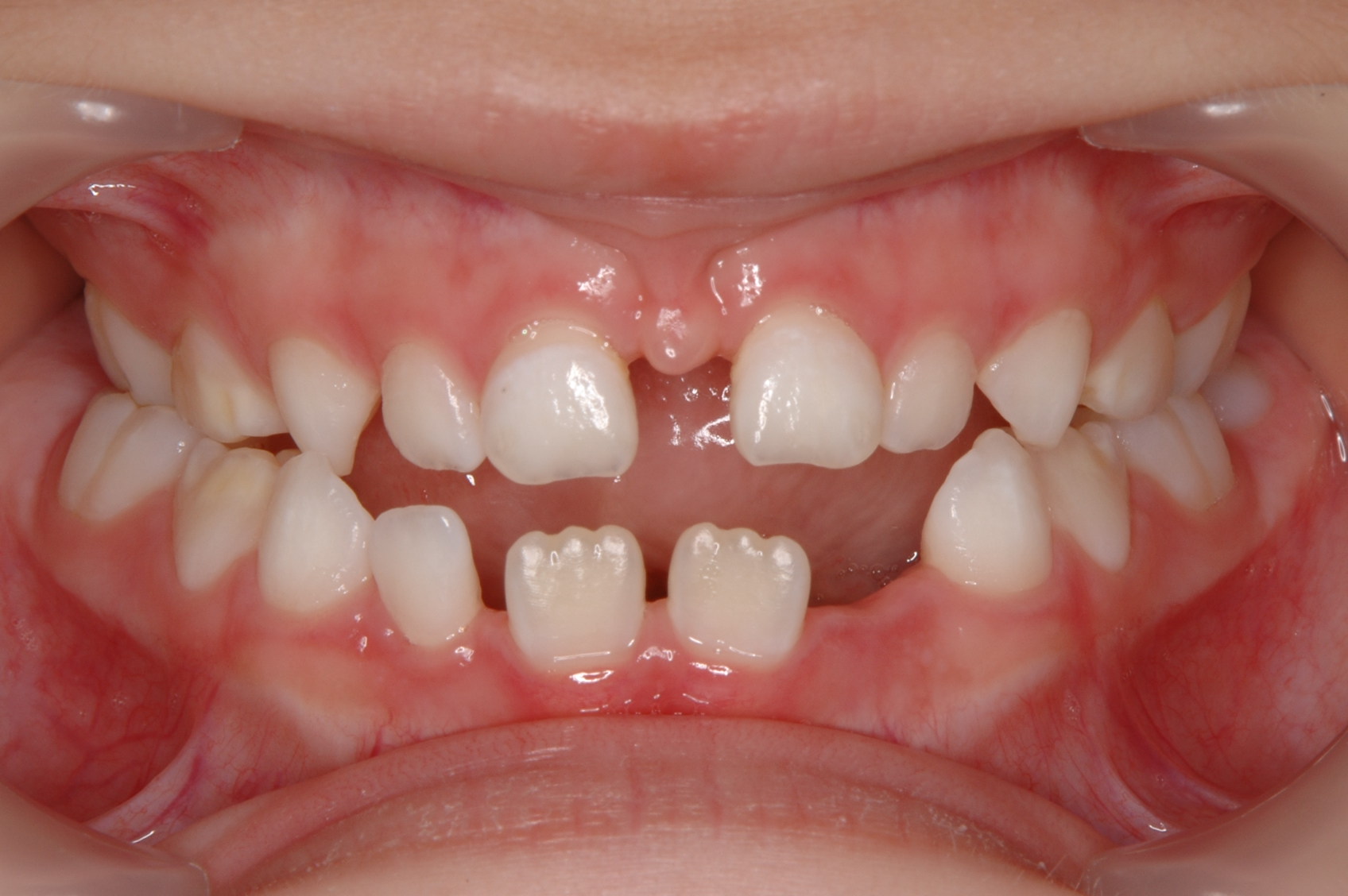
Alveolar cleft grafting is a surgical procedure, used to repair the defect in the

 One of the most controversial topics in alveolar cleft grafting is the timing of treatment, however, most centres recommend grafting around the ages of 6–8 years old as the
 In cleft lip and palate cases, the
 The goals of the surgery are to:
# Close any
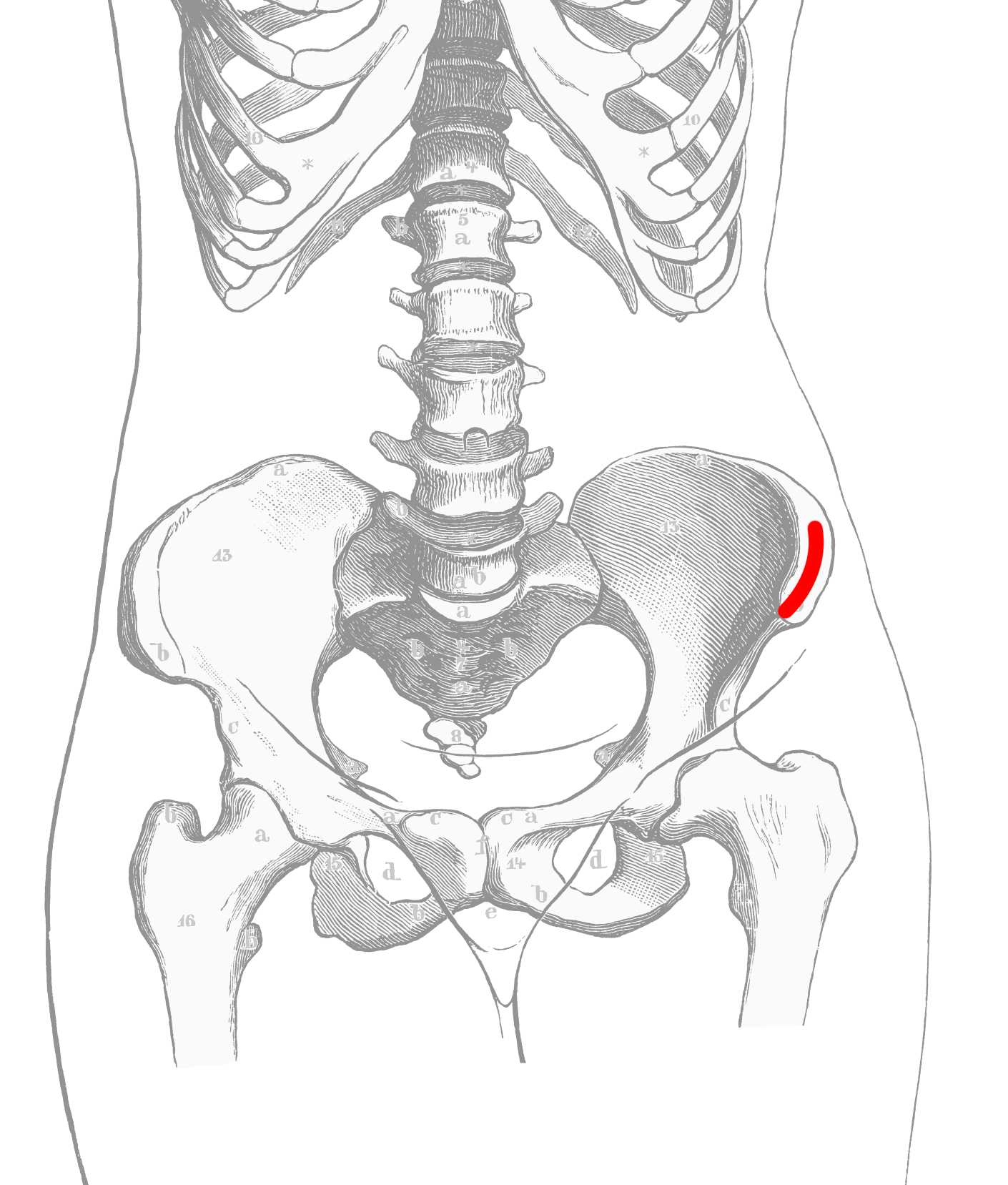
 The most common source of the bone graft is from the

upper jaw
The maxilla (plural: ''maxillae'' ) in vertebrates is the upper fixed (not fixed in Neopterygii) bone of the jaw formed from the fusion of two maxillary bones. In humans, the upper jaw includes the hard palate in the front of the mouth. The t ...
that is associated with cleft lip and palate
A cleft lip contains an opening in the upper lip that may extend into the nose. The opening may be on one side, both sides, or in the middle. A cleft palate occurs when the palate (the roof of the mouth) contains an opening into the nose. The ...
, where the bone defect is filled with bone or bone substitute, and any holes between the mouth and the nose are closed.
An alveolar cleft is a failure of the premaxilla
The premaxilla (or praemaxilla) is one of a pair of small cranial bones at the very tip of the upper jaw of many animals, usually, but not always, bearing teeth. In humans, they are fused with the maxilla. The "premaxilla" of therian mammal has ...
to fuse with the upper jaw
The maxilla (plural: ''maxillae'' ) in vertebrates is the upper fixed (not fixed in Neopterygii) bone of the jaw formed from the fusion of two maxillary bones. In humans, the upper jaw includes the hard palate in the front of the mouth. The t ...
leaving a defect in the bone. It is common in people with cleft palate
A cleft lip contains an opening in the upper lip that may extend into the nose. The opening may be on one side, both sides, or in the middle. A cleft palate occurs when the palate (the roof of the mouth) contains an opening into the nose. The ...
and is also associated with holes
A hole is an opening in or through a particular medium, usually a solid body. Holes occur through natural and artificial processes, and may be useful for various purposes, or may represent a problem needing to be addressed in many fields of en ...
between the mouth and the nose that affects speech, and allows fluid to move into the nose when eating and drinking.
Surgeries on the roof of the mouth early in life typically close the larger hole between the mouth and the nose (caused by the cleft in the palate) but do not repair the defect in the bone, or any holes further forward between the palate
The palate () is the roof of the mouth in humans and other mammals. It separates the oral cavity from the nasal cavity.
A similar structure is found in crocodilians, but in most other tetrapods, the oral and nasal cavities are not truly separ ...
and the upper lip. About the age of 8, just before the eye teeth are about to erupt into the bone defect (the cleft), braces are used to first widen the upper jaw, and position the premaxilla. Then, a surgery places bone or a bone substitute in the defect to allow the premaxilla to fuse to the rest of the maxilla, provide bone
A bone is a rigid organ that constitutes part of the skeleton in most vertebrate animals. Bones protect the various other organs of the body, produce red and white blood cells, store minerals, provide structure and support for the body, ...
for eruption of the canine, and close
Close may refer to:
Music
* ''Close'' (Kim Wilde album), 1988
* ''Close'' (Marvin Sapp album), 2017
* ''Close'' (Sean Bonniwell album), 1969
* "Close" (Sub Focus song), 2014
* "Close" (Nick Jonas song), 2016
* "Close" (Rae Sremmurd song), 201 ...
any remaining holes between the mouth and nose. If completed early, the canine teeth will erupt into the mouth with good bone support and remain healthy.
Medical uses
Alveolar cleft grafting is used primarily to allow the eruption of themaxillary canine
In human dentistry, the maxillary canine is the tooth located laterally (away from the midline of the face) from both maxillary lateral incisors of the mouth but mesial (toward the midline of the face) from both maxillary first premolars. Both t ...
s into the mouth between the ages of 8–13 years old. It is also used to close oranasal fistula
A fistula (plural: fistulas or fistulae ; from Latin ''fistula'', "tube, pipe") in anatomy is an abnormal connection between two hollow spaces (technically, two epithelialized surfaces), such as blood vessels, intestines, or other hollow or ...
s, stop fluid reflux into the nose, improve speech, support the maxillary lateral teeth, and stabilize the jaw for orthodontics or orthognathic surgery
Orthognathic surgery (), also known as corrective jaw surgery or simply jaw surgery, is surgery designed to correct conditions of the jaw and lower face related to structure, growth, airway issues including sleep apnea, TMJ disorders, malocclusi ...
.
Technique
In cleft palate patients bone grafting during the mixed dentition has been widely accepted since the mid-1960s. The goals of surgery are to stabilize themaxilla
The maxilla (plural: ''maxillae'' ) in vertebrates is the upper fixed (not fixed in Neopterygii) bone of the jaw formed from the fusion of two maxillary bones. In humans, the upper jaw includes the hard palate in the front of the mouth. T ...
, facilitate the healthy eruption of teeth that are adjacent the cleft, improving the esthetics of the base of the nose, create a bone base for dental implant
A dental implant (also known as an endosseous implant or fixture) is a prosthesis that interfaces with the bone of the jaw or skull to support a dental prosthesis such as a crown, bridge, denture, or facial prosthesis or to act as an orthodo ...
s, and to close any oro-nasal fistula
A fistula (plural: fistulas or fistulae ; from Latin ''fistula'', "tube, pipe") in anatomy is an abnormal connection between two hollow spaces (technically, two epithelialized surfaces), such as blood vessels, intestines, or other hollow or ...
s.
Pre-treatment monitoring and timing

 One of the most controversial topics in alveolar cleft grafting is the timing of treatment, however, most centres recommend grafting around the ages of 6–8 years old as the
 lateral incisor
Incisors (from Latin ''incidere'', "to cut") are the front teeth present in most mammals. They are located in the premaxilla above and on the mandible below. Humans have a total of eight (two on each side, top and bottom). Opossums have 18, wh ...
and maxillary canine
In human dentistry, the maxillary canine is the tooth located laterally (away from the midline of the face) from both maxillary lateral incisors of the mouth but mesial (toward the midline of the face) from both maxillary first premolars. Both t ...
near the cleft site. This is referred to as secondary grafting during mixed dentition (after eruption of the maxillary central incisors but before the eruption of the canine). A smaller proportion recommend primary grafting around the age of 2, but success rates are lower, and fewer patients are good candidates. Late secondary grafting (after eruption of the canine) has also been advocated but has been largely abandoned due to the loss of tooth support.
In secondary grafting, the dental age of the patient needs to be closely monitored for the eruption of the first maxillary molars in unilateral cases around the age of 6, and in bilateral cases the eruption of central incisors about the age of 8. This timing is used because there is minimal growth of the upper jaw after ages 6–7, the patient is more compliant with orthodontics that is required prior to surgery for expansion, the donor site is better developed, and the grafting precedes the eruption of teeth into the site.
In cases with a single cleft, 35-60% of lateral incisor
Incisors (from Latin ''incidere'', "to cut") are the front teeth present in most mammals. They are located in the premaxilla above and on the mandible below. Humans have a total of eight (two on each side, top and bottom). Opossums have 18, wh ...
s are congenitally missing, and cannot be relied on for timing. Instead, the eruption of the incisors and first molars is used as a queue to begin assessments. With bilateral cases, the premaxilla must be repositioned before grafting and special consideration must be given. During this time, the orthodontist must be wary of rotating teeth into the cleft site. Last, the size of the patient, defect, and social issues must all be taken into consideration and is best assessed with a CBCT scan as the patient enters the mixed dentition phase of dental development.
Pre-surgery Orthodontics
 In cleft lip and palate cases, the
 maxilla
The maxilla (plural: ''maxillae'' ) in vertebrates is the upper fixed (not fixed in Neopterygii) bone of the jaw formed from the fusion of two maxillary bones. In humans, the upper jaw includes the hard palate in the front of the mouth. T ...
is typically narrow compared to the lower jaw and must be expanded outward. An expansion appliance is placed in the maxilla 6–9 months prior to correct any crossbite
Crossbite is a form of malocclusion where a tooth (or teeth) has a more buccal or lingual position (that is, the tooth is either closer to the cheek or to the tongue) than its corresponding antagonist tooth in the upper or lower dental arch. In ...
or upper arch constriction. This will widen the cleft size, and so that parents and patients need to be warned the symptoms such as fluid reflux may worsen, although some centres will expand the jaw after surgery is complete. In the case of double clefts, expansion is typically before surgery because the premaxilla
The premaxilla (or praemaxilla) is one of a pair of small cranial bones at the very tip of the upper jaw of many animals, usually, but not always, bearing teeth. In humans, they are fused with the maxilla. The "premaxilla" of therian mammal has ...
needs to be repositioned forward, which cannot occur until the upper jaw is widened to allow room.
Surgical correction
 The goals of the surgery are to:
# Close any
 fistula
A fistula (plural: fistulas or fistulae ; from Latin ''fistula'', "tube, pipe") in anatomy is an abnormal connection between two hollow spaces (technically, two epithelialized surfaces), such as blood vessels, intestines, or other hollow or ...
s between the mouth and the nose
# Leave keratinized tissue around the tooth margins
# Create enough bone to stabilize the maxilla and allow teeth to erupt without a bone defect
At surgery, the gingiva is incised along the cleft margin to allow creation of pocket of tissue up towards the nose (recreating a nasal floor), and down towards the mouth (recreating a palate, and closing the palatal opening). The bony edges of the cleft are cleaned, and any extra teeth in the cleft are removed. The gingiva along the outside part of the upper jaw
The maxilla (plural: ''maxillae'' ) in vertebrates is the upper fixed (not fixed in Neopterygii) bone of the jaw formed from the fusion of two maxillary bones. In humans, the upper jaw includes the hard palate in the front of the mouth. The t ...
is raised, so that it can be drawn forward to close the cleft.
After creation of the pocket, but before suturing closed the gingiva a bone graft
Bone grafting is a surgical procedure that replaces missing bone in order to repair bone fractures that are extremely complex, pose a significant health risk to the patient, or fail to heal properly. Some small or acute fractures can be cured wit ...
is placed into the bony defect. This is most commonly harvested from the anterior hip but can also be obtained from other sites, donor bone, or bioactive materials such as bone morphogenic protein
Bone morphogenetic proteins (BMPs) are a group of growth factors also known as cytokines and as metabologens. Originally discovered by their ability to induce the formation of bone and cartilage, BMPs are now considered to constitute a group of pi ...
s. Once packed the cleft is filled with bone material the gingiva is sutured closed to create a water tight closure between the mouth and the nose.
Source of bone graft
 The most common source of the bone graft is from the
 iliac crest
The crest of the ilium (or iliac crest) is the superior border of the wing of ilium and the superiolateral margin of the greater pelvis.
Structure
The iliac crest stretches posteriorly from the anterior superior iliac spine (ASIS) to the poster ...
, harvested at the time of the cleft closure. Other sources such as the chin, and posterior iliac crest, or skull
The skull is a bone protective cavity for the brain. The skull is composed of four types of bone i.e., cranial bones, facial bones, ear ossicles and hyoid bone. However two parts are more prominent: the cranium and the mandible. In humans, th ...
can also be used. Artificial grafts such as demineralized bone, recombinent bone morphogenic protein or a mix of harvested bone and artificial grafts have also been used. Insufficient data exists to show that one is beneficial over the other.
Recovery
The mouth wounds recover in 7–10 days with precautions for fluid only diet for 5 days, and not to increase pressure in the nose or sinuses for 2–3 weeks. Evidence that the bone graft is forming will be seen on x-ray at about 8 weeks. Movement of teeth into the graft can begin at 3 months once bone graft consolidation is seen on xray. Recovery from the bone harvest will vary depending on the site (if harvested) with the anterior iliac crest being sore for 2–3 weeks. Success of the bone graft with the first surgery are approximately 85% but may vary with technique and bone grafting material. Orthodontics after surgery can close the space between thecentral incisor
Incisors (from Latin ''incidere'', "to cut") are the front teeth present in most mammals. They are located in the premaxilla above and on the mandible below. Humans have a total of eight (two on each side, top and bottom). Opossums have 18, wh ...
and maxillary canine
In human dentistry, the maxillary canine is the tooth located laterally (away from the midline of the face) from both maxillary lateral incisors of the mouth but mesial (toward the midline of the face) from both maxillary first premolars. Both t ...
in 50-75% of cases, and for those who can't have the space closed the gap can be filled with a dental implant once growth has finished.

History
The first attempts at bone grafting cleft patients were made by Lexer (1908) and Drachter (1914). Between 1921 and 1952 various attempts were made to graft patients using theturbinate
In anatomy, a nasal concha (), plural conchae (), also called a nasal turbinate or turbinal, is a long, narrow, curled shelf of bone that protrudes into the breathing passage of the nose in humans and various animals. The conchae are shaped lik ...
, rib and other harvest sites. Schmid (1954) at meetings in 1951 and 1952 reported on the treatment of patients using iliac crest bone grafts but stated, "The procedure has merely been presented for discussion". By 1964 the iliac crest bone graft had gained popularity and was presented at multiple meetings.
See also
* Cleft lip and cleft palate * Palatal expansionReferences
{{Reflist, 30em Surgical procedures and techniques Oral and maxillofacial surgery Dentistry procedures Tissue transplants Oral surgery