|
Xanthochromia
Xanthochromia, from the Greek ''xanthos'' (ξανθός) "yellow" and ''chroma'' (χρώμα) "colour", is the yellowish appearance of cerebrospinal fluid that occurs several hours after bleeding into the subarachnoid space caused by certain medical conditions, most commonly subarachnoid hemorrhage. Its presence can be determined by either spectrophotometry (measuring the absorption of particular wavelengths of light) or simple visual examination. It is unclear which method is superior. Physiology Cerebrospinal fluid, which fills the subarachnoid space between the arachnoid membrane and the pia mater surrounding the brain, is normally clear and colorless. When there has been bleeding into the subarachnoid space, the initial appearance of the cerebrospinal fluid can range from barely tinged with blood to frankly bloody, depending on the extent of bleeding. Within several hours, the red blood cells in the cerebrospinal fluid are destroyed, releasing their oxygen-carrying molecule he ... [...More Info...] [...Related Items...] OR: [Wikipedia] [Google] [Baidu] |
Subarachnoid Hemorrhage
Subarachnoid hemorrhage (SAH) is bleeding into the subarachnoid space—the area between the arachnoid membrane and the pia mater surrounding the brain. Symptoms may include a severe headache of rapid onset, vomiting, decreased level of consciousness, fever, and sometimes seizures. Neck stiffness or neck pain are also relatively common. In about a quarter of people a small bleed with resolving symptoms occurs within a month of a larger bleed. SAH may occur as a result of a head injury or spontaneously, usually from a ruptured cerebral aneurysm. Risk factors for spontaneous cases include high blood pressure, smoking, family history, alcoholism, and cocaine use. Generally, the diagnosis can be determined by a CT scan of the head if done within six hours of symptom onset. Occasionally, a lumbar puncture is also required. After confirmation further tests are usually performed to determine the underlying cause. Treatment is by prompt neurosurgery or endovascular coiling. Medic ... [...More Info...] [...Related Items...] OR: [Wikipedia] [Google] [Baidu] |
Cerebrospinal Fluid
Cerebrospinal fluid (CSF) is a clear, colorless body fluid found within the tissue that surrounds the brain and spinal cord of all vertebrates. CSF is produced by specialised ependymal cells in the choroid plexus of the ventricles of the brain, and absorbed in the arachnoid granulations. There is about 125 mL of CSF at any one time, and about 500 mL is generated every day. CSF acts as a shock absorber, cushion or buffer, providing basic mechanical and immunological protection to the brain inside the skull. CSF also serves a vital function in the cerebral autoregulation of cerebral blood flow. CSF occupies the subarachnoid space (between the arachnoid mater and the pia mater) and the ventricular system around and inside the brain and spinal cord. It fills the ventricles of the brain, cisterns, and sulci, as well as the central canal of the spinal cord. There is also a connection from the subarachnoid space to the bony labyrinth of the inner ear via the per ... [...More Info...] [...Related Items...] OR: [Wikipedia] [Google] [Baidu] |
Cerebral Aneurysm
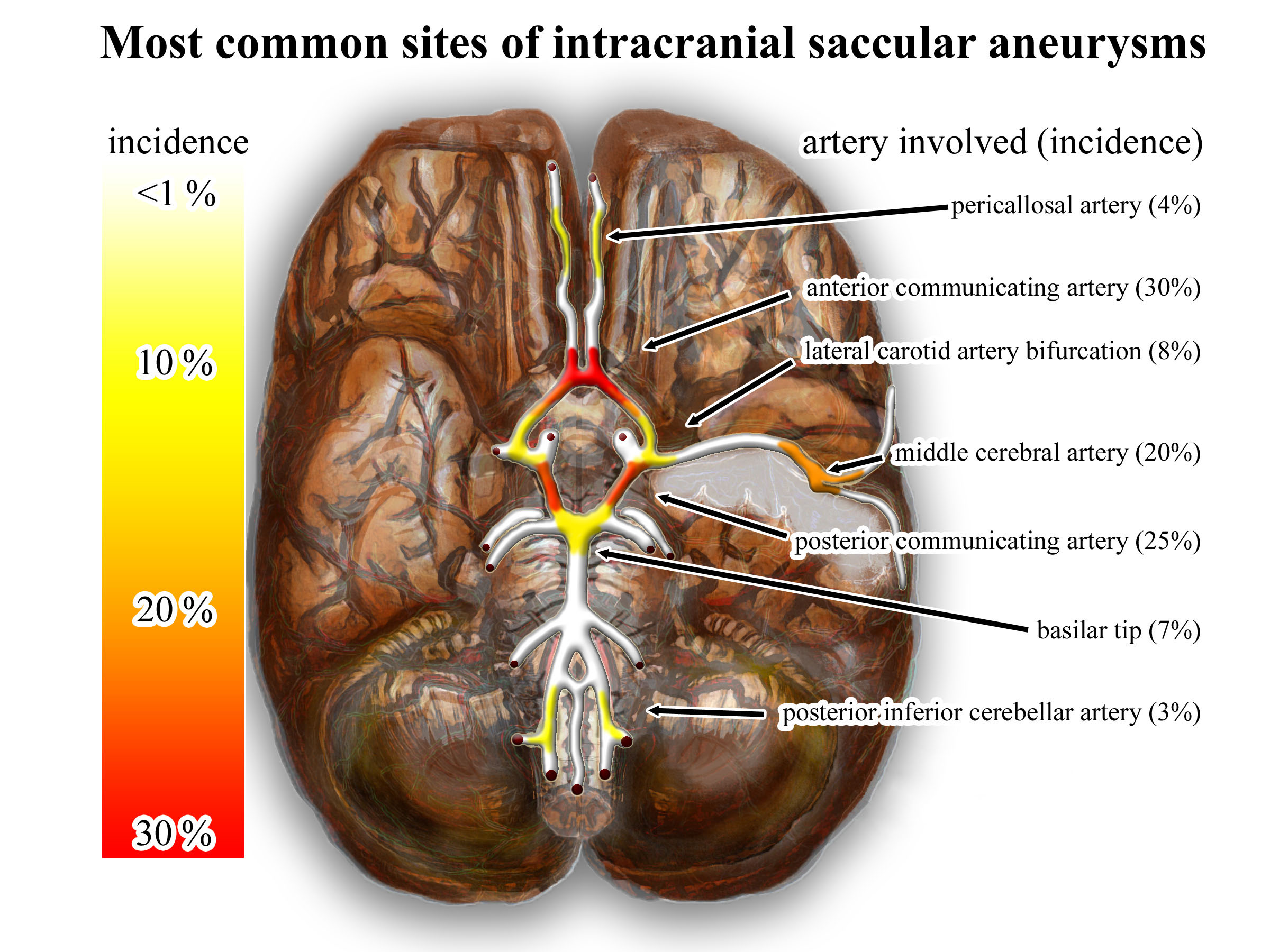
An intracranial aneurysm, also known as a brain aneurysm, is a cerebrovascular disorder in which weakness in the wall of a cerebral artery or vein causes a localized dilation or ballooning of the blood vessel. Aneurysms in the posterior circulation (basilar artery, vertebral arteries and posterior communicating artery) have a higher risk of rupture. Basilar artery aneurysms represent only 3–5% of all intracranial aneurysms but are the most common aneurysms in the posterior circulation. Classification Cerebral aneurysms are classified both by size and shape. Small aneurysms have a diameter of less than 15 mm. Larger aneurysms include those classified as large (15 to 25 mm), giant (25 to 50 mm), and super-giant (over 50 mm). Berry (saccular) aneurysms Saccular aneurysms, also known as berry aneurysms, appear as a round outpouching and are the most common form of cerebral aneurysm. Causes include connective tissue disorders, polycystic kidney disease, ... [...More Info...] [...Related Items...] OR: [Wikipedia] [Google] [Baidu] |
Thunderclap Headache
A thunderclap headache is a headache that is severe and has a sudden onset. It is defined as a severe headache that takes seconds to minutes to reach maximum intensity. Although approximately 75% are attributed to "primary" headaches—headache disorder, non-specific headache, idiopathic thunderclap headache, or uncertain headache disorder—the remainder are secondary to other causes, which can include some extremely dangerous acute conditions, as well as infections and other conditions. Usually, further investigations are performed to identify the underlying cause. Signs and symptoms A headache is called "thunderclap headache" if it is severe in character and reaches maximum severity within seconds to minutes of onset. In many cases, there are no other abnormalities, but the various causes of thunderclap headaches may lead to a number of neurological symptoms. Causes Approximately 75% are attributed to "primary" headaches: headache disorder, non-specific headache, idiopathic th ... [...More Info...] [...Related Items...] OR: [Wikipedia] [Google] [Baidu] |
Arachnoid (brain)
The arachnoid mater (or simply arachnoid) is one of the three meninges, the protective membranes that cover the brain and spinal cord. It is so named because of its resemblance to a spider web. The arachnoid mater is a derivative of the neural crest mesoectoderm in the embryo. Structure It is interposed between the two other meninges, the more superficial and much thicker dura mater and the deeper pia mater, from which it is separated by the subarachnoid space. The delicate arachnoid layer is not attached to the inside of the dura but against it and surrounds the brain and spinal cord. It does not line the brain down into its sulci (folds), as does the pia mater, with the exception of the longitudinal fissure, which divides the left and right cerebral hemispheres. Cerebrospinal fluid (CSF) flows under the arachnoid in the subarachnoid space, within a meshwork of trabeculae which span between the arachnoid and the pia. The arachnoid mater makes arachnoid villi, small protrusions ... [...More Info...] [...Related Items...] OR: [Wikipedia] [Google] [Baidu] |
Confusion
In medicine, confusion is the quality or state of being bewildered or unclear. The term "acute mental confusion"''Confusion Definition'' on Oxford Dictionaries. is often used interchangeably with in the '' International Statistical Classification of Diseases and Related He ...
[...More Info...] [...Related Items...] OR: [Wikipedia] [Google] [Baidu] |
Standard Illuminant
A standard illuminant is a theoretical source of visible light with a spectral power distribution that is published. Standard illuminants provide a basis for comparing images or colors recorded under different lighting. CIE illuminants The International Commission on Illumination (usually abbreviated CIE for its French name) is the body responsible for publishing all of the well-known standard illuminants. Each of these is known by a letter or by a letter-number combination. Illuminants A, B, and C were introduced in 1931, with the intention of respectively representing average incandescent light, direct sunlight, and average daylight. Illuminants D represent variations of daylight, illuminant E is the equal-energy illuminant, while illuminants F represent fluorescent lamps of various composition. There are instructions on how to experimentally produce light sources ("standard sources") corresponding to the older illuminants. For the relatively newer ones (such as series D), e ... [...More Info...] [...Related Items...] OR: [Wikipedia] [Google] [Baidu] |
International Commission On Illumination
The International Commission on Illumination (usually abbreviated CIE for its French name, Commission internationale de l'éclairage) is the international authority on light, illumination, colour, and colour spaces. It was established in 1913 as a successor to the Commission Internationale de Photométrie, which was founded in 1900, and is today based in Vienna, Austria. Organization The CIE has six active divisions, each of which establishes technical committees to carry out its program: * Division 1: Vision and Colour * Division 2: Physical Measurement of Light and Radiation * Division 3: Interior Environment and Lighting Design * Division 4: Transportation and Exterior Applications * Division 6: Photobiology and Photochemistry * Division 8: Image Technology Two divisions are no longer active: * Division 5: Exterior Lighting and Other Applications * Division 7: General Aspects of Lighting The President of the CIE from 2019 is Dr Peter Blattner from Switzerland. CIE publish ... [...More Info...] [...Related Items...] OR: [Wikipedia] [Google] [Baidu] |
Methemoglobin
Methemoglobin (British: methaemoglobin) (pronounced "met-hemoglobin") is a hemoglobin ''in the form of metalloprotein'', in which the iron in the heme group is in the Fe3+ (ferric) state, not the Fe2+ (ferrous) of normal hemoglobin. Sometimes, it is also referred to as ferrihemoglobin. Methemoglobin cannot bind oxygen, which means it cannot carry oxygen to tissues. It is bluish chocolate-brown in color. In human blood a trace amount of methemoglobin is normally produced spontaneously, but when present in excess the blood becomes abnormally dark bluish brown. The NADH-dependent enzyme methemoglobin reductase ( a type of diaphorase) is responsible for converting methemoglobin back to hemoglobin. Normally one to two percent of a person's hemoglobin is methemoglobin; a higher percentage than this can be genetic or caused by exposure to various chemicals and depending on the level can cause health problems known as methemoglobinemia. A higher level of methemoglobin will tend to cause a p ... [...More Info...] [...Related Items...] OR: [Wikipedia] [Google] [Baidu] |
Oxyhemoglobin
Hemoglobin (haemoglobin BrE) (from the Greek word αἷμα, ''haîma'' 'blood' + Latin ''globus'' 'ball, sphere' + ''-in'') (), abbreviated Hb or Hgb, is the iron-containing oxygen-transport metalloprotein present in red blood cells (erythrocytes) of almost all vertebrates (the exception being the fish family Channichthyidae) as well as the tissues of some invertebrates. Hemoglobin in blood carries oxygen from the respiratory organs (''e.g.'' lungs or gills) to the rest of the body (''i.e.'' tissues). There it releases the oxygen to permit aerobic respiration to provide energy to power functions of an organism in the process called metabolism. A healthy individual human has 12to 20grams of hemoglobin in every 100mL of blood. In mammals, the chromoprotein makes up about 96% of the red blood cells' dry content (by weight), and around 35% of the total content (including water). Hemoglobin has an oxygen-binding capacity of 1.34mL O2 per gram, which increases the total blood oxygen ... [...More Info...] [...Related Items...] OR: [Wikipedia] [Google] [Baidu] |
Absorption (electromagnetic Radiation)
In physics, absorption of electromagnetic radiation is how matter (typically electrons bound in atoms) takes up a photon's energy — and so transforms electromagnetic energy into internal energy of the absorber (for example, thermal energy). A notable effect is attenuation, or the gradual reduction of the intensity of light waves as they propagate through a medium. Although the absorption of waves does not usually depend on their intensity (linear absorption), in certain conditions ( optics) the medium's transparency changes by a factor that varies as a function of wave intensity, and saturable absorption (or nonlinear absorption) occurs. Quantifying absorption Many approaches can potentially quantify radiation absorption, with key examples following. * The absorption coefficient along with some closely related derived quantities * The attenuation coefficient (NB used infrequently with meaning synonymous with "absorption coefficient") * The Molar attenuation co ... [...More Info...] [...Related Items...] OR: [Wikipedia] [Google] [Baidu] |
Beer–Lambert Law
The Beer–Lambert law, also known as Beer's law, the Lambert–Beer law, or the Beer–Lambert–Bouguer law relates the attenuation of light to the properties of the material through which the light is travelling. The law is commonly applied to chemical analysis measurements and used in understanding attenuation in physical optics, for photons, neutrons, or rarefied gases. In mathematical physics, this law arises as a solution of the BGK equation. History The law was discovered by Pierre Bouguer before 1729, while looking at red wine, during a brief vacation in Alentejo, Portugal. It is often attributed to Johann Heinrich Lambert, who cited Bouguer's ''Essai d'optique sur la gradation de la lumière'' (Claude Jombert, Paris, 1729) – and even quoted from it – in his '' Photometria'' in 1760. Lambert's law stated that the loss of light intensity when it propagates in a medium is directly proportional to intensity and path length. Much later, the German scientist August ... [...More Info...] [...Related Items...] OR: [Wikipedia] [Google] [Baidu] |


