|
White Blood Cell Differential
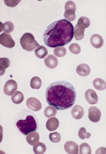
A white blood cell differential is a medical laboratory test that provides information about the types and amounts of white blood cells in a person's blood. The test, which is usually ordered as part of a complete blood count (CBC), measures the amounts of the five normal white blood cell typesneutrophils, lymphocytes, monocytes, eosinophils and basophilsas well as abnormal cell types if they are present. These results are reported as percentages and absolute values, and compared against reference ranges to determine whether the values are normal, low, or high. Changes in the amounts of white blood cells can aid in the diagnosis of many health conditions, including viral, bacterial, and parasitic infections and blood disorders such as leukemia. White blood cell differentials may be performed by an automated analyzera machine designed to run laboratory tests – or manually, by examining blood smears under a microscope. The test was performed manually until white blood cell dif ... [...More Info...] [...Related Items...] OR: [Wikipedia] [Google] [Baidu] |
Neutrophil
Neutrophils (also known as neutrocytes or heterophils) are the most abundant type of granulocytes and make up 40% to 70% of all white blood cells in humans. They form an essential part of the innate immune system, with their functions varying in different animals. They are formed from stem cells in the bone marrow and Cellular differentiation, differentiated into #Subpopulations, subpopulations of neutrophil-killers and neutrophil-cagers. They are short-lived and highly mobile, as they can enter parts of tissue where other cells/molecules cannot. Neutrophils may be subdivided into segmented neutrophils and banded neutrophils (or Band cell, bands). They form part of the polymorphonuclear cells family (PMNs) together with basophils and eosinophils. The name ''neutrophil'' derives from staining characteristics on hematoxylin and eosin (H&E stain, H&E) histology, histological or cell biology, cytological preparations. Whereas basophilic white blood cells stain dark blue and eosinoph ... [...More Info...] [...Related Items...] OR: [Wikipedia] [Google] [Baidu] |
Automated Analyzer
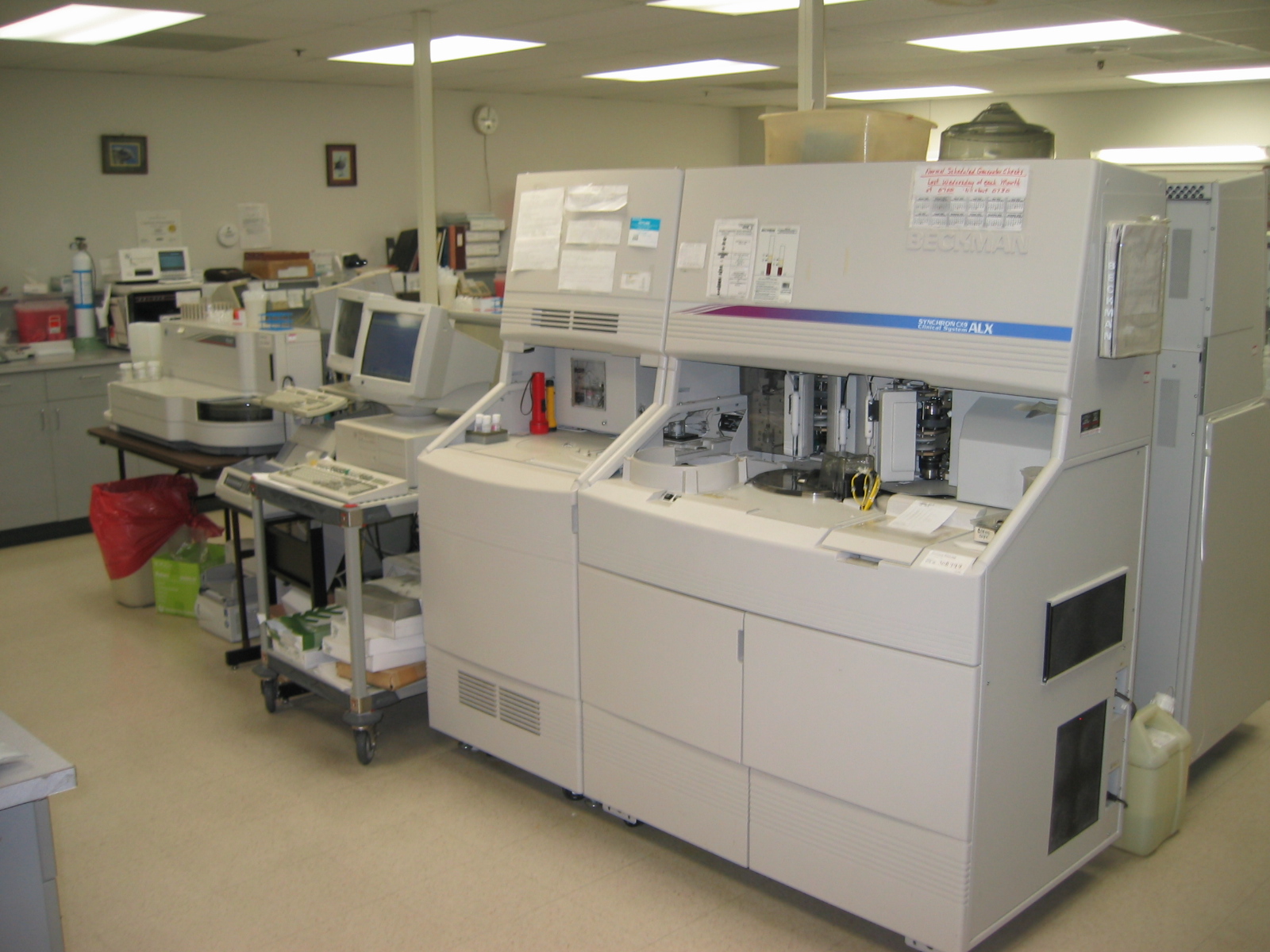
An automated analyser is a medical laboratory instrument designed to measure different chemicals and other characteristics in a number of biological samples quickly, with minimal human assistance. These measured properties of blood and other fluids may be useful in the diagnosis of disease. Photometry is the most common method for testing the amount of a specific analyte in a sample. In this technique, the sample undergoes a reaction to produce a color change. Then, a photometer measures the absorbance of the sample to indirectly measure the concentration of analyte present in the sample. The use of an Ion Selective Electrode (ISE) is another common analytical method that specifically measures ion concentrations. This typically measures the concentrations of sodium, calcium or potassium present in the sample. There are various methods of introducing samples into the analyser. Test tubes of samples are often loaded into racks. These racks can be inserted directly into some analy ... [...More Info...] [...Related Items...] OR: [Wikipedia] [Google] [Baidu] |
Reactive Lymphocyte
In immunology, reactive lymphocytes or variant lymphocytes are cytotoxic (CD8+) lymphocytes that become large as a result of antigen stimulation. Typically, they can be more than 30 μm in diameter with varying size and shape. The nucleus of a reactive lymphocyte can be round, elliptic, indented, cleft, or folded. The cytoplasm is often abundant and can be basophilic. Vacuoles and/or azurophilic granules are also sometimes present. Most often, the cytoplasm is gray, pale blue, or deep blue in colour. The distinctive cell associated with EBV or CMV is known as a "Downey cell", after Hal Downey, who contributed to the characterization of it in 1923. __TOC__ Causes Reactive lymphocytes are usually associated with viral illnesses, but they can also be present as a result of drug reactions (such as phenytoin), immunizations, radiation, and hormonal causes (such as stress and Addison's disease), as well as some autoimmune disorders (such as rheumatoid arthritis). Some pathog ... [...More Info...] [...Related Items...] OR: [Wikipedia] [Google] [Baidu] |
Blast Cell
In cell biology, a precursor cell, also called a blast cell or simply blast, is a partially differentiated cell, usually referred to as a unipotent cell that has lost most of its stem cell properties. A precursor cell is also known as a progenitor cell but progenitor cells are multipotent. Precursor cells are known as the intermediate cell before they become differentiated after being a stem cell. Usually, a precursor cell is a stem cell with the capacity to differentiate into only one cell type. Sometimes, ''precursor cell'' is used as an alternative term for unipotent stem cells. In embryology, precursor cells are a group of cells that later differentiate into one organ. A blastoma is any cancer created by malignancies of precursor cells. Precursor cells, and progenitor cells, have many potential uses in medicine. , there is research being done to use these cells to build heart valves, blood vessels and other tissues, by using blood and muscle precursor, or progenitor cel ... [...More Info...] [...Related Items...] OR: [Wikipedia] [Google] [Baidu] |
Hematological Disorder
Hematologic diseases are disorders which primarily affect the blood & blood-forming organs. Hematologic diseases include rare genetic disorders, anemia, HIV, sickle cell disease & complications from chemotherapy or transfusions. Myeloid * Hemoglobinopathies (congenital abnormality of the hemoglobin molecule or of the rate of hemoglobin synthesis) ** Sickle cell disease ** Thalassemia ** Methemoglobinemia * Anemias (lack of red blood cells or hemoglobin) ** Iron-deficiency anemia ** Megaloblastic anemia *** Vitamin B12 deficiency **** Pernicious anemia *** Folate deficiency ** Hemolytic anemias (destruction of red blood cells) *** Genetic disorders of RBC membrane **** Hereditary spherocytosis **** Hereditary elliptocytosis **** Congenital dyserythropoietic anemia *** Genetic disorders of RBC metabolism **** Glucose-6-phosphate dehydrogenase deficiency (G6PD) **** Pyruvate kinase deficiency *** Immune mediated hemolytic anemia (direct Coombs test is positive) **** Autoimmune hemol ... [...More Info...] [...Related Items...] OR: [Wikipedia] [Google] [Baidu] |
Infection
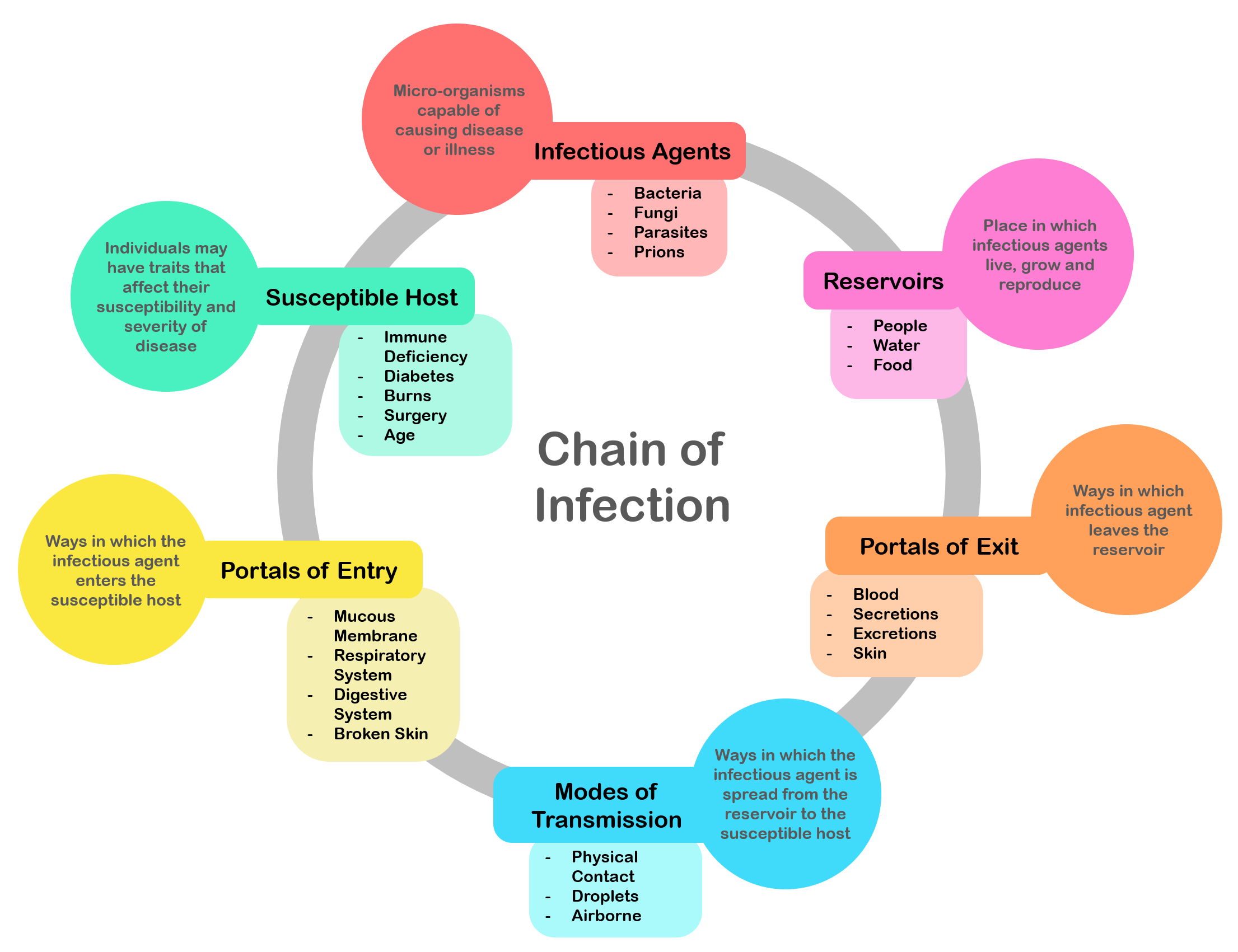
An infection is the invasion of tissues by pathogens, their multiplication, and the reaction of host tissues to the infectious agent and the toxins they produce. An infectious disease, also known as a transmissible disease or communicable disease, is an illness resulting from an infection. Infections can be caused by a wide range of pathogens, most prominently bacteria and viruses. Hosts can fight infections using their immune system. Mammalian hosts react to infections with an innate response, often involving inflammation, followed by an adaptive response. Specific medications used to treat infections include antibiotics, antivirals, antifungals, antiprotozoals, and antihelminthics. Infectious diseases resulted in 9.2 million deaths in 2013 (about 17% of all deaths). The branch of medicine that focuses on infections is referred to as infectious disease. Types Infections are caused by infectious agents (pathogens) including: * Bacteria (e.g. ''Mycobacterium tuberculosis'', ... [...More Info...] [...Related Items...] OR: [Wikipedia] [Google] [Baidu] |
Medical Examination
In a physical examination, medical examination, or clinical examination, a medical practitioner examines a patient for any possible medical signs or symptoms of a medical condition. It generally consists of a series of questions about the patient's medical history followed by an examination based on the reported symptoms. Together, the medical history and the physical examination help to determine a diagnosis and devise the treatment plan. These data then become part of the medical record. Types Routine The ''routine physical'', also known as ''general medical examination'', ''periodic health evaluation'', ''annual physical'', ''comprehensive medical exam'', ''general health check'', ''preventive health examination'', ''medical check-up'', or simply ''medical'', is a physical examination performed on an asymptomatic patient for medical screening purposes. These are normally performed by a pediatrician, family practice physician, physician assistant, a certified nurse prac ... [...More Info...] [...Related Items...] OR: [Wikipedia] [Google] [Baidu] |
Flow Cytometry
Flow cytometry (FC) is a technique used to detect and measure physical and chemical characteristics of a population of cells or particles. In this process, a sample containing cells or particles is suspended in a fluid and injected into the flow cytometer instrument. The sample is focused to ideally flow one cell at a time through a laser beam, where the light scattered is characteristic to the cells and their components. Cells are often labeled with fluorescent markers so light is absorbed and then emitted in a band of wavelengths. Tens of thousands of cells can be quickly examined and the data gathered are processed by a computer. Flow cytometry is routinely used in basic research, clinical practice, and clinical trials. Uses for flow cytometry include: * Cell counting * Cell sorting * Determining cell characteristics and function * Detecting microorganisms * Biomarker detection * Protein engineering detection * Diagnosis of health disorders such as blood cancers * Measuring ... [...More Info...] [...Related Items...] OR: [Wikipedia] [Google] [Baidu] |
Hematology Analyzer
Hematology analyzers ( also spelled haematology analysers in British English) are used to count and identify blood cells at high speed with accuracy. During the 1950s, laboratory technicians counted each individual blood cell underneath a microscope. Tedious and inconsistent, this was replaced with the first, very basic hematology analyzer, engineered by Wallace H. Coulter. The early hematology analyzers relied on Coulter's Principle (see Coulter counter). However, they have evolved to encompass numerous techniques. Uses Hematology analyzers are used to conduct a complete blood count (CBC), which is usually the first test requested by physicians to determine a patients general health status. A complete blood count includes red blood cell (RBC), white blood cell (WBC), hemoglobin, and platelet counts, as well as hematocrit levels. Other analyses include: * RBC distribution width * Mean corpuscular volume * Mean corpuscular hemoglobin * Mean corpuscular hemoglobin concentrations ... [...More Info...] [...Related Items...] OR: [Wikipedia] [Google] [Baidu] |
Coulter Counter
A Coulter counter is an apparatus for counting and sizing particles suspended in electrolytes. The Coulter counter is the commercial term for the technique known as resistive pulse sensing or electrical zone sensing, the apparatus is based on The Coulter principle named after its inventor, Wallace H. Coulter. A typical Coulter counter has one or more microchannels that separate two chambers containing electrolyte solutions. As fluid-containing particles or cells are drawn through each microchannel, each particle causes a brief change to the electrical resistance of the liquid. The counter detects these changes in the electrical resistance. Coulter principle The ''Coulter principle'' states that particles pulled through an orifice, concurrent with an electric current, produce a change in impedance that is proportional to the volume of the particle traversing the orifice. This pulse in impedance originates from the displacement of electrolyte caused by the particle. The princi ... [...More Info...] [...Related Items...] OR: [Wikipedia] [Google] [Baidu] |
Romanowsky Stain
Romanowsky staining, also known as Romanowsky–Giemsa staining, is a prototypical staining technique that was the forerunner of several distinct but similar stains widely used in hematology (the study of blood) and cytopathology (the study of diseased cells). Romanowsky-type stains are used to differentiate cells for microscopic examination in pathological specimens, especially blood and bone marrow films, and to detect parasites such as malaria within the blood. Stains that are related to or derived from the Romanowsky-type stains include Giemsa, Jenner, Wright, Field, May–Grünwald and Leishman stains. The staining technique is named after the Russian physician Dmitri Leonidovich Romanowsky (1861–1921), who was one of the first to recognize its potential for use as a blood stain. Mechanism The value of Romanowsky staining lies in its ability to produce a wide range of hues, allowing cellular components to be easily differentiated. This phenomenon is referred to as the '' ... [...More Info...] [...Related Items...] OR: [Wikipedia] [Google] [Baidu] |
Dmitri Leonidovich Romanowsky
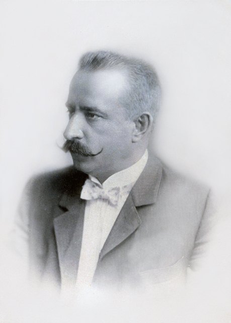
Dmitri Leonidovich Romanowsky (sometimes spelled Dmitry and Romanowski, russian: Дмитрий Леонидович Романовский; 1861–1921) was a Russian physician who is best known for his invention of an eponymous histological stain called Romanowsky stain. It paved the way for the discovery and diagnosis of microscopic pathogens, such as malarial parasites. Romanowsky was born in 1861 in Pskov Governorate, Russia. He attended the 6th Saint Petersburg Gymnasium. In 1880, he enrolled at the St. Petersburg University. He enrolled for two courses: natural science (physics and mathematics) and medicine. He concentrated on medicine in 1882 for a preparatory course to the Military Medical Academy. He graduated with honors in 1886. On 30 November 1886, he was appointed as a junior resident of the Ivangorod Ivangorod ( rus, Иванго́род, p=ɪvɐnˈɡorət; et, Jaanilinn; vot, Jaanilidna) is a town in Kingiseppsky District of Leningrad Oblast, Russia, loca ... [...More Info...] [...Related Items...] OR: [Wikipedia] [Google] [Baidu] |


.jpg)
