|
Venom-induced Consumption Coagulopathy
Venom-induced consumption coagulopathy (VICC) is a medical condition caused by the effects of some snake and caterpillar venoms on the blood. Important coagulation factors are activated by the specific serine proteases in the venom and as they become exhausted, coagulopathy develops. Symptoms are consistent with uncontrolled bleeding. Diagnosis is made using blood tests that assess clotting ability along with recent history of envenomation. Treatment generally involves pressure dressing, confirmatory blood testing, and antivenom administration. Signs and symptoms Symptoms are similar to those seen in other consumptive coagulopathies. These include obvious bleeding from the nose, gums, intravenous lines, or puncture sites. More serious symptoms such as vomiting blood, intestinal bleeding, and hemorrhage of internal organs may also be seen. Pathophysiology Venom-induced coagulopathy is caused by over-activation of the body's natural clotting system. This decreases clotting factor ... [...More Info...] [...Related Items...] OR: [Wikipedia] [Google] [Baidu] |
Snake
Snakes are elongated, Limbless vertebrate, limbless, carnivore, carnivorous reptiles of the suborder Serpentes . Like all other Squamata, squamates, snakes are ectothermic, amniote vertebrates covered in overlapping Scale (zoology), scales. Many species of snakes have skulls with several more joints than their lizard ancestors, enabling them to swallow prey much larger than their heads (cranial kinesis). To accommodate their narrow bodies, snakes' paired organs (such as kidneys) appear one in front of the other instead of side by side, and most have only one functional lung. Some species retain a pelvic girdle with a pair of vestigial claws on either side of the cloaca. Lizards have evolved elongate bodies without limbs or with greatly reduced limbs about twenty-five times independently via convergent evolution, leading to many lineages of legless lizards. These resemble snakes, but several common groups of legless lizards have eyelids and external ears, which snakes lack, altho ... [...More Info...] [...Related Items...] OR: [Wikipedia] [Google] [Baidu] |
Thrombotic Microangiopathy
Thrombotic microangiopathy (TMA) is a pathology that results in thrombosis in capillaries and arterioles, due to an endothelial injury. It may be seen in association with thrombocytopenia, anemia, purpura and kidney failure. The classic TMAs are hemolytic uremic syndrome and thrombotic thrombocytopenic purpura. Other conditions with TMA include atypical hemolytic uremic syndrome, disseminated intravascular coagulation, scleroderma renal crisis, malignant hypertension, antiphospholipid antibody syndrome, and drug toxicities, e.g. calcineurin inhibitor toxicity. Signs and symptoms The clinical presentation of TMA, although dependent on the type, typically includes: fever, microangiopathic hemolytic anemia (see schistocytes in a blood smear), kidney failure, thrombocytopenia and neurological manifestations. Generally, renal complications are particularly predominant with Shiga-toxin-associated hemolytic uremic syndrome (STx-HUS) and atypical HUS, whereas neurologic complicatio ... [...More Info...] [...Related Items...] OR: [Wikipedia] [Google] [Baidu] |
Clinical Urine Tests
A urine test is any medical test performed on a urine specimen. The analysis of urine is a valuable diagnostic tool because its composition reflects the functioning of many body systems, particularly the kidneys and urinary system, and specimens are easy to obtain. Common urine tests include the routine urinalysis, which examines the physical, chemical, and microscopic properties of the urine; urine drug screening; and urine pregnancy testing. Background The value of urine for diagnostic purposes has been recognized since ancient times. Urine examination was practiced in Sumer and Babylonia as early as 4000 BC, and is described in ancient Greek and Sanskrit texts. Contemporary urine testing uses a range of methods to investigate the physical and biochemical properties of the urine. For instance, the results of the routine urinalysis can provide information about the functioning of the kidneys and urinary system; suggest the presence of a urinary tract infection (UTI); and screen f ... [...More Info...] [...Related Items...] OR: [Wikipedia] [Google] [Baidu] |
Anticoagulation
Anticoagulants, commonly known as blood thinners, are chemical substances that prevent or reduce coagulation of blood, prolonging the clotting time. Some of them occur naturally in blood-eating animals such as leeches and mosquitoes, where they help keep the bite area unclotted long enough for the animal to obtain some blood. As a class of medications, anticoagulants are used in therapy for thrombotic disorders. Oral anticoagulants (OACs) are taken by many people in pill or tablet form, and various intravenous anticoagulant dosage forms are used in hospitals. Some anticoagulants are used in medical equipment, such as sample tubes, blood transfusion bags, heart–lung machines, and dialysis equipment. One of the first anticoagulants, warfarin, was initially approved as a rodenticide. Anticoagulants are closely related to antiplatelet drugs and thrombolytic drugs by manipulating the various pathways of blood coagulation. Specifically, antiplatelet drugs inhibit platelet aggre ... [...More Info...] [...Related Items...] OR: [Wikipedia] [Google] [Baidu] |
D-dimer
D-dimer (or D dimer) is a fibrin degradation product (or FDP), a small protein fragment present in the blood after a blood clot is degraded by fibrinolysis. It is so named because it contains two D fragments of the fibrin protein joined by a cross-link, hence forming a protein dimer. D-dimer concentration may be determined by a blood test to help diagnose thrombosis. Since its introduction in the 1990s, it has become an important test performed in people with suspected thrombotic disorders, such as venous thromboembolism. While a negative result practically rules out thrombosis, a positive result can indicate thrombosis, but does not exclude other potential causes. Its main use, therefore, is to exclude thromboembolic disease where the probability is low. D-dimer levels are used as a predictive biomarker for the blood disorder, disseminated intravascular coagulation and in the coagulation disorders associated with COVID-19 infection. A four-fold increase in the protein is an ind ... [...More Info...] [...Related Items...] OR: [Wikipedia] [Google] [Baidu] |
Fibrinogen
Fibrinogen (factor I) is a glycoprotein complex, produced in the liver, that circulates in the blood of all vertebrates. During tissue and vascular injury, it is converted enzymatically by thrombin to fibrin and then to a fibrin-based blood clot. Fibrin clots function primarily to occlude blood vessels to stop bleeding. Fibrin also binds and reduces the activity of thrombin. This activity, sometimes referred to as antithrombin I, limits clotting. Fibrin also mediates blood platelet and endothelial cell spreading, tissue fibroblast proliferation, capillary tube formation, and angiogenesis and thereby promotes revascularization and wound healing. Reduced and/or dysfunctional fibrinogens occur in various congenital and acquired human fibrinogen-related disorders. These disorders represent a group of rare conditions in which individuals may present with severe episodes of pathological bleeding and thrombosis; these conditions are treated by supplementing blood fibrinogen levels an ... [...More Info...] [...Related Items...] OR: [Wikipedia] [Google] [Baidu] |
Activated Partial Thromboplastin Time
The partial thromboplastin time (PTT), also known as the activated partial thromboplastin time (aPTT or APTT), is a blood test that characterizes coagulation of the blood. A historical name for this measure is the kaolin-cephalin clotting time (KCCT), reflecting kaolin and cephalin as materials historically used in the test. Apart from detecting abnormalities in blood clotting, partial thromboplastin time is also used to monitor the treatment effect of heparin, a widely prescribed drug that reduces blood's tendency to clot. The PTT measures the overall speed at which blood clots form by means of two consecutive series of biochemical reactions known as the ''intrinsic'' pathway and common pathway of coagulation. The PTT indirectly measures action of the following coagulation factors: I (fibrinogen), II (prothrombin), V (proaccelerin), VIII (anti-hemophilic factor), X (Stuart–Prower factor), XI (plasma thromboplastin antecedent), and XII (Hageman factor). The PTT is of ... [...More Info...] [...Related Items...] OR: [Wikipedia] [Google] [Baidu] |
International Normalized Ratio
The prothrombin time (PT) – along with its derived measures of prothrombin ratio (PR) and international normalized ratio (INR) – is an assay for evaluating the ''extrinsic'' pathway and common pathway of coagulation. This blood test is also called ''protime INR'' and ''PT/INR''. They are used to determine the clotting tendency of blood, in such things as the measure of warfarin dosage, liver damage, and vitamin K status. PT measures the following coagulation factors: I (fibrinogen), II (prothrombin), V (proaccelerin), VII (proconvertin), and X (Stuart–Prower factor). PT is often used in conjunction with the activated partial thromboplastin time (aPTT) which measures the ''intrinsic'' pathway and common pathway of coagulation. Laboratory measurement The reference range for prothrombin time depends on the analytical method used, but is usually around 12–13 seconds (results should always be interpreted using the reference range from the laboratory that performed ... [...More Info...] [...Related Items...] OR: [Wikipedia] [Google] [Baidu] |
Prothrombin Time
The prothrombin time (PT) – along with its derived measures of prothrombin ratio (PR) and international normalized ratio (INR) – is an assay for evaluating the ''extrinsic'' pathway and common pathway of coagulation. This blood test is also called ''protime INR'' and ''PT/INR''. They are used to determine the clotting tendency of blood, in such things as the measure of warfarin dosage, liver damage, and vitamin K status. PT measures the following coagulation factors: I (fibrinogen), II (prothrombin), V (proaccelerin), VII (proconvertin), and X (Stuart–Prower factor). PT is often used in conjunction with the activated partial thromboplastin time (aPTT) which measures the ''intrinsic'' pathway and common pathway of coagulation. Laboratory measurement The reference range for prothrombin time depends on the analytical method used, but is usually around 12–13 seconds (results should always be interpreted using the reference range from the laboratory that performed ... [...More Info...] [...Related Items...] OR: [Wikipedia] [Google] [Baidu] |
Complete Blood Count
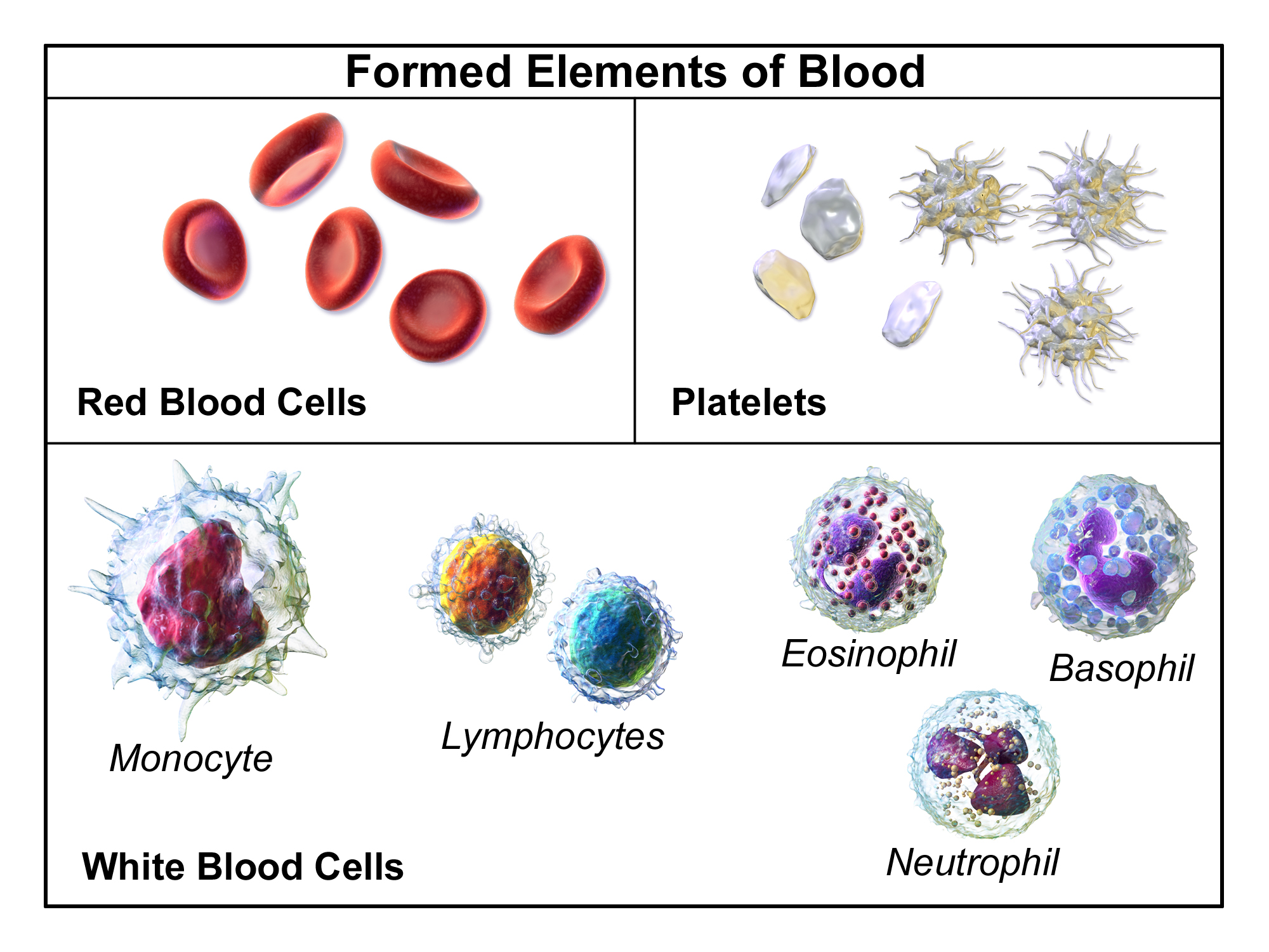
A complete blood count (CBC), also known as a full blood count (FBC), is a set of medical laboratory tests that provide cytometry, information about the cells in a person's blood. The CBC indicates the counts of white blood cells, red blood cells and platelets, the concentration of hemoglobin, and the hematocrit (the volume percentage of red blood cells). The red blood cell indices, which indicate the average size and hemoglobin content of red blood cells, are also reported, and a white blood cell differential, which counts the different types of white blood cells, may be included. The CBC is often carried out as part of a medical assessment and can be used to monitor health or diagnose diseases. The results are interpreted by comparing them to Reference ranges for blood tests, reference ranges, which vary with sex and age. Conditions like anemia and thrombocytopenia are defined by abnormal complete blood count results. The red blood cell indices can provide information about the ... [...More Info...] [...Related Items...] OR: [Wikipedia] [Google] [Baidu] |
Acute Kidney Injury
Acute kidney injury (AKI), previously called acute renal failure (ARF), is a sudden decrease in kidney function that develops within 7 days, as shown by an increase in serum creatinine or a decrease in urine output, or both. Causes of AKI are classified as either prerenal (due to decreased blood flow to the kidney), intrinsic renal (due to damage to the kidney itself), or postrenal (due to blockage of urine flow). Prerenal causes of AKI include sepsis, dehydration, excessive blood loss, cardiogenic shock, heart failure, cirrhosis, and certain medications like ACE inhibitors or NSAIDs. Intrinsic renal causes of AKI include glomerulonephritis, lupus nephritis, acute tubular necrosis, certain antibiotics, and chemotherapeutic agents. Postrenal causes of AKI include kidney stones, bladder cancer, neurogenic bladder, enlargement of the prostate, narrowing of the urethra, and certain medications like anticholinergics. The diagnosis of AKI is made based on a person's signs and sympto ... [...More Info...] [...Related Items...] OR: [Wikipedia] [Google] [Baidu] |
Microangiopathic Hemolytic Anemia
Microangiopathic hemolytic anemia (MAHA) is a microangiopathic subgroup of hemolytic anemia (loss of red blood cells through destruction) caused by factors in the small blood vessels. It is identified by the finding of anemia and schistocytes on microscopy of the blood film. Signs and symptoms In diseases such as hemolytic uremic syndrome, disseminated intravascular coagulation, thrombotic thrombocytopenic purpura, and malignant hypertension, the endothelial layer of small vessels is damaged with resulting fibrin deposition and platelet aggregation. As red blood cells travel through these damaged vessels, they are fragmented resulting in intravascular hemolysis. The resulting schistocytes (red cell fragments) are also increasingly targeted for destruction by the reticuloendothelial system in the spleen, due to their narrow passage through obstructed vessel lumina. It is seen in systemic lupus erythematosus, where immune complexes aggregate with platelets, forming intravascular ... [...More Info...] [...Related Items...] OR: [Wikipedia] [Google] [Baidu] |


