|
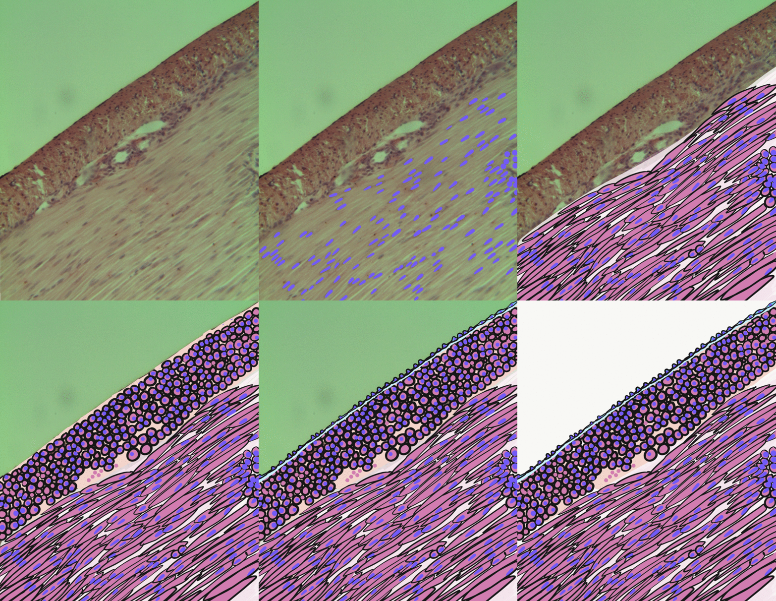
Vein Graft Disease
In medicine, vein graft failure (VGF) is a condition in which vein grafts, which are used as alternative conduits in bypass surgeries (e.g. CABG), get occluded. Veins, mainly the great saphenous vein (GSV) are the most frequently used conduits in bypass surgeries ( CABG or PABG), due to their ease of use and availability. Some structural changes of intima thickening and vein wall remodeling are necessary for vein graft adaptation to the arterial environment. The reasons why some of the grafts progress to clinical stenosis is unknown. The patency rates of saphenous vein grafts after CABG at 1 year is approximately 80%. After 5 years the patency rate drops to 65% and at 10 years GSV patency rates are approximately 50%, with only half of the veins are atherosclerosis free. VGF may be identified in asymptomatic patients, but can also produce symptoms of ischemia, depending on the area of the supplied territory of the heart, and the function of native arteries and other grafts. VGF ... [...More Info...] [...Related Items...] OR: [Wikipedia] [Google] [Baidu] |
Vein
Veins are blood vessels in humans and most other animals that carry blood towards the heart. Most veins carry deoxygenated blood from the tissues back to the heart; exceptions are the pulmonary and umbilical veins, both of which carry oxygenated blood to the heart. In contrast to veins, arteries carry blood away from the heart. Veins are less muscular than arteries and are often closer to the skin. There are valves (called ''pocket valves'') in most veins to prevent backflow. Structure Veins are present throughout the body as tubes that carry blood back to the heart. Veins are classified in a number of ways, including superficial vs. deep, pulmonary vs. systemic, and large vs. small. * Superficial veins are those closer to the surface of the body, and have no corresponding arteries. *Deep veins are deeper in the body and have corresponding arteries. *Perforator veins drain from the superficial to the deep veins. These are usually referred to in the lower limbs and feet. *Communic ... [...More Info...] [...Related Items...] OR: [Wikipedia] [Google] [Baidu] |
Smooth Muscle Cells
Smooth muscle is an involuntary non-striated muscle, so-called because it has no sarcomeres and therefore no striations (''bands'' or ''stripes''). It is divided into two subgroups, single-unit and multiunit smooth muscle. Within single-unit muscle, the whole bundle or sheet of smooth muscle cells contracts as a syncytium. Smooth muscle is found in the walls of hollow organs, including the stomach, intestines, bladder and uterus; in the walls of passageways, such as blood, and lymph vessels, and in the tracts of the respiratory, urinary, and reproductive systems. In the eyes, the ciliary muscles, a type of smooth muscle, dilate and contract the iris and alter the shape of the lens. In the skin, smooth muscle cells such as those of the arrector pili cause hair to stand erect in response to cold temperature or fear. Structure Gross anatomy Smooth muscle is grouped into two types: single-unit smooth muscle, also known as visceral smooth muscle, and multiunit smooth muscle. M ... [...More Info...] [...Related Items...] OR: [Wikipedia] [Google] [Baidu] |

