|
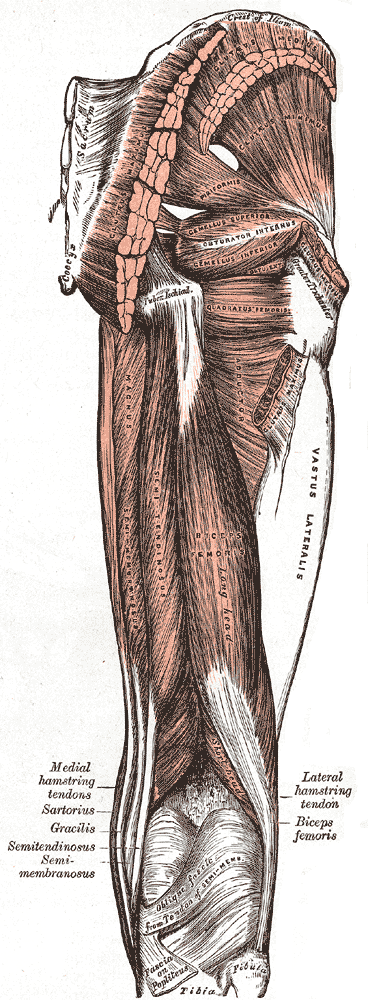
Vastus Lateralis
The vastus lateralis (), also called the vastus externus, is the largest and most powerful part of the quadriceps femoris, a muscle in the thigh. Together with other muscles of the quadriceps group, it serves to extend the knee joint, moving the lower leg forward. It arises from a series of flat, broad tendons attached to the femur, and attaches to the outer border of the patella. It ultimately joins with the other muscles that make up the quadriceps in the quadriceps tendon, which travels over the knee to connect to the tibia. The vastus lateralis is the recommended site for intramuscular injection in infants less than 7 months old and those unable to walk, with loss of muscular tone.Mann, E. (2016). ''Injection (Intramuscular): Clinician Information.'' The Johanna Briggs Institute. Structure The vastus lateralis muscle arises from several areas of the femur, including the upper part of the intertrochanteric line; the lower, anterior borders of the greater trochanter, to the out ... [...More Info...] [...Related Items...] OR: [Wikipedia] [Google] [Baidu] |
Rectus Femoris Muscle
The rectus femoris muscle is one of the four quadriceps muscles of the human body. The others are the vastus medialis, the vastus intermedius (deep to the rectus femoris), and the vastus lateralis. All four parts of the quadriceps muscle attach to the patella (knee cap) by the quadriceps tendon. The rectus femoris is situated in the middle of the front of the thigh; it is fusiform in shape, and its superficial fibers are arranged in a bipenniform manner, the deep fibers running straight ( la, rectus) down to the deep aponeurosis. Its functions are to flex the thigh at the hip joint and to extend the leg at the knee joint. Structure It arises by two tendons: one, the anterior or straight, from the anterior inferior iliac spine; the other, the posterior or reflected, from a groove above the rim of the acetabulum. The two unite at an acute angle and spread into an aponeurosis that is prolonged downward on the anterior surface of the muscle, and from this the muscular fibers ... [...More Info...] [...Related Items...] OR: [Wikipedia] [Google] [Baidu] |
Femur
The femur (; ), or thigh bone, is the proximal bone of the hindlimb in tetrapod vertebrates. The head of the femur articulates with the acetabulum in the pelvic bone forming the hip joint, while the distal part of the femur articulates with the tibia (shinbone) and patella (kneecap), forming the knee joint. By most measures the two (left and right) femurs are the strongest bones of the body, and in humans, the largest and thickest. Structure The femur is the only bone in the upper leg. The two femurs converge medially toward the knees, where they articulate with the proximal ends of the tibiae. The angle of convergence of the femora is a major factor in determining the femoral-tibial angle. Human females have thicker pelvic bones, causing their femora to converge more than in males. In the condition ''genu valgum'' (knock knee) the femurs converge so much that the knees touch one another. The opposite extreme is ''genu varum'' (bow-leggedness). In the general populatio ... [...More Info...] [...Related Items...] OR: [Wikipedia] [Google] [Baidu] |
Knee Extensors
In humans and other primates, the knee joins the thigh with the leg and consists of two joints: one between the femur and tibia (tibiofemoral joint), and one between the femur and patella (patellofemoral joint). It is the largest joint in the human body. The knee is a modified hinge joint, which permits flexion and extension as well as slight internal and external rotation. The knee is vulnerable to injury and to the development of osteoarthritis. It is often termed a ''compound joint'' having tibiofemoral and patellofemoral components. (The fibular collateral ligament is often considered with tibiofemoral components.) Structure The knee is a modified hinge joint, a type of synovial joint, which is composed of three functional compartments: the patellofemoral articulation, consisting of the patella, or "kneecap", and the patellar groove on the front of the femur through which it slides; and the medial and lateral tibiofemoral articulations linking the femur, or thigh b ... [...More Info...] [...Related Items...] OR: [Wikipedia] [Google] [Baidu] |
Knee-joint
In humans and other primates, the knee joins the thigh with the leg and consists of two joints: one between the femur and tibia (tibiofemoral joint), and one between the femur and patella (patellofemoral joint). It is the largest joint in the human body. The knee is a modified hinge joint, which permits flexion and extension as well as slight internal and external rotation. The knee is vulnerable to injury and to the development of osteoarthritis. It is often termed a ''compound joint'' having tibiofemoral and patellofemoral components. (The fibular collateral ligament is often considered with tibiofemoral components.) Structure The knee is a modified hinge joint, a type of synovial joint, which is composed of three functional compartments: the patellofemoral articulation, consisting of the patella, or "kneecap", and the patellar groove on the front of the femur through which it slides; and the medial and lateral tibiofemoral articulations linking the femur, or thigh bo ... [...More Info...] [...Related Items...] OR: [Wikipedia] [Google] [Baidu] |
Biceps Femoris
The biceps femoris () is a muscle of the thigh located to the posterior, or back. As its name implies, it has two parts, one of which (the long head) forms part of the hamstrings muscle group. Structure It has two heads of origin: *the ''long head'' arises from the lower and inner impression on the posterior part of the tuberosity of the ischium. This is a common tendon origin with the semitendinosus muscle, and from the lower part of the sacrotuberous ligament. *the ''short head'', arises from the lateral lip of the linea aspera, between the adductor magnus and vastus lateralis extending up almost as high as the insertion of the gluteus maximus, from the lateral prolongation of the linea aspera to within 5 cm. of the lateral condyle; and from the lateral intermuscular septum. The two muscle heads joint together distally and unite in an intricate fashion. The fibers of the long head form a fusiform belly, which passes obliquely downward and lateralward across the sciat ... [...More Info...] [...Related Items...] OR: [Wikipedia] [Google] [Baidu] |
Lateral Intermuscular Septum Of Thigh
The lateral intermuscular septum of thigh is a fold of deep fascia in the thigh. It is between the vastus lateralis and biceps femoris. It separates the anterior compartment of the thigh from the posterior compartment of the thigh. See also *Medial intermuscular septum of thigh * Anterior compartment of thigh *Posterior compartment of thigh The posterior compartment of the thigh is one of the fascial compartments that contains the knee flexors and hip extensors known as the hamstring muscles, as well as vascular and nervous elements, particularly the sciatic nerve. Structure The ... References External links Topographical Anatomy of the Lower Limb - Listed Alphabeticallyfrom UAMS Department of Neurobiology and Developmental Sciences from anatomy.med.umich.edu Lower limb anatomy {{musculoskeletal-stub ... [...More Info...] [...Related Items...] OR: [Wikipedia] [Google] [Baidu] |
Gluteus Maximus
The gluteus maximus is the main extensor muscle of the hip. It is the largest and outermost of the three gluteal muscles and makes up a large part of the shape and appearance of each side of the hips. It is the single largest muscle in the human body. Its thick fleshy mass, in a quadrilateral shape, forms the prominence of the buttocks. The other gluteal muscles are the medius and minimus, and sometimes informally these are collectively referred to as the glutes. Its large size is one of the most characteristic features of the muscular system in humans,Norman Eizenberg et al., ''General Anatomy: Principles and Applications'' (2008), p. 17. connected as it is with the power of maintaining the trunk in the erect posture. Other primates have much flatter hips and cannot sustain standing erectly. The muscle is made up of muscle fascicles lying parallel with one another, and are collected together into larger bundles separated by fibrous septa. Structure The gluteus maximus is the ... [...More Info...] [...Related Items...] OR: [Wikipedia] [Google] [Baidu] |
Linea Aspera
The linea aspera ( la, rough line) is a ridge of roughened surface on the posterior surface of the shaft of the femur. It is the site of attachments of muscles and the intermuscular septum. Its margins diverge above and below. The linea aspera is a prominent longitudinal ridge or crest, on the middle third of the bone, presenting a medial and a lateral lip, and a narrow rough, intermediate line. It is an important insertion point for the adductors and the lateral and medial intermuscular septa that divides the thigh into three compartments. The tension generated by muscle attached to the bones is responsible for the formation of the ridges. Structure Above Above, the linea aspera is prolonged by three ridges. * The lateral ridge is very rough, and runs almost vertically upward to the base of the greater trochanter. It is termed the gluteal tuberosity, and gives attachment to part of the gluteus maximus: its upper part is often elongated into a roughened crest, on which a mor ... [...More Info...] [...Related Items...] OR: [Wikipedia] [Google] [Baidu] |
Gluteal Tuberosity
The gluteal tuberosity is the lateral one of the three upward prolongations of the linea aspera of the femur, extending to the base of the greater trochanter. It serves as the principal insertion site for the gluteus maximus muscle. Structure The gluteal tuberosity is the lateral prolongation of three prolongations of the linea aspera that extending superior-ward from the superior extremity of the linea aspera on the posterior surface of the femur. The gluteal tuberosity takes the form of either an elongated depression or a rough ridge. It extends from the linea aspera nearly vertically superior-ward to the base of the greater trochanter. Its superior part is often elongated to form a roughened crest, upon which a more or less prominent rounded tubercle - the third trochanter - is occasionally developed. Attachments The gluteal tuberosity is the principal site of insertion of the gluteus maximus muscle, accepting the muscle's tendon (the gluteus maximus muscle additionally al ... [...More Info...] [...Related Items...] OR: [Wikipedia] [Google] [Baidu] |
Greater Trochanter
The greater trochanter of the femur is a large, irregular, quadrilateral eminence and a part of the skeletal system. It is directed lateral and medially and slightly posterior. In the adult it is about 2–4 cm lower than the femoral head.Standring, Susan, editor. ''Gray’s Anatomy: The Anatomical Basis of Clinical Practice''. Forty-First edition, Elsevier Limited, 2016, p. 1327. Because the pelvic outlet in the female is larger than in the male, there is a greater distance between the greater trochanters in the female. It has two surfaces and four borders. It is a traction epiphysis. Surfaces The ''lateral surface'', quadrilateral in form, is broad, rough, convex, and marked by a diagonal impression, which extends from the postero-superior to the antero-inferior angle, and serves for the insertion of the tendon of the gluteus medius. Above the impression is a triangular surface, sometimes rough for part of the tendon of the same muscle, sometimes smooth for the interposi ... [...More Info...] [...Related Items...] OR: [Wikipedia] [Google] [Baidu] |
Intertrochanteric Line
The intertrochanteric line (or ''spiral line of the femur''White (2005), p 256 ) is a line located on the anterior side of the proximal end of the femur. Structure The rough, variable ridge stretches between the lesser trochanter and the greater trochanter forming the base of the neck of the femur, roughly following the direction of the shaft of the femur. The iliofemoral ligament — the largest ligament of the human body — attaches above the line which also strengthens the capsule of the hip joint. The lower half, less prominent than the upper half, gives origin to the upper part of the Vastus medialis. Just like the intertrochanteric crest on the posterior side of the femoral head, the intertrochanteric line marks the transition between the femoral neck and shaft.Platzer (2004), p 192 The distal capsular attachment on the femur follows the shape of the irregular rim between the head and the neck. As a consequence, the capsule of the hip joint attaches in the reg ... [...More Info...] [...Related Items...] OR: [Wikipedia] [Google] [Baidu] |
Intramuscular Injection
Intramuscular injection, often abbreviated IM, is the injection of a substance into a muscle. In medicine, it is one of several methods for parenteral administration of medications. Intramuscular injection may be preferred because muscles have larger and more numerous blood vessels than subcutaneous tissue, leading to faster absorption than subcutaneous or intradermal injections. Medication administered via intramuscular injection is not subject to the first-pass metabolism effect which affects oral medications. Common sites for intramuscular injections include the deltoid muscle of the upper arm and the gluteal muscle of the buttock. In infants, the vastus lateralis muscle of the thigh is commonly used. The injection site must be cleaned before administering the injection, and the injection is then administered in a fast, darting motion to decrease the discomfort to the individual. The volume to be injected in the muscle is usually limited to 2–5 milliliters, depending on in ... [...More Info...] [...Related Items...] OR: [Wikipedia] [Google] [Baidu] |


