|
Vascular Tumor
A vascular tumor is a tumor of vascular origin; a soft tissue growth that can be either benign or malignant, formed from blood vessels or lymph vessels. Examples of vascular tumors include hemangiomas, lymphangiomas, hemangioendotheliomas, Kaposi's sarcomas, angiosarcomas, and hemangioblastomas. An angioma refers to any type of benign vascular tumor. Some vascular tumors can be associated with serious blood-clotting disorders, making correct diagnosis critical. A vascular tumor may be described in terms of being ''highly vascularized'', or ''poorly vascularized'', referring to the degree of blood supply to the tumor. Classification Vascular tumors make up one of the classifications of vascular anomalies. The other grouping is vascular malformations. Vascular tumors can be further subclassified as being benign, borderline or aggressive, and malignant. Vascular tumors are described as ''proliferative'', and vascular malformations as ''nonproliferative''. Types A vascular tumor ... [...More Info...] [...Related Items...] OR: [Wikipedia] [Google] [Baidu] |
Hemangioma
A hemangioma or haemangioma is a usually benign vascular tumor derived from blood vessel cell types. The most common form, seen in infants, is an infantile hemangioma, known colloquially as a "strawberry mark", most commonly presenting on the skin at birth or in the first weeks of life. A hemangioma can occur anywhere on the body, but most commonly appears on the face, scalp, chest or back. They tend to grow for up to a year before gradually shrinking as the child gets older. A hemangioma may need to be treated if it interferes with vision or breathing or is likely to cause long-term disfigurement. In rare cases internal hemangiomas can cause or contribute to other medical problems. Most of the time they tend to disappear in 10 years. The first line treatment option is beta blockers, which are highly effective in the majority of cases. Ones that form at birth are called congenital hemangiomas while ones that form later in life are called infantile hemangiomas. Types Hema ... [...More Info...] [...Related Items...] OR: [Wikipedia] [Google] [Baidu] |
Endothelial Cell
The endothelium is a single layer of squamous endothelial cells that line the interior surface of blood vessels and lymphatic vessels. The endothelium forms an interface between circulating blood or lymph in the lumen and the rest of the vessel wall. Endothelial cells form the barrier between vessels and tissue and control the flow of substances and fluid into and out of a tissue. Endothelial cells in direct contact with blood are called vascular endothelial cells whereas those in direct contact with lymph are known as lymphatic endothelial cells. Vascular endothelial cells line the entire circulatory system, from the heart to the smallest capillaries. These cells have unique functions that include fluid filtration, such as in the glomerulus of the kidney, blood vessel tone, hemostasis, neutrophil recruitment, and hormone trafficking. Endothelium of the interior surfaces of the heart chambers is called endocardium. An impaired function can lead to serious health issues throug ... [...More Info...] [...Related Items...] OR: [Wikipedia] [Google] [Baidu] |
Tufted Angioma
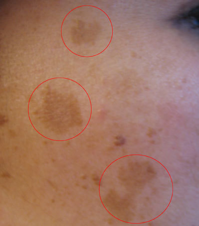
A tufted angioma (also known as an "Acquired tufted angioma," "Angioblastoma," "Angioblastoma of Nakagawa," "Hypertrophic hemangioma," "Progressive capillary hemangioma," and "Tufted hemangioma") usually develops in infancy or early childhood on the neck and upper trunk, and is an ill-defined, dull red macule with a mottled appearance, varying from 2 to 5 cm in diameter.James, William; Berger, Timothy; Elston, Dirk (2005). ''Andrews' Diseases of the Skin: Clinical Dermatology''. (10th ed.). Saunders. . See also * List of cutaneous conditions *Skin lesion A skin condition, also known as cutaneous condition, is any medical condition that affects the integumentary system—the organ system that encloses the body and includes skin, nails, and related muscle and glands. The major function of this s ... References External links Dermal and subcutaneous growths {{Dermal-growth-stub ... [...More Info...] [...Related Items...] OR: [Wikipedia] [Google] [Baidu] |
Pyogenic Granuloma
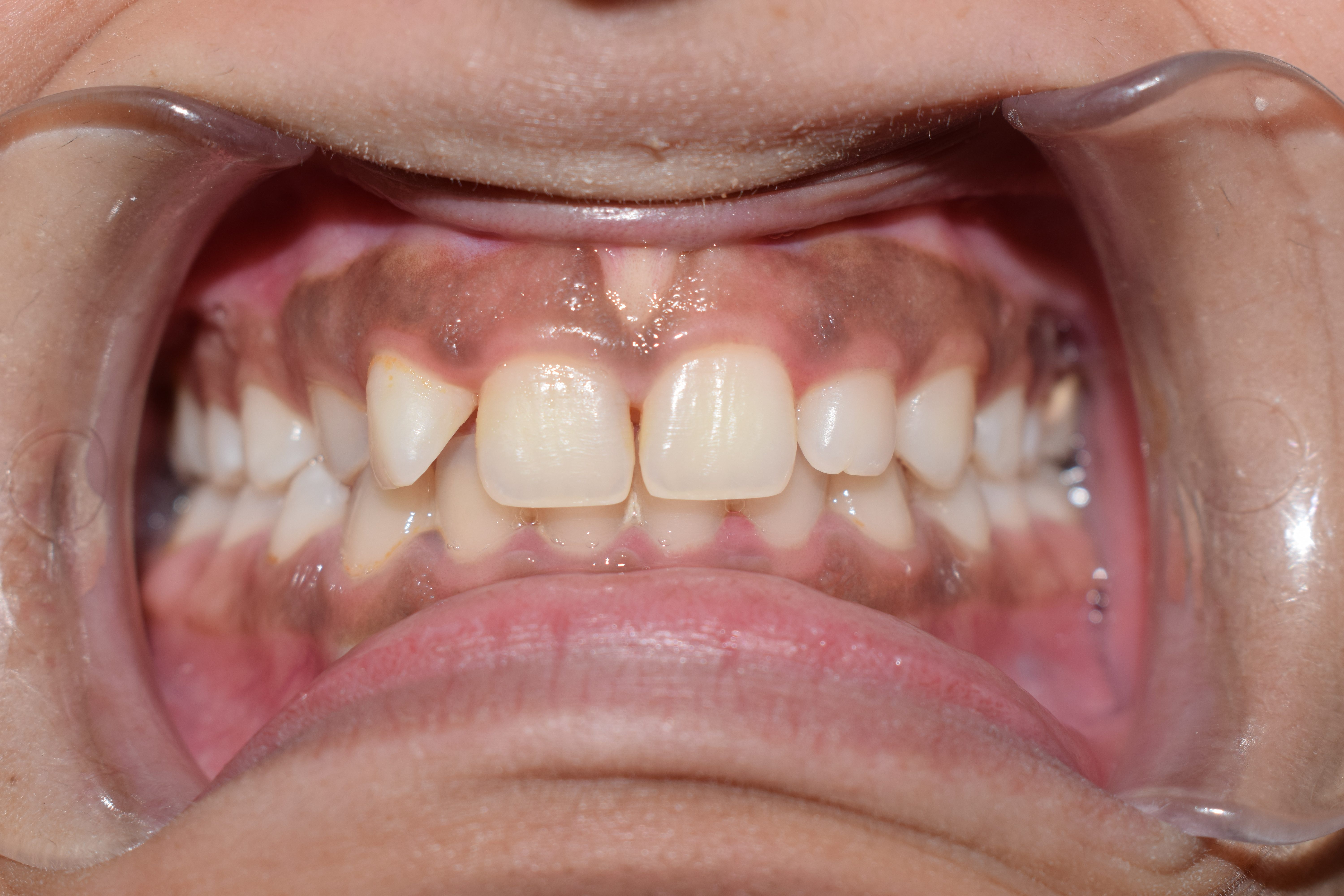
A pyogenic granuloma or lobular capillary hemangioma is a vascular tumor that occurs on both mucosa and skin, and appears as an overgrowth of tissue due to irritation, physical trauma, or hormonal factors. It is often found to involve the gums, skin, or nasal septum, and has also been found far from the head, such as in the thigh. Pyogenic granulomas may be seen at any age, and are more common in females than males. In pregnant women, lesions may occur in the first trimester with an increasing incidence until the seventh month, and are often seen on the gums. Signs and symptoms The appearance of pyogenic granuloma is usually a color ranging from red/pink to purple, grows rapidly, and can be smooth or mushroom-shaped. Younger lesions are more likely to be red because of their high number of blood vessels. Older lesions begin to change into a pink color. Size commonly ranges from a few millimeters to centimeters, though smaller or larger lesions may occur. A pyogenic granuloma can ... [...More Info...] [...Related Items...] OR: [Wikipedia] [Google] [Baidu] |
Gums
The gums or gingiva (plural: ''gingivae'') consist of the mucosal tissue that lies over the mandible and maxilla inside the mouth. Gum health and disease can have an effect on general health. Structure The gums are part of the soft tissue lining of the mouth. They surround the teeth and provide a seal around them. Unlike the soft tissue linings of the lips and cheeks, most of the gums are tightly bound to the underlying bone which helps resist the friction of food passing over them. Thus when healthy, it presents an effective barrier to the barrage of periodontal insults to deeper tissue. Healthy gums are usually coral pink in light skinned people, and may be naturally darker with melanin pigmentation. Changes in color, particularly increased redness, together with swelling and an increased tendency to bleed, suggest an inflammation that is possibly due to the accumulation of bacterial plaque. Overall, the clinical appearance of the tissue reflects the underlying histology, bo ... [...More Info...] [...Related Items...] OR: [Wikipedia] [Google] [Baidu] |
Pregnancy
Pregnancy is the time during which one or more offspring develops ( gestates) inside a woman's uterus (womb). A multiple pregnancy involves more than one offspring, such as with twins. Pregnancy usually occurs by sexual intercourse, but can also occur through assisted reproductive technology procedures. A pregnancy may end in a live birth, a miscarriage, an induced abortion, or a stillbirth. Childbirth typically occurs around 40 weeks from the start of the last menstrual period (LMP), a span known as the gestational age. This is just over nine months. Counting by fertilization age, the length is about 38 weeks. Pregnancy is "the presence of an implanted human embryo or fetus in the uterus"; implantation occurs on average 8–9 days after fertilization. An '' embryo'' is the term for the developing offspring during the first seven weeks following implantation (i.e. ten weeks' gestational age), after which the term ''fetus'' is used until birth. Signs an ... [...More Info...] [...Related Items...] OR: [Wikipedia] [Google] [Baidu] |
Thrombosis
Thrombosis (from Ancient Greek "clotting") is the formation of a blood clot inside a blood vessel, obstructing the flow of blood through the circulatory system. When a blood vessel (a vein or an artery) is injured, the body uses platelets (thrombocytes) and fibrin to form a blood clot to prevent blood loss. Even when a blood vessel is not injured, blood clots may form in the body under certain conditions. A clot, or a piece of the clot, that breaks free and begins to travel around the body is known as an embolus. Thrombosis may occur in veins (venous thrombosis) or in arteries (arterial thrombosis). Venous thrombosis (sometimes called DVT, deep vein thrombosis) leads to a blood clot in the affected part of the body, while arterial thrombosis (and, rarely, severe venous thrombosis) affects the blood supply and leads to damage of the tissue supplied by that artery (ischemia and necrosis). A piece of either an arterial or a venous thrombus can break off as an embolus, which could ... [...More Info...] [...Related Items...] OR: [Wikipedia] [Google] [Baidu] |
Central Nervous System
The central nervous system (CNS) is the part of the nervous system consisting primarily of the brain and spinal cord. The CNS is so named because the brain integrates the received information and coordinates and influences the activity of all parts of the bodies of bilaterally symmetric and triploblastic animals—that is, all multicellular animals except sponges and diploblasts. It is a structure composed of nervous tissue positioned along the rostral (nose end) to caudal (tail end) axis of the body and may have an enlarged section at the rostral end which is a brain. Only arthropods, cephalopods and vertebrates have a true brain (precursor structures exist in onychophorans, gastropods and lancelets). The rest of this article exclusively discusses the vertebrate central nervous system, which is radically distinct from all other animals. Overview In vertebrates, the brain and spinal cord are both enclosed in the meninges. The meninges provide a barrier to chemicals dissolv ... [...More Info...] [...Related Items...] OR: [Wikipedia] [Google] [Baidu] |
Hemangioblastoma
Hemangioblastomas, or haemangioblastomas, are vascular tumors of the central nervous system that originate from the vascular system, usually during middle age. Sometimes, these tumors occur in other sites such as the spinal cord and retina. They may be associated with other diseases such as polycythemia (increased blood cell count), pancreatic cysts and Von Hippel–Lindau syndrome (VHL syndrome). Hemangioblastomas are most commonly composed of stromal cells in small blood vessels and usually occur in the cerebellum, brainstem or spinal cord. They are classed as grade I tumors under the World Health Organization's classification system. Presentation Complications Hemangioblastomas can cause an abnormally high number of red blood cells in the bloodstream due to ectopic production of the hormone erythropoietin as a paraneoplastic syndrome. Pathogenesis Hemangioblastomas are composed of endothelial cells, pericytes and stromal cells. In VHL syndrome the von Hippel-Lindau protein ... [...More Info...] [...Related Items...] OR: [Wikipedia] [Google] [Baidu] |
Thrombocytopenia
Thrombocytopenia is a condition characterized by abnormally low levels of platelets, also known as thrombocytes, in the blood. It is the most common coagulation disorder among intensive care patients and is seen in a fifth of medical patients and a third of surgical patients. A normal human platelet count ranges from 150,000 to 450,000 platelets/microliter (μl) of blood. Values outside this range do not necessarily indicate disease. One common definition of thrombocytopenia requiring emergency treatment is a platelet count below 50,000/μl. Thrombocytopenia can be contrasted with the conditions associated with an abnormally ''high'' level of platelets in the blood - thrombocythemia (when the cause is unknown), and thrombocytosis (when the cause is known). Signs and symptoms Thrombocytopenia usually has no symptoms and is picked up on a routine complete blood count. Some individuals with thrombocytopenia may experience external bleeding, such as nosebleeds or bleeding gums. S ... [...More Info...] [...Related Items...] OR: [Wikipedia] [Google] [Baidu] |
GLUT 1
Glucose transporter 1 (or GLUT1), also known as solute carrier family 2, facilitated glucose transporter member 1 (SLC2A1), is a uniporter protein that in humans is encoded by the ''SLC2A1'' gene. GLUT1 facilitated diffusion, facilitates the transport of glucose across the cell membrane, plasma membranes of mammalian cells. This gene encodes a major glucose transporter in the mammalian blood–brain barrier. The encoded protein is found primarily in the cell membrane and on the cell surface, where it can also function as a Cell surface receptor, receptor for Human T-lymphotropic virus, human T-cell leukemia virus (HTLV) Human T-lymphotropic virus 1, I and Human T-lymphotropic virus 2, II. One good source of GLUT1 is erythrocyte membranes. GLUT1 accounts for 2 percent of the protein in the plasma membrane of erythrocytes. GLUT1, found in the plasma membrane of erythrocytes, is a classic example of a uniporter. After glucose is transported into the erythrocyte, it is rapidly phosphory ... [...More Info...] [...Related Items...] OR: [Wikipedia] [Google] [Baidu] |
Hypoxia (medical)
Hypoxia is a condition in which the body or a region of the body is deprived of adequate oxygen supply at the tissue level. Hypoxia may be classified as either '' generalized'', affecting the whole body, or ''local'', affecting a region of the body. Although hypoxia is often a pathological condition, variations in arterial oxygen concentrations can be part of the normal physiology, for example, during strenuous physical exercise.. Hypoxia differs from hypoxemia and anoxemia, in that hypoxia refers to a state in which oxygen present in a tissue or the whole body is insufficient, whereas hypoxemia and anoxemia refer specifically to states that have low or no oxygen in the blood. Hypoxia in which there is complete absence of oxygen supply is referred to as anoxia. Hypoxia can be due to external causes, when the breathing gas is hypoxic, or internal causes, such as reduced effectiveness of gas transfer in the lungs, reduced capacity of the blood to carry oxygen, compromised general ... [...More Info...] [...Related Items...] OR: [Wikipedia] [Google] [Baidu] |


