|
Vaginal Agenesis
Vaginal atresia is a condition in which the vagina is abnormally closed or absent. The main causes can either be complete vaginal hypoplasia, or a vaginal obstruction, often caused by an imperforate hymen or, less commonly, a transverse vaginal septum. It results in uterovaginal outflow tract obstruction. This condition does not usually occur by itself within an individual, but coupled with other developmental disorders within the female. The disorders that are usually coupled with a female who has vaginal atresia are Mayer-Rokitansky-Küster-Hauser syndrome, Bardet-Biedl syndrome, or Fraser syndrome. One out of every 5,000 women have this abnormality. Symptoms and signs Symptoms and signs in the newborn can be sepsis, abdominal mass, and respiratory distress. Other abdominopelvic or perineal congenital anomalies frequently prompt radiographic evaluation in the newborn, resulting in a diagnosis of coincident vaginal atresia. Symptoms for vaginal atresia include cyclical abdominal ... [...More Info...] [...Related Items...] OR: [Wikipedia] [Google] [Baidu] |
Vagina
In mammals, the vagina is the elastic, muscular part of the female genital tract. In humans, it extends from the vestibule to the cervix. The outer vaginal opening is normally partly covered by a thin layer of mucosal tissue called the hymen. At the deep end, the cervix (neck of the uterus) bulges into the vagina. The vagina allows for sexual intercourse and birth. It also channels menstrual flow, which occurs in humans and closely related primates as part of the menstrual cycle. Although research on the vagina is especially lacking for different animals, its location, structure and size are documented as varying among species. Female mammals usually have two external openings in the vulva; these are the urethral opening for the urinary tract and the vaginal opening for the genital tract. This is different from male mammals, who usually have a single urethral opening for both urination and reproduction. The vaginal opening is much larger than the nearby urethral opening, an ... [...More Info...] [...Related Items...] OR: [Wikipedia] [Google] [Baidu] |
Short Arm
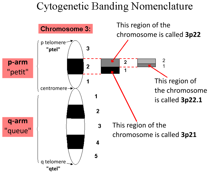
In genetics, a locus (plural loci) is a specific, fixed position on a chromosome where a particular gene or genetic marker A genetic marker is a gene or DNA sequence with a known location on a chromosome that can be used to identify individuals or species. It can be described as a variation (which may arise due to mutation or alteration in the genomic loci) that can be ... is located. Each chromosome carries many genes, with each gene occupying a different position or locus; in humans, the total number of Human genome#Coding sequences (protein-coding genes), protein-coding genes in a complete haploid set of 23 chromosomes is estimated at 19,000–20,000. Genes may possess multiple variants known as alleles, and an allele may also be said to reside at a particular locus. Diploid and polyploid cells whose chromosomes have the same allele at a given locus are called homozygote, homozygous with respect to that locus, while those that have different alleles at a given locus are called ... [...More Info...] [...Related Items...] OR: [Wikipedia] [Google] [Baidu] |
Hypoplasia
Hypoplasia (from Ancient Greek ὑπo- ''hypo-'' 'under' + πλάσις ''plasis'' 'formation'; adjective form ''hypoplastic'') is underdevelopment or incomplete development of a tissue or organ. Dictionary of Cell and Molecular Biology (11 March 2008) Although the term is not always used precisely, it properly refers to an inadequate or below-normal number of cells.Hypoplasia Stedman's Medical Dictionary. lww.com Hypoplasia is similar to |
Septate Vagina
A uterine malformation is a type of female genital malformation resulting from an abnormal development of the Müllerian duct(s) during embryogenesis. Symptoms range from amenorrhea, infertility, recurrent pregnancy loss, and pain, to normal functioning depending on the nature of the defect. Types The American Fertility Society (now American Society of Reproductive Medicine) Classification distinguishes: ; Class I—Müllerian agenesis (absent uterus). : This condition is represented by the hypoplasia or the agenesis (total absence) of the different parts of the uterus: :* Vaginal hypoplasia or agenesis :* Cervical hypoplasia or agenesis :* Fundal hypoplasia or agenesis (absence or hypoplasia of the fundus of the uterus) :* Tubal hypoplasia or agenesis (absence or hypoplasia of the Fallopian tubes) :* Combined hypoplasia the agenesis of different part of the uterus :This condition is also called Mayer-Rokitansky-Kuster-Hauser syndrome. The patient with MRKH syndrome will hav ... [...More Info...] [...Related Items...] OR: [Wikipedia] [Google] [Baidu] |
Hypogonadism
Hypogonadism means diminished functional activity of the gonads—the testes or the ovaries—that may result in diminished production of sex hormones. Low androgen (e.g., testosterone) levels are referred to as hypoandrogenism and low estrogen (e.g., estradiol) as hypoestrogenism. These are responsible for the observed signs and symptoms in both males and females. Hypogonadism, commonly referred to by the symptom "low testosterone" or "Low T", can also decrease other hormones secreted by the gonads including progesterone, DHEA, anti-Müllerian hormone, activin, and inhibin. Sperm development (spermatogenesis) and release of the egg from the ovaries (ovulation) may be impaired by hypogonadism, which, depending on the degree of severity, may result in partial or complete infertility. In January 2020, the American College of Physicians issued clinical guidelines for testosterone treatment in adult men with age-related low levels of testosterone. The guidelines are supported b ... [...More Info...] [...Related Items...] OR: [Wikipedia] [Google] [Baidu] |
Uterus Duplex
A uterine malformation is a type of female genital malformation resulting from an abnormal development of the Müllerian duct(s) during embryogenesis. Symptoms range from amenorrhea, infertility, recurrent pregnancy loss, and pain, to normal functioning depending on the nature of the defect. Types The American Fertility Society (now American Society of Reproductive Medicine) Classification distinguishes: ; Class I—Müllerian agenesis (absent uterus). : This condition is represented by the hypoplasia or the agenesis (total absence) of the different parts of the uterus: :* Vaginal hypoplasia or agenesis :* Cervical hypoplasia or agenesis :* Fundal hypoplasia or agenesis (absence or hypoplasia of the fundus of the uterus) :* Tubal hypoplasia or agenesis (absence or hypoplasia of the Fallopian tubes) :* Combined hypoplasia the agenesis of different part of the uterus :This condition is also called Mayer-Rokitansky-Kuster-Hauser syndrome. The patient with MRKH syndrome will h ... [...More Info...] [...Related Items...] OR: [Wikipedia] [Google] [Baidu] |
Kidney Failure
Kidney failure, also known as end-stage kidney disease, is a medical condition in which the kidneys can no longer adequately filter waste products from the blood, functioning at less than 15% of normal levels. Kidney failure is classified as either acute kidney failure, which develops rapidly and may resolve; and chronic kidney failure, which develops slowly and can often be irreversible. Symptoms may include leg swelling, feeling tired, vomiting, loss of appetite, and confusion. Complications of acute and chronic failure include uremia, high blood potassium, and volume overload. Complications of chronic failure also include heart disease, high blood pressure, and anemia. Causes of acute kidney failure include low blood pressure, blockage of the urinary tract, certain medications, muscle breakdown, and hemolytic uremic syndrome. Causes of chronic kidney failure include diabetes, high blood pressure, nephrotic syndrome, and polycystic kidney disease. Diagnosis of acute failure ... [...More Info...] [...Related Items...] OR: [Wikipedia] [Google] [Baidu] |
Ectopic Ureter
Ectopic ureter (or ureteral ectopia) is a medical condition where the ureter, rather than terminating at the urinary bladder, terminates at a different site. In males this site is usually the urethra, in females this is usually the urethra or vagina. It can be associated with renal dysplasia, frequent urinary tract infections, and urinary incontinence (usually continuous drip incontinence). Ectopic ureters are found in 1 of every 2000–4000 patients, and can be difficult to diagnose, but are most often seen on CT scans. Ectopic ureter is commonly a result of a duplicated renal collecting system, a duplex kidney with 2 ureters. In this case, usually one ureter drains correctly to the bladder, with the duplicated ureter presenting as ectopic. The embryology that explains the pathology of an ectopic ureter is a cephalad origin of the ureteral bud on the mesonephric duct. With an abnormally long common excretory duct, the ureter never becomes incorporated into the bladder, and, t ... [...More Info...] [...Related Items...] OR: [Wikipedia] [Google] [Baidu] |
Ciliopathic
A ciliopathy is any genetic disorder that affects the cellular cilia or the cilia anchoring structures, the basal bodies, or ciliary function. Primary cilia are important in guiding the process of development, so abnormal ciliary function while an embryo is developing can lead to a set of malformations that can occur regardless of the particular genetic problem. The similarity of the clinical features of these developmental disorders means that they form a recognizable cluster of syndromes, loosely attributed to abnormal ciliary function and hence called ciliopathies. Regardless of the actual genetic cause, it is clustering of a set of characteristic physiological features which define whether a syndrome is a ciliopathy. Although ciliopathies are usually considered to involve proteins that localize to motile and/or immotile (primary) cilia or centrosomes, it is possible for ciliopathies to be associated with unexpected proteins such as XPNPEP3, which localizes to mitochondria bu ... [...More Info...] [...Related Items...] OR: [Wikipedia] [Google] [Baidu] |
Paramesonephric Duct
Paramesonephric ducts (or Müllerian ducts) are paired ducts of the embryo that run down the lateral sides of the genital ridge and terminate at the sinus tubercle in the primitive urogenital sinus. In the female, they will develop to form the fallopian tubes, uterus, cervix, and the upper one-third of the vagina. Development The female reproductive system is composed of two embryological segments: the urogenital sinus and the paramesonephric ducts. The two are conjoined at the sinus tubercle. Paramesonephric ducts are present on the embryo of both sexes. Only in females do they develop into reproductive organs. They degenerate in males of certain species, but the adjoining mesonephric ducts develop into male reproductive organs. The sex based differences in the contributions of the paramesonephric ducts to reproductive organs is based on the presence, and degree of presence, of Anti-Müllerian hormone. During the formation of the reproductive system, the paramesonephric ducts ar ... [...More Info...] [...Related Items...] OR: [Wikipedia] [Google] [Baidu] |
Androgen
An androgen (from Greek ''andr-'', the stem of the word meaning "man") is any natural or synthetic steroid hormone that regulates the development and maintenance of male characteristics in vertebrates by binding to androgen receptors. This includes the embryological development of the primary male sex organs, and the development of male secondary sex characteristics at puberty. Androgens are synthesized in the testes, the ovaries, and the adrenal glands. Androgens increase in both males and females during puberty. The major androgen in males is testosterone. Dihydrotestosterone (DHT) and androstenedione are of equal importance in male development. DHT ''in utero'' causes differentiation of the penis, scrotum and prostate. In adulthood, DHT contributes to balding, prostate growth, and sebaceous gland activity. Although androgens are commonly thought of only as male sex hormones, females also have them, but at lower levels: they function in libido and sexual arousal. Also, an ... [...More Info...] [...Related Items...] OR: [Wikipedia] [Google] [Baidu] |


