|
Vagal Trigone
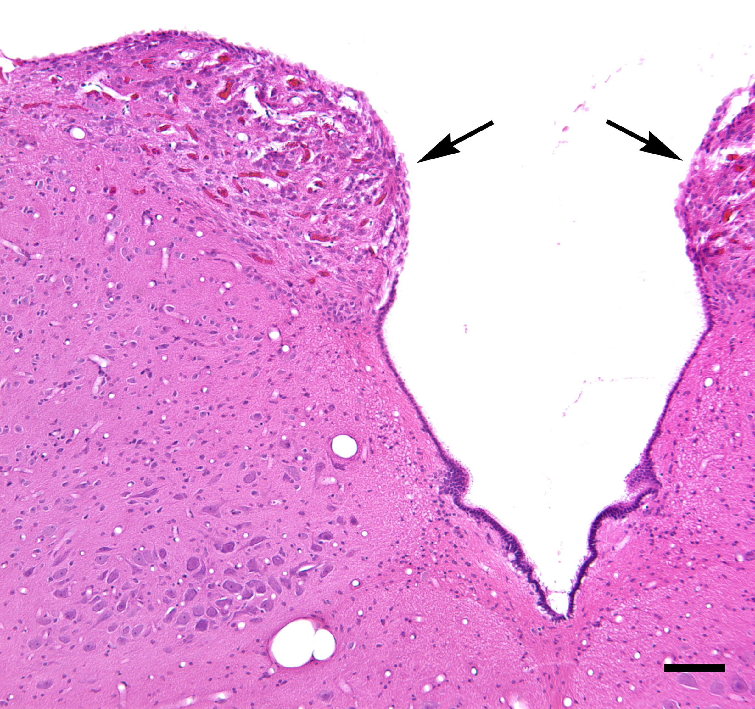
The cells of the dorsal nucleus of vagus nerve are spindle-shaped, like those of the posterior column of the spinal cord, and the nucleus is usually considered as representing the base of the posterior column. It measures about 2 cm. in length, and in the lower, closed part of the medulla oblongata is situated behind the dorsal nucleus of the vagus; whereas in the upper, open part it lies lateral to that nucleus, and corresponds to an eminence, named the vagal trigone (ala cinerea), in the rhomboid fossa. The vagal trigone is separated from the area postrema by a narrow strip of thickened ependyma The ependyma is the thin Neuroepithelial cell, neuroepithelial (Simple columnar epithelium, simple columnar ciliated epithelium) lining of the ventricular system of the brain and the central canal of the spinal cord. The ependyma is one of the fou ... – the funiculus separans. References External links * https://web.archive.org/web/20070927162218/http://www.ib.amwaw.edu.pl/an ... [...More Info...] [...Related Items...] OR: [Wikipedia] [Google] [Baidu] |
Dorsal Nucleus Of Vagus Nerve
The dorsal nucleus of vagus nerve (or posterior nucleus of vagus nerve or dorsal vagal nucleus or nucleus dorsalis nervi vagi or nucleus posterior nervi vagi) is a cranial nerve nucleus for the vagus nerve in the medulla that lies ventral to the floor of the fourth ventricle. It mostly serves parasympathetic vagal functions in the gastrointestinal tract, lungs, and other thoracic and abdominal vagal innervations. These functions include, among others, bronchoconstriction and gland secretion. The cell bodies for the preganglionic parasympathetic vagal neurons that innervate the heart reside in the nucleus ambiguus. Additional cell bodies are found in the nucleus ambiguus, which give rise to the branchial efferent motor fibers of the vagus nerve (CN X) terminating in the laryngeal, pharyngeal muscles, and musculus uvulae. Additional images File:Gray694.png, Section of the medulla oblongata at about the middle of the olive. File:Gray696.png, The cranial nerve nuclei schematically ... [...More Info...] [...Related Items...] OR: [Wikipedia] [Google] [Baidu] |
Posterior Column , a relative future tense
{{disambiguation ...
Posterior may refer to: * Posterior (anatomy), the end of an organism opposite to its head ** Buttocks, as a euphemism * Posterior horn (other) * Posterior probability, the conditional probability that is assigned when the relevant evidence is taken into account * Posterior tense Relative tense and absolute tense are distinct possible uses of the grammatical category of Grammatical tense, tense. Absolute tense means the grammatical expression of time reference (usually past tense, past, present tense, present or future tense ... [...More Info...] [...Related Items...] OR: [Wikipedia] [Google] [Baidu] |
Spinal Cord
The spinal cord is a long, thin, tubular structure made up of nervous tissue, which extends from the medulla oblongata in the brainstem to the lumbar region of the vertebral column (backbone). The backbone encloses the central canal of the spinal cord, which contains cerebrospinal fluid. The brain and spinal cord together make up the central nervous system (CNS). In humans, the spinal cord begins at the occipital bone, passing through the foramen magnum and then enters the spinal canal at the beginning of the cervical vertebrae. The spinal cord extends down to between the first and second lumbar vertebrae, where it ends. The enclosing bony vertebral column protects the relatively shorter spinal cord. It is around long in adult men and around long in adult women. The diameter of the spinal cord ranges from in the cervical and lumbar regions to in the thoracic area. The spinal cord functions primarily in the transmission of nerve signals from the motor cortex to the body, ... [...More Info...] [...Related Items...] OR: [Wikipedia] [Google] [Baidu] |
Medulla Oblongata
The medulla oblongata or simply medulla is a long stem-like structure which makes up the lower part of the brainstem. It is anterior and partially inferior to the cerebellum. It is a cone-shaped neuronal mass responsible for autonomic (involuntary) functions, ranging from vomiting to sneezing. The medulla contains the cardiac, respiratory, vomiting and vasomotor centers, and therefore deals with the autonomic functions of breathing, heart rate and blood pressure as well as the sleep–wake cycle. During embryonic development, the medulla oblongata develops from the myelencephalon. The myelencephalon is a secondary vesicle which forms during the maturation of the rhombencephalon, also referred to as the hindbrain. The bulb is an archaic term for the medulla oblongata. In modern clinical usage, the word bulbar (as in bulbar palsy) is retained for terms that relate to the medulla oblongata, particularly in reference to medical conditions. The word bulbar can refer to the nerves ... [...More Info...] [...Related Items...] OR: [Wikipedia] [Google] [Baidu] |
Rhomboid Fossa
The rhomboid fossa is a rhombus-shaped depression that is the anterior part of the fourth ventricle. Its anterior wall, formed by the back of the pons and the medulla oblongata, constitutes the floor of the fourth ventricle. It is covered by a thin layer of grey matter continuous with that of the spinal cord; superficial to this is a thin lamina of neuroglia which constitutes the ependyma of the ventricle and supports a layer of ciliated epithelium. Parts The fossa consists of three parts, superior, intermediate, and inferior: ;The superior part :The superior part is triangular in shape and limited laterally by the superior cerebellar peduncle; its apex, directed upward, is continuous with the cerebral aqueduct; its base is represented by an imaginary line at the level of the upper ends of the superior foveae. ;The intermediate part :The intermediate part extends from this level to that of the horizontal portions of the taeniae of the ventricle; it is narrow above where it is li ... [...More Info...] [...Related Items...] OR: [Wikipedia] [Google] [Baidu] |
Area Postrema
The area postrema, a paired structure in the medulla oblongata of the brainstem, is a circumventricular organ having permeable capillaries and sensory neurons that enable its dual role to detect circulating chemical messengers in the blood and transduce them into neural signals and networks. Its position adjacent to the bilateral nuclei of the solitary tract and role as a sensory transducer allow it to integrate blood-to-brain autonomic functions. Such roles of the area postrema include its detection of circulating hormones involved in vomiting, thirst, hunger, and blood pressure control. Structure The area postrema is a paired protuberance found at the inferoposterior limit of the fourth ventricle. Specialized ependymal cells are found within the area postrema. These cells differ slightly from the majority of ependymal cells (ependymocytes), forming a unicellular epithelial lining of the ventricles and central canal. The area postrema is separated from the vagal trigone by ... [...More Info...] [...Related Items...] OR: [Wikipedia] [Google] [Baidu] |
Ependyma
The ependyma is the thin Neuroepithelial cell, neuroepithelial (Simple columnar epithelium, simple columnar ciliated epithelium) lining of the ventricular system of the brain and the central canal of the spinal cord. The ependyma is one of the four types of neuroglia in the central nervous system (CNS). It is involved in the production of cerebrospinal fluid (CSF), and is shown to serve as a reservoir for neuroregeneration. Structure The ependyma is made up of ependymal Cell (biology), cells called ependymocytes, a type of glial cell. These cells line the Ventricular system, ventricles in the brain and the central canal of the spinal cord, which become filled with cerebrospinal fluid. These are nervous tissue cells with simple columnar shape, much like that of some mucosal epithelial cells. Early monociliated ependymal cells are differentiated to multiciliated ependymal cells for their function in circulating cerebrospinal fluid. The epithelial polarity#Basolateral membranes, bas ... [...More Info...] [...Related Items...] OR: [Wikipedia] [Google] [Baidu] |

