|
Vestibule Of The Ear
The vestibule is the central part of the bony labyrinth in the inner ear, and is situated medial to the eardrum, behind the cochlea, and in front of the three semicircular canals. The name comes from the Latin ', literally an entrance hall. Structure The vestibule is somewhat oval in shape, but flattened transversely; it measures about 5 mm from front to back, the same from top to bottom, and about 3 mm across. In its lateral or tympanic wall is the oval window, closed, in the fresh state, by the base of the stapes and annular ligament. On its medial wall, at the forepart, is a small circular depression, the recessus sphæricus, which is perforated, at its anterior and inferior part, by several minute holes (macula cribrosa media) for the passage of filaments of the acoustic nerve to the saccule; and behind this depression is an oblique ridge, the crista vestibuli, the anterior end of which is named the pyramid of the vestibule. This ridge bifurcates below to ... [...More Info...] [...Related Items...] OR: [Wikipedia] [Google] [Baidu] |
Bony Labyrinth
The bony labyrinth (also osseous labyrinth or otic capsule) is the rigid, bony outer wall of the inner ear in the temporal bone. It consists of three parts: the vestibule, semicircular canals, and cochlea. These are cavities hollowed out of the substance of the bone, and lined by periosteum. They contain a clear fluid, the perilymph, in which the membranous labyrinth is situated. A fracture classification system in which temporal bone fractures detected by computed tomography are delineated based on disruption of the otic capsule has been found to be predictive for complications of temporal bone trauma such as facial nerve injury, sensorineural deafness and cerebrospinal fluid otorrhea. On radiographic images, the otic capsule is the densest portion of the temporal bone. In otospongiosis, a leading cause of adult-onset hearing loss, the otic capsule is exclusively affected. This area normally undergoes no remodeling in adult life and is extremely dense. With otospong ... [...More Info...] [...Related Items...] OR: [Wikipedia] [Google] [Baidu] |
Crista Vestibuli
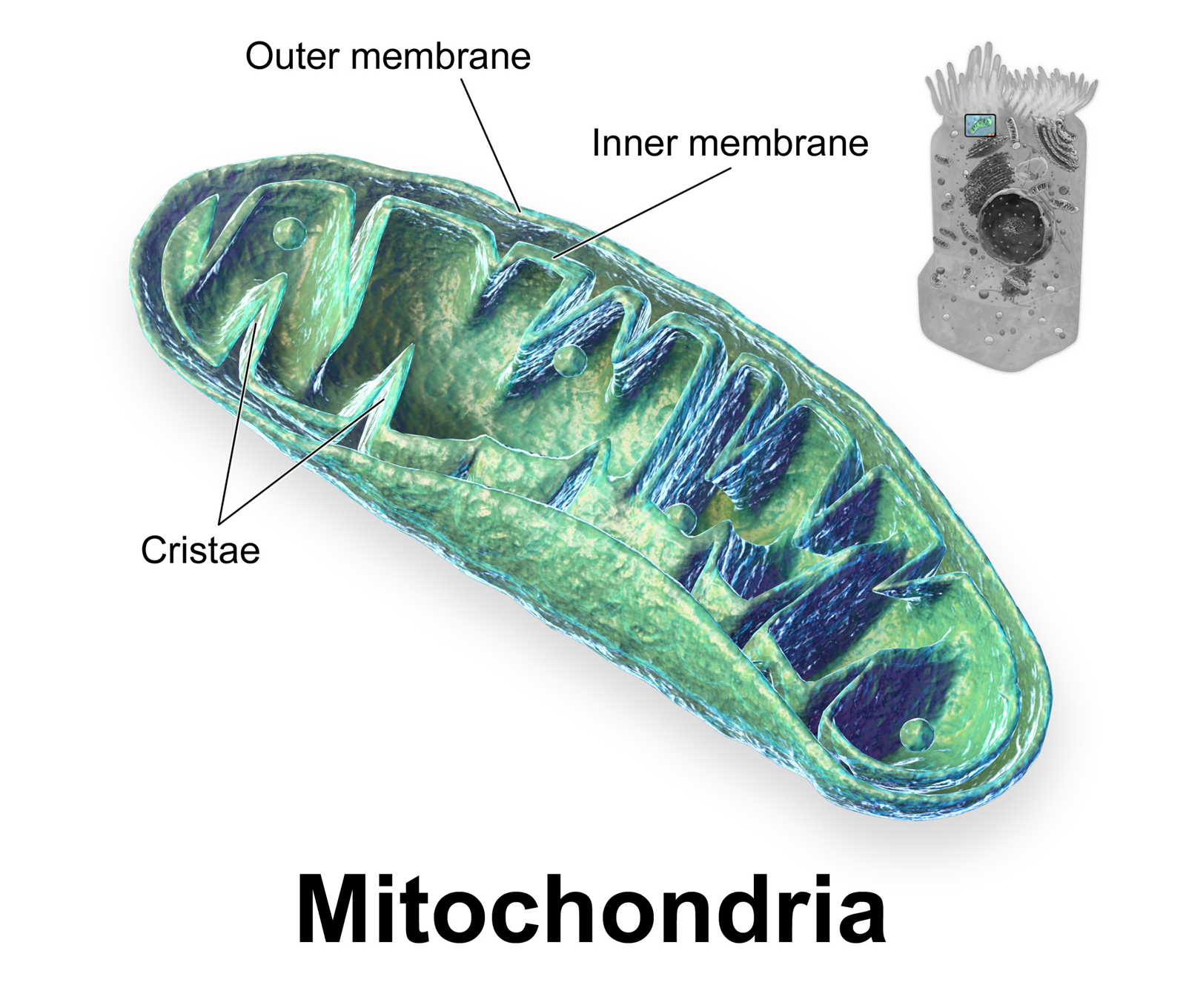
A crista (; plural cristae) is a fold in the inner membrane of a mitochondrion. The name is from the Latin for ''crest'' or ''plume'', and it gives the inner membrane its characteristic wrinkled shape, providing a large amount of surface area for chemical reactions to occur on. This aids aerobic cellular respiration, because the mitochondrion requires oxygen. Cristae are studded with proteins, including ATP synthase and a variety of cytochromes. Background With the discovery of the dual-membrane nature of mitochondria, the pioneers of mitochondrial ultrastructural research proposed different models for the organization of the mitochondrial inner membrane. Three models proposed were: *Baffle model – According to Palade (1953), the mitochondrial inner membrane is convoluted in a baffle-like manner with broad openings towards the intra-cristal space. This model entered most textbooks and was widely believed for a long time. *Septa model – Sjöstrand (1953) suggested that sh ... [...More Info...] [...Related Items...] OR: [Wikipedia] [Google] [Baidu] |
Vestibular Duct
The vestibular duct or scala vestibuli is a perilymph-filled cavity inside the cochlea of the inner ear that conducts sound vibrations to the cochlear duct. It is separated from the cochlear duct by Reissner's membrane and extends from the vestibule of the ear to the helicotrema where it joins the tympanic duct. Additional images Image:Gray923.png, The cochlea and vestibule, viewed from above. Image:Gray903.png, Transverse section of the cochlear duct of a fetal cat. Image:Gray921.png, Interior of right osseous labyrinth. Image:Gray928.png, Diagrammatic longitudinal section of the cochlea. See also * Tympanic duct References internal websites Slidefrom University of Kansas Diagramat Indiana University – Purdue University Indianapolis Imageat University of New England (United States) The University of New England (UNE) is a private research university in Maine with campuses in Portland and Biddeford, as well as a study abroad campus in Tangier, Morocco. Dur ... [...More Info...] [...Related Items...] OR: [Wikipedia] [Google] [Baidu] |
Utricle (ear)
The utricle and saccule are the two otolith organs in the vertebrate inner ear. They are part of the balancing system ( membranous labyrinth) in the vestibule of the bony labyrinth (small oval chamber). They use small stones and a viscous fluid to stimulate hair cells to detect motion and orientation. The utricle detects linear accelerations and head-tilts in the horizontal plane. The word utricle comes . Structure The utricle is larger than the saccule and is of an oblong form, compressed transversely, and occupies the upper and back part of the vestibule Vestibule or Vestibulum can have the following meanings, each primarily based upon a common origin, from early 17th century French, derived from Latin ''vestibulum, -i n.'' "entrance court". Anatomy In general, vestibule is a small space or cavity ..., lying in contact with the recessus ellipticus and the part below it. Macula The macula of utricle (macula acustica utriculi) is a small (2 by 3 mm) thickening lying h ... [...More Info...] [...Related Items...] OR: [Wikipedia] [Google] [Baidu] |
Dura Mater
In neuroanatomy, dura mater is a thick membrane made of dense irregular connective tissue that surrounds the brain and spinal cord. It is the outermost of the three layers of membrane called the meninges that protect the central nervous system. The other two meningeal layers are the arachnoid mater and the pia mater. It envelops the arachnoid mater, which is responsible for keeping in the cerebrospinal fluid. It is derived primarily from the neural crest cell population, with postnatal contributions of the paraxial mesoderm. Structure The dura mater has several functions and layers. The dura mater is a membrane that envelops the arachnoid mater. It surrounds and supports the dural sinuses (also called dural venous sinuses, cerebral sinuses, or cranial sinuses) and carries blood from the brain toward the heart. Cranial dura mater has two layers called '' lamellae'', a superficial layer (also called the periosteal layer), which serves as the skull's inner periosteum, ca ... [...More Info...] [...Related Items...] OR: [Wikipedia] [Google] [Baidu] |
Endolymphatic Duct
From the posterior wall of the saccule a canal, the endolymphatic duct, is given off; this duct is joined by the ductus utriculosaccularis, and then passes along the aquaeductus vestibuli and ends in a blind pouch (endolymphatic sac) on the posterior surface of the petrous portion of the temporal bone, where it is in contact with the dura mater In neuroanatomy, dura mater is a thick membrane made of dense irregular connective tissue that surrounds the brain and spinal cord. It is the outermost of the three layers of membrane called the meninges that protect the central nervous syste .... Disorders of the endolymphatic duct include Meniere's Disease and Enlarged Vestibular Aqueduct. Additional images File:Gray902.png, Transverse section through head of fetal sheep, in the region of the labyrinth. X 30. File:Gray927.png, Transverse section of a human semicircular canal and duct References External links *The Endolymphatic Duct and Sac Vestibular system ... [...More Info...] [...Related Items...] OR: [Wikipedia] [Google] [Baidu] |
Membranous Labyrinth
The membranous labyrinth is a collection of fluid filled tubes and chambers which contain the receptors for the senses of equilibrium and hearing. It is lodged within the bony labyrinth in the inner ear and has the same general form; it is, however, considerably smaller and is partly separated from the bony walls by a quantity of fluid, the perilymph. In certain places, it is fixed to the walls of the cavity. The membranous labyrinth contains fluid called endolymph. The walls of the membranous labyrinth are lined with distributions of the cochlear nerve, one of the two branches of the vestibulocochlear nerve. The other branch is the vestibular nerve. Within the vestibule, the membranous labyrinth does not quite preserve the form of the bony labyrinth, but consists of two membranous sacs, the utricle, and the saccule The saccule is a bed of sensory cells in the inner ear The inner ear (internal ear, auris interna) is the innermost part of the vertebrate ear. In verteb ... [...More Info...] [...Related Items...] OR: [Wikipedia] [Google] [Baidu] |
Temporal Bone
The temporal bones are situated at the sides and base of the skull, and lateral to the temporal lobes of the cerebral cortex. The temporal bones are overlaid by the sides of the head known as the temples, and house the structures of the ears. The lower seven cranial nerves and the major vessels to and from the brain traverse the temporal bone. Structure The temporal bone consists of four parts— the squamous, mastoid, petrous and tympanic parts. The squamous part is the largest and most superiorly positioned relative to the rest of the bone. The zygomatic process is a long, arched process projecting from the lower region of the squamous part and it articulates with the zygomatic bone. Posteroinferior to the squamous is the mastoid part. Fused with the squamous and mastoid parts and between the sphenoid and occipital bones lies the petrous part, which is shaped like a pyramid. The tympanic part is relatively small and lies inferior to the squamous part, anterior to t ... [...More Info...] [...Related Items...] OR: [Wikipedia] [Google] [Baidu] |
Vestibular Aqueduct
At the hinder part of the medial wall of the vestibule is the orifice of the vestibular aqueduct, which extends to the posterior surface of the petrous portion of the temporal bone. It transmits a small vein, and contains a tubular prolongation of the membranous labyrinth, the ductus endolymphaticus, which ends in a cul-de-sac, the endolymphatic sac, between the layers of the dura mater within the cranial cavity. Pathology Enlargement of the vestibular aqueduct to greater than 2 mm is associated with enlarged vestibular aqueduct syndrome, a disease entity that is associated with one-sided hearing loss in children. The diagnosis can be made by high resolution CT or MRI, with comparison to the adjacent posterior semicircular canal. If the vestibular aqueduct is larger in size, and the clinical presentation is consistent, the diagnosis can be made. Treatment is with mechanical hearing implants. There is an association with Pendred syndrome Pendred syndrome is a genetic d ... [...More Info...] [...Related Items...] OR: [Wikipedia] [Google] [Baidu] |
Cochlear Duct
The cochlear duct (bounded by the scala media) is an endolymph filled cavity inside the cochlea, located between the tympanic duct and the vestibular duct, separated by the basilar membrane and the vestibular membrane (Reissner's membrane) respectively. The cochlear duct houses the organ of Corti. Structure The cochlear duct is part of the cochlea. It is separated from the tympanic duct (scala tympani) by the basilar membrane. It is separated from the vestibular duct (scala vestibuli) by the vestibular membrane (Reissner's membrane). The stria vascularis is located in the wall of the cochlear duct. Development The cochlear duct develops from the ventral otic vesicle (otocyst). It grows slightly flattened between the middle and outside of the body. This development may be regulated by the genes EYA1, SIX1, GATA3, and TBX1. The organ of Corti develops inside the cochlear duct. Function The cochlear duct contains the organ of Corti. This is attached to the basi ... [...More Info...] [...Related Items...] OR: [Wikipedia] [Google] [Baidu] |
Saccule
The saccule is a bed of sensory cells in the inner ear The inner ear (internal ear, auris interna) is the innermost part of the vertebrate ear. In vertebrates, the inner ear is mainly responsible for sound detection and balance. In mammals, it consists of the bony labyrinth, a hollow cavity in t .... It translates head movements into neural impulses for the brain to interpret. The saccule detects linear accelerations and head tilts in the vertical plane. When the head moves vertically, the sensory cells of the saccule are disturbed and the neurons connected to them begin transmitting impulses to the brain. These impulses travel along the vestibular portion of the eighth cranial nerve to the vestibular nuclei in the brainstem. The vestibular system is important in maintaining balance, or equilibrium. The vestibular system includes the saccule, utricle, and the three semicircular canals. The Vestibule of the ear, vestibule is the name of the fluid-filled, membrano ... [...More Info...] [...Related Items...] OR: [Wikipedia] [Google] [Baidu] |
Inner Ear
The inner ear (internal ear, auris interna) is the innermost part of the vertebrate ear. In vertebrates, the inner ear is mainly responsible for sound detection and balance. In mammals, it consists of the bony labyrinth, a hollow cavity in the temporal bone of the skull with a system of passages comprising two main functional parts: * The cochlea, dedicated to hearing; converting sound pressure patterns from the outer ear into electrochemical impulses which are passed on to the brain via the auditory nerve. * The vestibular system, dedicated to balance The inner ear is found in all vertebrates, with substantial variations in form and function. The inner ear is innervated by the eighth cranial nerve in all vertebrates. Structure The labyrinth can be divided by layer or by region. Bony and membranous labyrinths The bony labyrinth, or osseous labyrinth, is the network of passages with bony walls lined with periosteum. The three major parts of the bony labyrinth are the ... [...More Info...] [...Related Items...] OR: [Wikipedia] [Google] [Baidu] |


