|
Ventilation Perfusion Scan
A ventilation/perfusion lung scan, also called a V/Q lung scan, or ventilation/perfusion scintigraphy, is a type of medical imaging using scintigraphy and medical isotopes to evaluate the circulation of air and blood within a patient's lungs, in order to determine the ventilation/perfusion ratio. The ventilation part of the test looks at the ability of air to reach all parts of the lungs, while the perfusion part evaluates how well blood circulates within the lungs. As Q in physiology is the letter used to describe bloodflow the term V/Q scan emerged. Uses This test is most commonly done in order to check for the presence of a blood clot or abnormal blood flow inside the lungs (such as a pulmonary embolism (PE) although computed tomography with radiocontrast is now more commonly used for this purpose. The V/Q scan may be used in some circumstances where radiocontrast would be inappropriate, as in Allergy to contrast agent or kidney failure. A V/Q lung scan may be performed in t ... [...More Info...] [...Related Items...] OR: [Wikipedia] [Google] [Baidu] |
Medical Imaging
Medical imaging is the technique and process of imaging the interior of a body for clinical analysis and medical intervention, as well as visual representation of the function of some organs or tissues ( physiology). Medical imaging seeks to reveal internal structures hidden by the skin and bones, as well as to diagnose and treat disease. Medical imaging also establishes a database of normal anatomy and physiology to make it possible to identify abnormalities. Although imaging of removed organs and tissues can be performed for medical reasons, such procedures are usually considered part of pathology instead of medical imaging. Measurement and recording techniques that are not primarily designed to produce images, such as electroencephalography (EEG), magnetoencephalography (MEG), electrocardiography (ECG), and others, represent other technologies that produce data susceptible to representation as a parameter graph versus time or maps that contain data about the measurement ... [...More Info...] [...Related Items...] OR: [Wikipedia] [Google] [Baidu] |
Pulmonary Embolism Scintigraphy PLoS
The lungs are the primary organs of the respiratory system in humans and most other animals, including some snails and a small number of fish. In mammals and most other vertebrates, two lungs are located near the backbone on either side of the heart. Their function in the respiratory system is to extract oxygen from the air and transfer it into the bloodstream, and to release carbon dioxide from the bloodstream into the atmosphere, in a process of gas exchange. Respiration is driven by different muscular systems in different species. Mammals, reptiles and birds use their different muscles to support and foster breathing. In earlier tetrapods, air was driven into the lungs by the pharyngeal muscles via buccal pumping, a mechanism still seen in amphibians. In humans, the main muscle of respiration that drives breathing is the diaphragm. The lungs also provide airflow that makes vocal sounds including human speech possible. Humans have two lungs, one on the left and one on ... [...More Info...] [...Related Items...] OR: [Wikipedia] [Google] [Baidu] |
Ventilation/perfusion Ratio
In respiratory physiology, the ventilation/perfusion ratio (V/Q ratio) is a ratio used to assess the efficiency and adequacy of the matching of two variables: * V – ventilation – the air that reaches the alveoli * Q – perfusion – the blood that reaches the alveoli via the capillaries The V/Q ratio can therefore be defined as the ratio of the amount of air reaching the alveoli per minute to the amount of blood reaching the alveoli per minute—a ratio of volumetric flow rates. These two variables, V and Q, constitute the main determinants of the blood oxygen (O2) and carbon dioxide (CO2) concentration. The V/Q ratio can be measured with a ventilation/perfusion scan. A V/Q mismatch can cause Type 1 respiratory failure. Physiology Ideally, the oxygen provided via ventilation would be just enough to saturate the blood fully. In the typical adult, 1 litre of blood can hold about 200 mL of oxygen; 1 litre of dry air has about 210 mL of oxygen. Therefore, under these conditio ... [...More Info...] [...Related Items...] OR: [Wikipedia] [Google] [Baidu] |
Positron Emission Tomography
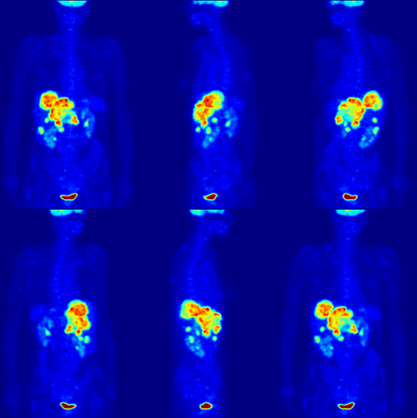
Positron emission tomography (PET) is a functional imaging technique that uses radioactive substances known as radiotracers to visualize and measure changes in metabolic processes, and in other physiological activities including blood flow, regional chemical composition, and absorption. Different tracers are used for various imaging purposes, depending on the target process within the body. For example: * Fluorodeoxyglucose ( 18F">sup>18FDG or FDG) is commonly used to detect cancer; * 18Fodium fluoride">sup>18Fodium fluoride (Na18F) is widely used for detecting bone formation; * Oxygen-15 (15O) is sometimes used to measure blood flow. PET is a common imaging technique, a medical scintillography technique used in nuclear medicine. A radiopharmaceutical – a radioisotope attached to a drug – is injected into the body as a radioactive tracer, tracer. When the radiopharmaceutical undergoes beta plus decay, a positron is emitted, and when the positron interacts with an or ... [...More Info...] [...Related Items...] OR: [Wikipedia] [Google] [Baidu] |
Gamma Camera
A gamma camera (γ-camera), also called a scintillation camera or Anger camera, is a device used to image gamma radiation emitting radioisotopes, a technique known as scintigraphy. The applications of scintigraphy include early drug development and nuclear medical imaging to view and analyse images of the human body or the distribution of medically injected, inhaled, or ingested radionuclides emitting gamma rays. Imaging techniques Scintigraphy ("scint") is the use of gamma cameras to capture emitted radiation from internal radioisotopes to create two-dimensional images. SPECT (single photon emission computed tomography) imaging, as used in nuclear cardiac stress testing, is performed using gamma cameras. Usually one, two or three detectors or heads, are slowly rotated around the patient's torso. Multi-headed gamma cameras can also be used for positron emission tomography (PET) scanning, provided that their hardware and software can be configured to detect "coincidences" ... [...More Info...] [...Related Items...] OR: [Wikipedia] [Google] [Baidu] |
Technetium Tc-99m Albumin Aggregated
Technetium 99mTc albumin aggregated (99mTc-MAA) is an injectable radiopharmaceutical used in nuclear medicine. It consists of a sterile aqueous suspension of Technetium-99m (99mTc) labeled to human albumin aggregate particles. It is commonly used for lung perfusion scanning. It is also less commonly used to visualise a peritoneovenous shunt and for isotope venography. Preparation DraxImage MAA kits for preparing 99mTc-MAA are available in the United States from only a single manufacturer; Jubilant DraxImage Inc. The kits are delivered to nuclear pharmacies as lyophilized powders of non-radioactive ingredients sealed under nitrogen. A nuclear pharmacist adds anywhere from 50 - 100 mCi of Na 99mTcO4">sup>99mTcO4to the reaction vial to make the final product, in the pH range of 3.8 to 8.0. After being allowed to react at room temperature for 15 minutes to ensure maximum labeling of the human albumin with 99mTc, the kit can then be diluted with sterile normal saline as needed. ... [...More Info...] [...Related Items...] OR: [Wikipedia] [Google] [Baidu] |
Radioactive Nanoparticle
A radioactive nanoparticle is a nanoparticle that contains radioactive materials. Radioactive nanoparticles have applications in medical diagnostics, medical imaging, toxicokinetics, and environmental health, and are being investigated for applications in nuclear nanomedicine. Radioactive nanoparticles present special challenges in operational health physics and internal dosimetry that are not present for other substances, although existing radiation protection measures and hazard controls for nanoparticles generally apply. Types and applications Engineered Engineered radioactive nanoparticles are used in medical imaging techniques such as positron emission tomography and single-photon emission computed tomography, and an aerosol of carbon nanoparticles containing technetium-99m are used in a commercially available procedure for ventilation/perfusion scintigraphy of the lungs. Engineered radioactive nanoparticles are also used as a radiolabel to detect the presence of ... [...More Info...] [...Related Items...] OR: [Wikipedia] [Google] [Baidu] |
DTPA
Pentetic acid or diethylenetriaminepentaacetic acid (DTPA) is an aminopolycarboxylic acid consisting of a diethylenetriamine backbone with five carboxymethyl groups. The molecule can be viewed as an expanded version of EDTA and is used similarly. It is a white solid with limited solubility in water. Coordination properties The conjugate base of DTPA has a high affinity for metal cations. Thus, the penta-anion DTPA5− is potentially an octadentate ligand assuming that each nitrogen centre and each –COO− group counts as a centre for coordination. The formation constants for its complexes are about 100 greater than those for EDTA.J. Roger Hart "Ethylenediaminetetraacetic Acid and Related Chelating Agents" in Ullmann's Encyclopedia of Industrial Chemistry, Wiley-VCH, Weinheim, 2005. As a chelating agent, DTPA wraps around a metal ion by forming up to eight bonds. Its complexes can also have an extra water molecule that coordinates the metal ion. Transition metals, however, ... [...More Info...] [...Related Items...] OR: [Wikipedia] [Google] [Baidu] |
Technetium-99m
Technetium-99m (99mTc) is a metastable nuclear isomer of technetium-99 (itself an isotope of technetium), symbolized as 99mTc, that is used in tens of millions of medical diagnostic procedures annually, making it the most commonly used medical radioisotope in the world. Technetium-99m is used as a radioactive tracer and can be detected in the body by medical equipment ( gamma cameras). It is well suited to the role, because it emits readily detectable gamma rays with a photon energy of 140 keV (these 8.8 pm photons are about the same wavelength as emitted by conventional X-ray diagnostic equipment) and its half-life for gamma emission is 6.0058 hours (meaning 93.7% of it decays to 99Tc in 24 hours). The relatively "short" physical half-life of the isotope and its biological half-life of 1 day (in terms of human activity and metabolism) allows for scanning procedures which collect data rapidly but keep total patient radiation exposure low. The same characteristics ma ... [...More Info...] [...Related Items...] OR: [Wikipedia] [Google] [Baidu] |
Krypton-81m
There are 34 known isotopes of krypton (36Kr) with atomic mass numbers from 69 through 102. Naturally occurring krypton is made of five stable isotopes and one () which is slightly radioactive with an extremely long half-life, plus traces of radioisotopes that are produced by cosmic rays in the atmosphere. List of isotopes , - , 69Kr , style="text-align:right" , 36 , style="text-align:right" , 33 , 68.96518(43)# , 32(10) ms , β+ , 69Br , 5/2−# , , , - , 70Kr , style="text-align:right" , 36 , style="text-align:right" , 34 , 69.95526(41)# , 52(17) ms , β+ , 70Br , 0+ , , , - , rowspan=2, 71Kr , rowspan=2 style="text-align:right" , 36 , rowspan=2 style="text-align:right" , 35 , rowspan=2, 70.94963(70) , rowspan=2, 100(3) ms , β+ (94.8%) , 71Br , rowspan=2, (5/2)− , rowspan=2, , rowspan=2, , - , β+, p (5.2%) , 70Se , - , 72Kr , style="text-align:right" , 36 , style="text-align:right" , 36 , 71.942092(9) , 17.16 ... [...More Info...] [...Related Items...] OR: [Wikipedia] [Google] [Baidu] |
Xenon-133
Naturally occurring xenon (54Xe) consists of seven stable isotopes and two very long-lived isotopes. Double electron capture has been observed in 124Xe (half-life ) and double beta decay in 136Xe (half-life ), which are among the longest measured half-lives of all nuclides. The isotopes 126Xe and 134Xe are also predicted to undergo double beta decay, but this has never been observed in these isotopes, so they are considered to be stable. Beyond these stable forms, 32 artificial unstable isotopes and various isomers have been studied, the longest-lived of which is 127Xe with a half-life of 36.345 days. All other isotopes have half-lives less than 12 days, most less than 20 hours. The shortest-lived isotope, 108Xe, has a half-life of 58 μs, and is the heaviest known nuclide with equal numbers of protons and neutrons. Of known isomers, the longest-lived is 131mXe with a half-life of 11.934 days. 129Xe is produced by beta decay of 129I (half-life: 16 million years); 131mXe, 133X ... [...More Info...] [...Related Items...] OR: [Wikipedia] [Google] [Baidu] |


.jpg)

