|
Utero
The uterus (from Latin ''uterus'', plural ''uteri'') or womb () is the organ in the reproductive system of most female mammals, including humans that accommodates the embryonic and fetal development of one or more embryos until birth. The uterus is a hormone-responsive sex organ that contains glands in its lining that secrete uterine milk for embryonic nourishment. In the human, the lower end of the uterus, is a narrow part known as the isthmus that connects to the cervix, leading to the vagina. The upper end, the body of the uterus, is connected to the fallopian tubes, at the uterine horns, and the rounded part above the openings to the fallopian tubes is the fundus. The connection of the uterine cavity with a fallopian tube is called the uterotubal junction. The fertilized egg is carried to the uterus along the fallopian tube. It will have divided on its journey to form a blastocyst that will implant itself into the lining of the uterus – the endometrium, where it will rece ... [...More Info...] [...Related Items...] OR: [Wikipedia] [Google] [Baidu] |
Uterotubal Junction
The uterotubal junction is the connection between the endometrial cavity of the uterus and the fallopian tube (uterine tube) at the proximal tubal opening, the beginning of the intramural part of the fallopian tube. Histologically, the endometrial epithelium changes over to ciliated tubal epithelium. Function Patency of the uterotubal junction is necessary for normal reproduction. The tubes can get blocked here by infection (salpingitis) and surgical intervention may be necessary. Mouse studies have indicated that selective passage of individual spermatozoa may occur at this junction, with abnormal morphology being identified as a significant selection criterion, leading to predominantly normal sperm passing towards the ovum. Absence of the protein calmegin has also been suggested as a critical factor for reliable sperm passage. Other The uterotubal junction is accessible by hysteroscopy and the entry point for tubal cannulation and falloposcopy Falloposcopy (occasionally also ... [...More Info...] [...Related Items...] OR: [Wikipedia] [Google] [Baidu] |
Cleavage (embryo)
In embryology, cleavage is the division of cells in the early development of the embryo, following fertilization. The zygotes of many species undergo rapid cell cycles with no significant overall growth, producing a cluster of cells the same size as the original zygote. The different cells derived from cleavage are called blastomeres and form a compact mass called the morula. Cleavage ends with the formation of the blastula, or of the blastocyst in mammals. Depending mostly on the concentration of yolk in the egg, the cleavage can be holoblastic (total or entire cleavage) or meroblastic (partial cleavage). The pole of the egg with the highest concentration of yolk is referred to as the vegetal pole while the opposite is referred to as the animal pole. Cleavage differs from other forms of cell division in that it increases the number of cells and nuclear mass without increasing the cytoplasmic mass. This means that with each successive subdivision, there is roughly half the cyto ... [...More Info...] [...Related Items...] OR: [Wikipedia] [Google] [Baidu] |
Paramesonephric Duct
Paramesonephric ducts (or Müllerian ducts) are paired ducts of the embryo that run down the lateral sides of the genital ridge and terminate at the sinus tubercle in the primitive urogenital sinus. In the female, they will develop to form the fallopian tubes, uterus, cervix, and the upper one-third of the vagina. Development The female reproductive system is composed of two embryological segments: the urogenital sinus and the paramesonephric ducts. The two are conjoined at the sinus tubercle. Paramesonephric ducts are present on the embryo of both sexes. Only in females do they develop into reproductive organs. They degenerate in males of certain species, but the adjoining mesonephric ducts develop into male reproductive organs. The sex based differences in the contributions of the paramesonephric ducts to reproductive organs is based on the presence, and degree of presence, of Anti-Müllerian hormone. During the formation of the reproductive system, the paramesonephric ducts ar ... [...More Info...] [...Related Items...] OR: [Wikipedia] [Google] [Baidu] |
Cervix
The cervix or cervix uteri (Latin, 'neck of the uterus') is the lower part of the uterus (womb) in the human female reproductive system. The cervix is usually 2 to 3 cm long (~1 inch) and roughly cylindrical in shape, which changes during pregnancy. The narrow, central cervical canal runs along its entire length, connecting the uterine cavity and the lumen of the vagina. The opening into the uterus is called the internal os, and the opening into the vagina is called the external os. The lower part of the cervix, known as the vaginal portion of the cervix (or ectocervix), bulges into the top of the vagina. The cervix has been documented anatomically since at least the time of Hippocrates, over 2,000 years ago. The cervical canal is a passage through which sperm must travel to fertilize an egg cell after sexual intercourse. Several methods of contraception, including cervical caps and cervical diaphragms, aim to block or prevent the passage of sperm through the cervical ... [...More Info...] [...Related Items...] OR: [Wikipedia] [Google] [Baidu] |
Prenatal Development
Prenatal development () includes the development of the embryo and of the fetus during a viviparous animal's gestation. Prenatal development starts with fertilization, in the germinal stage of embryonic development, and continues in fetal development until birth. In human pregnancy, prenatal development is also called antenatal development. The development of the human embryo follows fertilization, and continues as fetal development. By the end of the tenth week of gestational age the embryo has acquired its basic form and is referred to as a fetus. The next period is that of fetal development where many organs become fully developed. This fetal period is described both topically (by organ) and chronologically (by time) with major occurrences being listed by gestational age. The very early stages of embryonic development are the same in all mammals, but later stages of development, and the length of gestation varies. Terminology In the human: Different terms are used to ... [...More Info...] [...Related Items...] OR: [Wikipedia] [Google] [Baidu] |
Female Reproductive System
The female reproductive system is made up of the internal and external sex organs that function in the reproduction of new offspring. In humans, the female reproductive system is immature at birth and develops to maturity at puberty to be able to produce gametes, and to carry a fetus to full term. The internal sex organs are the vagina, uterus, fallopian tubes, and ovaries. The female reproductive tract includes the vagina, uterus, and fallopian tubes and is prone to infections. The vagina allows for sexual intercourse and childbirth, and is connected to the uterus at the cervix. The uterus or womb accommodates the embryo which develops into the fetus. The uterus also produces secretions which help the transit of sperm to the fallopian tubes, where sperm fertilize ova (egg cells) produced by the ovaries. The external sex organs are also known as the ''genitals'' and these are the organs of the vulva including the labia, clitoris, and vaginal opening. During the menstrual cycle ... [...More Info...] [...Related Items...] OR: [Wikipedia] [Google] [Baidu] |
Vagina
In mammals, the vagina is the elastic, muscular part of the female genital tract. In humans, it extends from the vestibule to the cervix. The outer vaginal opening is normally partly covered by a thin layer of mucosal tissue called the hymen. At the deep end, the cervix (neck of the uterus) bulges into the vagina. The vagina allows for sexual intercourse and birth. It also channels menstrual flow, which occurs in humans and closely related primates as part of the menstrual cycle. Although research on the vagina is especially lacking for different animals, its location, structure and size are documented as varying among species. Female mammals usually have two external openings in the vulva; these are the urethral opening for the urinary tract and the vaginal opening for the genital tract. This is different from male mammals, who usually have a single urethral opening for both urination and reproduction. The vaginal opening is much larger than the nearby urethral opening, an ... [...More Info...] [...Related Items...] OR: [Wikipedia] [Google] [Baidu] |
Endometrium
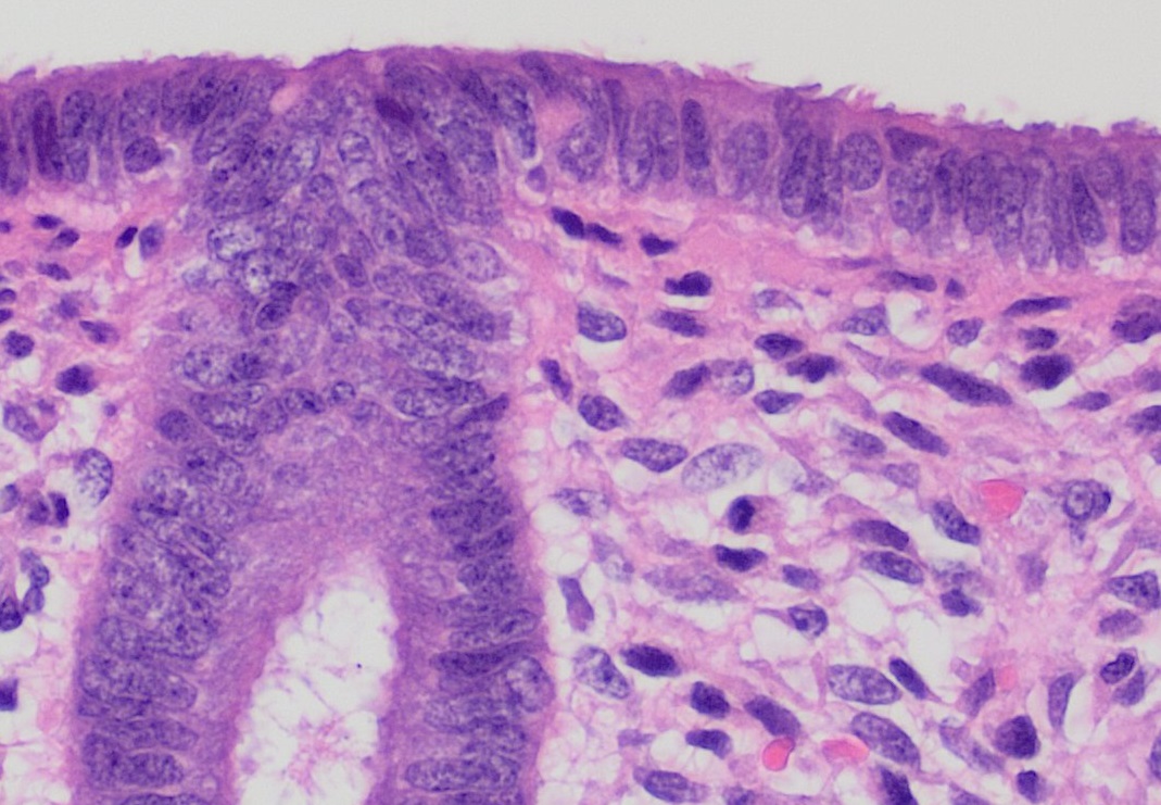
The endometrium is the inner epithelial layer, along with its mucous membrane, of the mammalian uterus. It has a basal layer and a functional layer: the basal layer contains stem cells which regenerate the functional layer. The functional layer thickens and then is shed during menstruation in humans and some other mammals, including apes, Old World monkeys, some species of bat, the elephant shrew and the Cairo spiny mouse. In most other mammals, the endometrium is reabsorbed in the estrous cycle. During pregnancy, the glands and blood vessels in the endometrium further increase in size and number. Vascular spaces fuse and become interconnected, forming the placenta, which supplies oxygen and nutrition to the embryo and fetus.Blue Histology - Female Reproductive System . School ... [...More Info...] [...Related Items...] OR: [Wikipedia] [Google] [Baidu] |
Implantation (embryology)
Implantation (nidation) is the stage in the embryonic development of mammals in which the blastocyst hatches as the embryo, adheres, and invades into the wall of the female's uterus. Implantation is the first stage of gestation, and when successful the female is considered to be pregnant. In a woman, an implanted embryo is detected by the presence of increased levels of human chorionic gonadotropin (hCG) in a pregnancy test. The implanted embryo will receive oxygen and nutrients in order to grow. There is an extensive variation in the type of trophoblast cells, and structures of the placenta across the different species of mammals. Of the five recognised stages of implantation including two pre-implantation stages that precede placentation, the first four are similar across the species. The five stages are migration and hatching, pre-contact, attachment, adhesion, and invasion. The two pre-implantation stages are associated with the pre-implantation embryo. In humans following ... [...More Info...] [...Related Items...] OR: [Wikipedia] [Google] [Baidu] |
Hollow Organ
In biology, an organ is a collection of tissues joined in a structural unit to serve a common function. In the hierarchy of life, an organ lies between tissue and an organ system. Tissues are formed from same type cells to act together in a function. Tissues of different types combine to form an organ which has a specific function. The intestinal wall for example is formed by epithelial tissue and smooth muscle tissue. Two or more organs working together in the execution of a specific body function form an organ system, also called a biological system or body system. An organ's tissues can be broadly categorized as parenchyma, the functional tissue, and stroma, the structural tissue with supportive, connective, or ancillary functions. For example, the gland's tissue that makes the hormones is the parenchyma, whereas the stroma includes the nerves that innervate the parenchyma, the blood vessels that oxygenate and nourish it and carry away its metabolic wastes, and the co ... [...More Info...] [...Related Items...] OR: [Wikipedia] [Google] [Baidu] |
Uterine Isthmus
The uterine isthmus is the inferior-posterior part of uterus, on its cervical end — here the uterine muscle (myometrium) is narrower and thinner. It connects the body and cervix. The uterine isthmus can become more compressibile in pregnancy, which is a finding known as Hegar's sign Hegar's sign is a non-sensitive indication of pregnancy in women—its absence does not exclude pregnancy. It pertains to the features of the cervix and the uterine isthmus. It is demonstrated as a softening in the consistency of the uterus, and th .... References Uterus {{genitourinary-stub, sex=female ... [...More Info...] [...Related Items...] OR: [Wikipedia] [Google] [Baidu] |
Blastocyst
The blastocyst is a structure formed in the early embryonic development of mammals. It possesses an inner cell mass (ICM) also known as the ''embryoblast'' which subsequently forms the embryo, and an outer layer of trophoblast cells called the trophectoderm. This layer surrounds the inner cell mass and a fluid-filled cavity known as the blastocoel. In the late blastocyst the trophectoderm is known as the trophoblast. The trophoblast gives rise to the chorion and amnion, the two fetal membranes that surround the embryo. The placenta derives from the embryonic chorion (the portion of the chorion that develops villi) and the underlying uterine tissue of the mother. The name "blastocyst" arises from the Greek ' ("a sprout") and ' ("bladder, capsule"). In other animals this is a structure consisting of an undifferentiated ball of cells and is called a blastula. In humans, blastocyst formation begins about five days after fertilization when a fluid-filled cavity opens up in the mor ... [...More Info...] [...Related Items...] OR: [Wikipedia] [Google] [Baidu] |


