|
Usual Interstitial Pneumonia
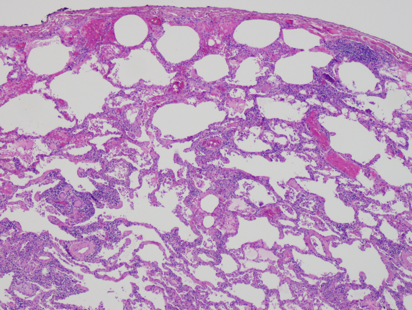
Usual interstitial pneumonia (UIP) is a form of lung disease characterized by progressive scarring of both lungs. The scarring (fibrosis) involves the pulmonary interstitium (the supporting framework of the lung). UIP is thus classified as a form of interstitial lung disease. Terminology The term "usual" refers to the fact that UIP is the most common form of interstitial fibrosis. "Pneumonia" indicates "lung abnormality", which includes fibrosis and inflammation. A term previously used for UIP in the British literature is cryptogenic fibrosing alveolitis (CFA), a term that has fallen out of favor since the basic underlying pathology is now thought to be fibrosis, not inflammation. The term usual interstitial pneumonitis (UIP) has also often been used, but again, the ''-itis'' part of that name may overemphasize inflammation. Signs and symptoms The typical symptoms of UIP are progressive shortness of breath and cough for a period of months. In some patients, UIP is diagnosed only ... [...More Info...] [...Related Items...] OR: [Wikipedia] [Google] [Baidu] |
European Respiratory Society
The European Respiratory Society, or ERS, is a non-profit organization with offices in Lausanne, Brussels and Sheffield. It was founded in 1990 in the field of respiratory medicine. The organization was formed with the merger of the Societas Europaea Physiologiae Clinicae Respoiratoriae (founded in 1966) and the European Society of Pneumology (founded 1981). The organization's membership is made up of medical professionals and scientists working in the area of respiratory medicine. ERS founded thEuropean Lung Foundation(ELF) in 2000. The foundation aims to bring together patients and the public with respiratory professionals to positively influence lung health. Activities The ERS publishes academic journals and books as well as hosting events aimed at educating health professionals, including a large annual congress. The organization's stated goal is to "promote lung health in order to alleviate suffering from disease and drive standards for respiratory medicine globally". Publi ... [...More Info...] [...Related Items...] OR: [Wikipedia] [Google] [Baidu] |
Pulmonary Langerhans Cell Histiocytosis
The lungs are the primary organs of the respiratory system in humans and most other animals, including some snails and a small number of fish. In mammals and most other vertebrates, two lungs are located near the backbone on either side of the heart. Their function in the respiratory system is to extract oxygen from the air and transfer it into the bloodstream, and to release carbon dioxide from the bloodstream into the atmosphere, in a process of gas exchange. Respiration is driven by different muscular systems in different species. Mammals, reptiles and birds use their different muscles to support and foster breathing. In earlier tetrapods, air was driven into the lungs by the pharyngeal muscles via buccal pumping, a mechanism still seen in amphibians. In humans, the main muscle of respiration that drives breathing is the diaphragm. The lungs also provide airflow that makes vocal sounds including human speech possible. Humans have two lungs, one on the left and one on the ... [...More Info...] [...Related Items...] OR: [Wikipedia] [Google] [Baidu] |
Sarcoidosis
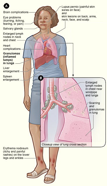
Sarcoidosis (also known as ''Besnier-Boeck-Schaumann disease'') is a disease involving abnormal collections of inflammatory cells that form lumps known as granulomata. The disease usually begins in the lungs, skin, or lymph nodes. Less commonly affected are the eyes, liver, heart, and brain. Any organ can be affected though. The signs and symptoms depend on the organ involved. Often, no, or only mild, symptoms are seen. When it affects the lungs, wheezing, coughing, shortness of breath, or chest pain may occur. Some may have Löfgren syndrome with fever, large lymph nodes, arthritis, and a rash known as erythema nodosum. The cause of sarcoidosis is unknown. Some believe it may be due to an immune reaction to a trigger such as an infection or chemicals in those who are genetically predisposed. Those with affected family members are at greater risk. Diagnosis is partly based on signs and symptoms, which may be supported by biopsy. Findings that make it likely include large lymph n ... [...More Info...] [...Related Items...] OR: [Wikipedia] [Google] [Baidu] |
Non-specific Interstitial Pneumonia
Non-specific interstitial pneumonia (NSIP) is a form of idiopathic interstitial pneumonia. Symptoms Symptoms include cough, difficulty breathing, and fatigue. Causes It has been suggested that idiopathic nonspecific interstitial pneumonia has an autoimmune mechanism, and is a possible complication of undifferentiated connective tissue disease; however, not enough research has been done at this time to find a cause. Patients with NSIP will often have other unrelated lung diseases like COPD or emphysema, along with other auto-immune disorders. Diagnosis A full clinical diagnosis can only be made from a lung biopsy of the tissue, fully best performed by a VATS, done by a cardio-thoracic surgeon. Some pulmonologists may first attempt a bronchoscopy; however, this frequently fails to give a full or correct diagnosis. Lung biopsies performed on patients with NSIP reveal two different disease patterns – cellular and fibrosing – which are associated with different prognoses. The ce ... [...More Info...] [...Related Items...] OR: [Wikipedia] [Google] [Baidu] |
Chronic Hypersensitivity Pneumonitis
Hypersensitivity pneumonitis (HP) or extrinsic allergic alveolitis (EAA) is a syndrome caused by the repetitive inhalation of antigens from the environment in susceptible or sensitized people. Common antigens include molds, bacteria, bird droppings, bird feathers, agricultural dusts, bioaerosols and chemicals from paints or plastics. People affected by this type of lung inflammation (pneumonitis) are commonly exposed to the antigens by their occupations, hobbies, the environment and animals. The inhaled antigens produce a hypersensitivity immune reaction causing inflammation of the airspaces (alveoli) and small airways (bronchioles) within the lung. Hypersensitivity pneumonitis may eventually lead to interstitial lung disease. Signs and symptoms Hypersensitivity pneumonitis (HP) can be categorized as acute, subacute, and chronic based on the duration of the illness. Acute In the acute form of HP dose of antigen exposure tends to be very high but only for a short duration. Symptoms ... [...More Info...] [...Related Items...] OR: [Wikipedia] [Google] [Baidu] |
Pleural Effusion
A pleural effusion is accumulation of excessive fluid in the pleural space, the potential space that surrounds each lung. Under normal conditions, pleural fluid is secreted by the parietal pleural capillaries at a rate of 0.6 millilitre per kilogram weight per hour, and is cleared by lymphatic absorption leaving behind only 5–15 millilitres of fluid, which helps to maintain a functional vacuum between the parietal and visceral pleurae. Excess fluid within the pleural space can impair inspiration by upsetting the functional vacuum and hydrostatically increasing the resistance against lung expansion, resulting in a fully or partially collapsed lung. Various kinds of fluid can accumulate in the pleural space, such as serous fluid (hydrothorax), blood (hemothorax), pus (pyothorax, more commonly known as pleural empyema), chyle ( chylothorax), or very rarely urine (urinothorax). When unspecified, the term "pleural effusion" normally refers to hydrothorax. A pleural effusion can a ... [...More Info...] [...Related Items...] OR: [Wikipedia] [Google] [Baidu] |
Pleural Plaques
Pleural thickening is an increase in the bulkiness of one or both of the pulmonary pleurae. Causes Pleural plaques Pleural plaques are patchy collections of hyalinized collagen in the parietal pleura. They have a holly leaf appearance on X-ray. They are indicators of asbestos exposure, and the most common asbestos-induced lesion. They usually appear after 20 years or more of exposure and never degenerate into mesothelioma. They appear as fibrous plaques on the parietal pleura, usually on both sides, and at the posterior and inferior part of the chest wall as well as the diaphragm. See also * Pleural disease Pleural disease occurs in the pleural space, which is the thin fluid-filled area in between the two pulmonary pleurae in the human body. There are several disorders and complications that can occur within the pleural area, and the surrounding tis ... References Asbestos Respiratory diseases {{respiratory-disease-stub ... [...More Info...] [...Related Items...] OR: [Wikipedia] [Google] [Baidu] |
Lung Nodule
A lung nodule or pulmonary nodule is a relatively small focal density in the lung. A solitary pulmonary nodule (SPN) or coin lesion, is a mass in the lung smaller than three centimeters in diameter. A pulmonary micronodule has a diameter of less than three millimetres. There may also be multiple nodules. One or more lung nodules can be an incidental finding found in up to 0.2% of chest X-rays and around 1% of CT scans. The nodule most commonly represents a benign tumor such as a granuloma or hamartoma, but in around 20% of cases it represents a malignant cancer, especially in older adults and smokers. Conversely, 10 to 20% of patients with lung cancer are diagnosed in this way. If the patient has a history of smoking or the nodule is growing, the possibility of cancer may need to be excluded through further radiological studies and interventions, possibly including surgical resection. The prognosis depends on the underlying condition. Causes Not every round spot on a radiolog ... [...More Info...] [...Related Items...] OR: [Wikipedia] [Google] [Baidu] |
Ground-glass Opacity
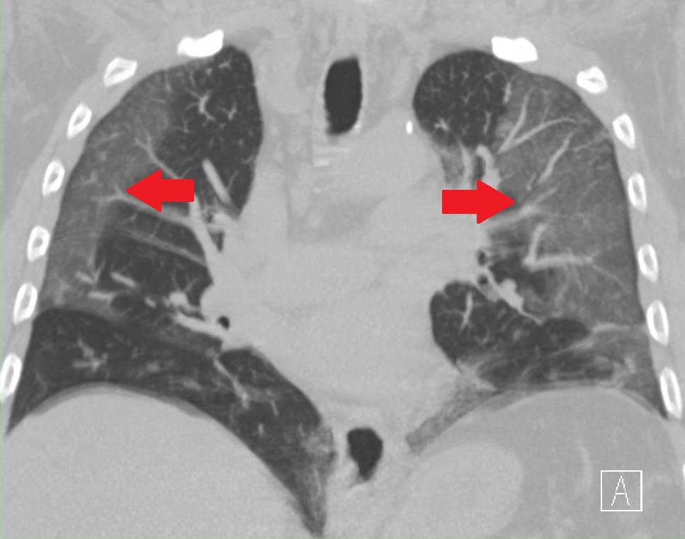
Ground-glass opacity (GGO) is a finding seen on chest x-ray (radiograph) or computed tomography (CT) imaging of the lungs. It is typically defined as an area of hazy opacification (x-ray) or increased attenuation (CT) due to air displacement by fluid, airway collapse, fibrosis, or a neoplastic process. When a substance other than air fills an area of the lung it increases that area's density. On both x-ray and CT, this appears more grey or hazy as opposed to the normally dark-appearing lungs. Although it can sometimes be seen in normal lungs, common pathologic causes include infections, interstitial lung disease, and pulmonary edema. Definition In both CT and chest radiographs, normal lungs appear dark due to the relative lower density of air compared to the surrounding tissues. When air is replaced by another substance (e.g. fluid or fibrosis), the density of the area increases, causing the tissue to appear lighter or more grey. Ground-glass opacity is most often used to d ... [...More Info...] [...Related Items...] OR: [Wikipedia] [Google] [Baidu] |
Bronchiectasis
Bronchiectasis is a disease in which there is permanent enlargement of parts of the bronchi, airways of the lung. Symptoms typically include a chronic cough with sputum, mucus production. Other symptoms include shortness of breath, hemoptysis, coughing up blood, and chest pain. Wheezing and nail clubbing may also occur. Those with the disease often get lung infections. Bronchiectasis may result from a number of infection, infectious and acquired causes, including measles, pneumonia, tuberculosis, immune system problems, as well as the genetic disorder cystic fibrosis. Cystic fibrosis eventually results in severe bronchiectasis in nearly all cases. The cause in 10–50% of those without cystic fibrosis is unknown. The mechanism of disease is breakdown of the airways due to an excessive inflammatory response. Involved airways (bronchi) become enlarged and thus less able to clear secretions. These secretions increase the amount of bacteria in the lungs, resulting in airway blockage ... [...More Info...] [...Related Items...] OR: [Wikipedia] [Google] [Baidu] |
-CT_scan.jpg)


