|
Ulnae
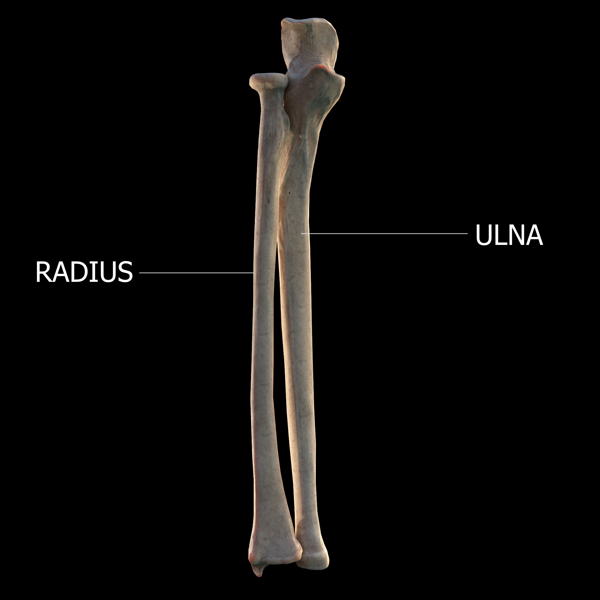
The ulna (''pl''. ulnae or ulnas) is a long bone found in the forearm that stretches from the elbow to the smallest finger, and when in anatomical position, is found on the medial side of the forearm. That is, the ulna is on the same side of the forearm as the little finger. It runs parallel to the radius, the other long bone in the forearm. The ulna is usually slightly longer than the radius, but the radius is thicker. Therefore, the radius is considered to be the larger of the two. Structure The ulna is a long bone found in the forearm that stretches from the elbow to the smallest finger, and when in anatomical position, is found on the medial side of the forearm. It is broader close to the elbow, and narrows as it approaches the wrist. Close to the elbow, the ulna has a bony process, the olecranon process, a hook-like structure that fits into the olecranon fossa of the humerus. This prevents hyperextension and forms a hinge joint with the trochlea of the humerus. There is als ... [...More Info...] [...Related Items...] OR: [Wikipedia] [Google] [Baidu] |
Semilunar Notch
The trochlear notch (), also known as semilunar notch and greater sigmoid cavity, is a large depression in the upper extremity of the ulna that fits the trochlea of the humerus (the bone directly above the ulna in the arm) as part of the elbow joint. It is formed by the olecranon and the coronoid process. About the middle of either side of this notch is an indentation, which contracts it somewhat, and indicates the junction of the olecranon and the coronoid process. The notch is concave from above downward, and divided into a medial and a lateral portion by a smooth ridge running from the summit of the olecranon to the tip of the coronoid process. The medial portion is the larger, and is slightly concave transversely; the lateral is convex above, slightly concave below. References External links * * *Upperextremity/arm/radiology/lat-elbowat the Dartmouth Medical School The Geisel School of Medicine at Dartmouth is the graduate medical school of Dartmouth College in Hano ... [...More Info...] [...Related Items...] OR: [Wikipedia] [Google] [Baidu] |
Coronoid Process Of The Ulna
The coronoid process of the ulna is a triangular process projecting forward from the anterior proximal portion of the ulna. Structure Its ''base'' is continuous with the body of the bone, and of considerable strength. Anatomy Its ''apex'' is pointed, slightly curved upward, and in flexion of the forearm is received into the coronoid fossa of the humerus. Its ''upper surface'' is smooth, convex, and forms the lower part of the semilunar notch. Its ''antero-inferior'' surface is concave, and marked by a rough impression for the insertion of the brachialis muscle. At the junction of this surface with the front of the body is a rough eminence, the tuberosity of the ulna, which gives insertion to a part of the brachialis; to the lateral border of this tuberosity the oblique cord is attached. Its ''lateral surface'' presents a narrow, oblong, articular depression, the radial notch. Its ''medial surface'', by its prominent, free margin, serves for the attachment of part of the uln ... [...More Info...] [...Related Items...] OR: [Wikipedia] [Google] [Baidu] |
Radial Notch
The radial notch of the ulna (lesser sigmoid cavity) is a narrow, oblong, articular depression on the lateral side of the coronoid process; it receives the circumferential articular surface of the head of the radius. It is concave from before backward, and its prominent extremities serve for the attachment of the annular ligament. Additional images File:Gray333.png, Annular ligament of radius, from above. References External links * *elbow/elbowbones/bones3at the Dartmouth Medical School The Geisel School of Medicine at Dartmouth is the graduate medical school of Dartmouth College in Hanover, New Hampshire. The fourth oldest medical school in the United States, it was founded in 1797 by New England physician Nathan Smith. It is o ...'s Department of Anatomy Upper limb anatomy Ulna {{musculoskeletal-stub ... [...More Info...] [...Related Items...] OR: [Wikipedia] [Google] [Baidu] |
Coronoid Process Of The Ulna
The coronoid process of the ulna is a triangular process projecting forward from the anterior proximal portion of the ulna. Structure Its ''base'' is continuous with the body of the bone, and of considerable strength. Anatomy Its ''apex'' is pointed, slightly curved upward, and in flexion of the forearm is received into the coronoid fossa of the humerus. Its ''upper surface'' is smooth, convex, and forms the lower part of the semilunar notch. Its ''antero-inferior'' surface is concave, and marked by a rough impression for the insertion of the brachialis muscle. At the junction of this surface with the front of the body is a rough eminence, the tuberosity of the ulna, which gives insertion to a part of the brachialis; to the lateral border of this tuberosity the oblique cord is attached. Its ''lateral surface'' presents a narrow, oblong, articular depression, the radial notch. Its ''medial surface'', by its prominent, free margin, serves for the attachment of part of the uln ... [...More Info...] [...Related Items...] OR: [Wikipedia] [Google] [Baidu] |
Ulnar Tuberosity
The tuberosity of the ulna is a rough eminence on the proximal end of the ulna. It occurs at the junction of the antero-inferior surface of the coronoid process with the front of the body. It provides an insertion point to a tendon of the brachialis (the oblique cord The oblique cord is a ligament between the ulnar and radius bones in the forearm near the elbow. It takes the form of a small, flattened band, extending downward and lateralward, from the lateral side of the Tuberosity of the ulna, ulnar tuberosity ... of the brachialis is attached to the lateral border). References External links * Additional images File:Ulna_ant_med_Arabic_YM.jpg, Tuberosity of the ulna. File:Gray329_Arabic_YM.png, Tuberosity of the ulna. File:MedialHumerusRadiusUlnaArticulated_Arabic_YM.jpg, Tuberosity of the ulna. Ulna {{musculoskeletal-stub ... [...More Info...] [...Related Items...] OR: [Wikipedia] [Google] [Baidu] |
Ulnar Styloid Process
The styloid process of the ulna is a bony prominence found at distal end of the ulna in the forearm. Structure The styloid process of the ulna projects from the medial and back part of the ulna. It descends a little lower than the head. The head is separated from the styloid process by a depression for the attachment of the apex of the triangular articular disk, and behind, by a shallow groove for the tendon of the extensor carpi ulnaris muscle. The styloid process of the ulnar varies in length between 2 mm and 6 mm. Function The rounded end of the styloid process of the ulna connects to the ulnar collateral ligament of the wrist. The radioulnar ligaments also attaches to the base of the styloid process of the ulna. Clinical significance Fractures of the styloid process of the ulna seldom require treatment when they occur in association with a distal radius fracture. The major exception is when the joint between these bones, the distal radioulnar joint (or DRUJ), is unst ... [...More Info...] [...Related Items...] OR: [Wikipedia] [Google] [Baidu] |
Process (anatomy)
In anatomy, a process ( la, processus) is a projection or outgrowth of tissue from a larger body. For instance, in a vertebra, a process may serve for muscle attachment and leverage (as in the case of the transverse and spinous processes), or to fit (forming a synovial joint), with another vertebra (as in the case of the articular processes).Moore, Keith L. et al. (2010) ''Clinically Oriented Anatomy'', 6th Ed, p.442 fig. 4.2 The word is used even at the microanatomic level, where cells can have processes such as cilia or pedicels. Depending on the tissue, processes may also be called by other terms, such as ''apophysis'', ''tubercle'', or ''protuberance''. Examples Examples of processes include: *The many processes of the human skull: ** The mastoid and styloid processes of the temporal bone ** The zygomatic process of the temporal bone ** The zygomatic process of the frontal bone ** The orbital, temporal, lateral, frontal, and maxillary processes of the zygomatic bone ... [...More Info...] [...Related Items...] OR: [Wikipedia] [Google] [Baidu] |
Forearm
The forearm is the region of the upper limb between the elbow and the wrist. The term forearm is used in anatomy to distinguish it from the arm, a word which is most often used to describe the entire appendage of the upper limb, but which in anatomy, technically, means only the region of the upper arm, whereas the lower "arm" is called the forearm. It is homologous to the region of the leg that lies between the knee and the ankle joints, the crus. The forearm contains two long bones, the radius and the ulna, forming the two radioulnar joints. The interosseous membrane connects these bones. Ultimately, the forearm is covered by skin, the anterior surface usually being less hairy than the posterior surface. The forearm contains many muscles, including the flexors and extensors of the wrist, flexors and extensors of the digits, a flexor of the elbow (brachioradialis), and pronators and supinators that turn the hand to face down or upwards, respectively. In cross-section, the for ... [...More Info...] [...Related Items...] OR: [Wikipedia] [Google] [Baidu] |
Flexor Pollicis Longus
The flexor pollicis longus (; FPL, Latin ''flexor'', bender; ''pollicis'', of the thumb; ''longus'', long) is a muscle in the forearm and hand that flexes the thumb. It lies in the same plane as the flexor digitorum profundus. This muscle is unique to humans, being either rudimentary or absent in other primates. A meta-analysis indicated accessory flexor pollicis longus is present in around 48% of the population. Human anatomy Origin and insertion It arises from the grooved anterior (side of palm) surface of the body of the radius, extending from immediately below the radial tuberosity and oblique line to within a short distance of the pronator quadratus muscle.Gray 1918, ''Flexor Pollicis Longus'', paras 20, 25 An occasionally present accessory long head of the flexor pollicis longus muscle is called 'Gantzer's muscle'. It may cause compression of the anterior interosseous nerve. It arises also from the adjacent part of the interosseous membrane of the forearm, and generally ... [...More Info...] [...Related Items...] OR: [Wikipedia] [Google] [Baidu] |
Pronator Teres
The pronator teres is a muscle (located mainly in the forearm) that, along with the pronator quadratus, serves to pronate the forearm (turning it so that the palm faces posteriorly when from the anatomical position). It has two attachments, to the medial humeral supracondylar ridge and the ulnar tuberosity, and inserts near the middle of the radius. Structure The pronator teres has two heads—humeral and ulnar. * The humeral head, the larger and more superficial, arises from the medial supracondylar ridge immediately superior to the medial epicondyle of the humerus, and from the common flexor tendon (which arises from the medial epicondyle). * The ulnar head (or ulnar tuberosity) is a thin fasciculus, which arises from the medial side of the coronoid process of the ulna, and joins the preceding at an acute angle. The median nerve enters the forearm between the two heads of the muscle, and is separated from the ulnar artery by the ulnar head. The muscle passes obliquely across ... [...More Info...] [...Related Items...] OR: [Wikipedia] [Google] [Baidu] |
Flexor Digitorum Profundus
The flexor digitorum profundus is a muscle in the forearm of humans that flexes the fingers (also known as digits). It is considered an extrinsic hand muscle because it acts on the hand while its muscle belly is located in the forearm. Together the flexor pollicis longus, pronator quadratus, and flexor digitorum profundus form the deep layer of ventral forearm muscles.Platzer 2004, p 162 The muscle is named . Structure Flexor digitorum profundus originates in the upper 3/4 of the anterior and medial surfaces of the ulna, interosseous membrane and deep fascia of the forearm. The muscle fans out into four tendons (one to each of the second to fifth fingers) to the palmar base of the distal phalanx. Along with the flexor digitorum superficialis, it has long tendons that run down the arm and through the carpal tunnel and attach to the palmar side of the phalanges of the fingers. Flexor digitorum profundus lies deep to the superficialis, but it attaches more distally. Therefore, ... [...More Info...] [...Related Items...] OR: [Wikipedia] [Google] [Baidu] |
Flexor Digitorum Superficialis Muscle
Flexor digitorum superficialis (''flexor digitorum sublimis'') is an extrinsic flexor muscle of the fingers at the proximal interphalangeal joints. It is in the anterior compartment of the forearm. It is sometimes considered to be the deepest part of the superficial layer of this compartment, and sometimes considered to be a distinct, "intermediate layer" of this compartment. It is relatively common for the Flexor digitorum superficialis to be missing from the little finger, bilaterally and unilaterally, which can cause problems when diagnosing a little finger injury. Structure The muscle has two classically described heads – the humeroulnar and radial – and it is between these heads that the median nerve and ulnar artery pass. The ulnar collateral ligament of elbow joint gives its origin to part of this muscle. Four long tendons come off this muscle near the wrist and travel through the carpal tunnel formed by the flexor retinaculum. These tendons, along with those of flex ... [...More Info...] [...Related Items...] OR: [Wikipedia] [Google] [Baidu] |

