|
Tumor-homing Bacteria
Tumor-homing bacteria are facultative or obligate anaerobic bacteria (capable of producing ATP when oxygen is absent or is destroyed in normal oxygen levels) that are able to target cancerous cells in the body, suppress tumor growth and survive in the body for a long time even after the infection. When this type of bacteria is administered into the body, it migrates to the cancerous tissues and starts to grow, and then deploys distinct mechanisms to destroy solid tumors. Each bacteria species uses a different process to eliminate the tumor. Some common tumor homing bacteria include '' Salmonella, Clostridium, Bifidobacterium, Listeria'', and ''Streptococcus''. The earliest research of this type of bacteria was highlighted in 1813 when scientists began observing that patients that had gas gangrene, an infection caused by the bacteria '' Clostridium'', were able to have tumor regressions. Tumor-inhibition mechanisms Different strains of tumor homing bacteria in distinct environme ... [...More Info...] [...Related Items...] OR: [Wikipedia] [Google] [Baidu] |
Facultative Anaerobic Organism
A facultative anaerobic organism is an organism that makes Adenosine triphosphate, ATP by aerobic respiration if oxygen is present, but is capable of switching to Fermentation (biochemistry), fermentation if oxygen is absent. Some examples of facultatively anaerobic bacteria are ''Staphylococcus'' Species, spp., ''Escherichia coli'', ''Salmonella'', ''Listeria'' spp., ''Shewanella oneidensis'' and ''Yersinia pestis''. Certain eukaryotes are also facultative anaerobes, including fungi such as ''Saccharomyces cerevisiae'' and many aquatic invertebrates such as Nereid (worm), nereid polychaetes. See also * Aerobic respiration * Anaerobic respiration * Fermentation * Obligate aerobe * Obligate anaerobe * Microaerophile References External links Facultative Anaerobic Bacteria {{Bacteria Anaerobic respiration Cellular respiration ... [...More Info...] [...Related Items...] OR: [Wikipedia] [Google] [Baidu] |
NADPH Oxidase
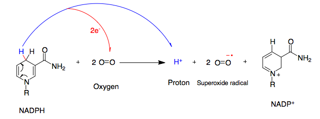
NADPH oxidase (nicotinamide adenine dinucleotide phosphate oxidase) is a membrane-bound enzyme complex that faces the extracellular space. It can be found in the plasma membrane as well as in the membranes of phagosomes used by neutrophil white blood cells to engulf microorganisms. Human isoforms of the catalytic component of the complex include NOX1, NOX2, NOX3, NOX4, NOX5, DUOX1, and DUOX2. Reaction NADPH oxidase catalyzes the production of a superoxide free radical by transferring one electron to oxygen from NADPH. : Types In mammals, NADPH oxidase is found in two types: one in white blood cells (neutrophilic) and the other in vascular cells, differing in biochemical structure and functions. Neutrophilic NADPH oxidase produces superoxide almost instantaneously, whereas the vascular enzyme produces superoxide in minutes to hours. Moreover, in white blood cells, superoxide has been found to transfer electrons across the membrane to extracellular oxygen, while in vascula ... [...More Info...] [...Related Items...] OR: [Wikipedia] [Google] [Baidu] |
Bladder Cancer
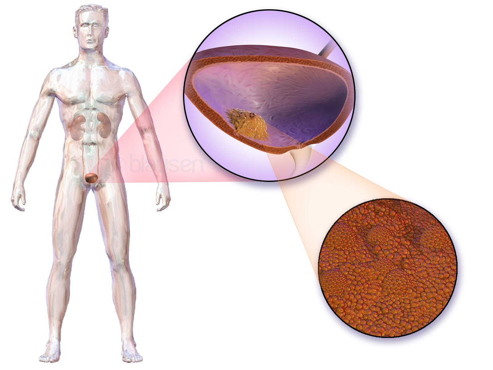
Bladder cancer is any of several types of cancer arising from the tissues of the urinary bladder. Symptoms include blood in the urine, pain with urination, and low back pain. It is caused when epithelial cells that line the bladder become malignant. Risk factors for bladder cancer include smoking, family history, prior radiation therapy, frequent bladder infections, and exposure to certain chemicals. The most common type is transitional cell carcinoma. Other types include squamous cell carcinoma and adenocarcinoma. Diagnosis is typically by cystoscopy with tissue biopsies. Staging of the cancer is determined by transurethral resection and medical imaging. Treatment depends on the stage of the cancer. It may include some combination of surgery, radiation therapy, chemotherapy, or immunotherapy. Surgical options may include transurethral resection, partial or complete removal of the bladder, or urinary diversion. The typical five-year survival rates in the United States i ... [...More Info...] [...Related Items...] OR: [Wikipedia] [Google] [Baidu] |
Mycobacterium Bovis
''Mycobacterium bovis'' is a slow-growing (16- to 20-hour generation time) aerobic bacterium and the causative agent of tuberculosis in cattle (known as bovine TB). It is related to ''Mycobacterium tuberculosis'', the bacterium which causes tuberculosis in humans. ''M. bovis'' can jump the species barrier and cause tuberculosis-like infection in humans and other mammals. Bacterium morphology and staining The bacteria are curved or straight rods. They sometimes form filaments, which fragment into bacilli or cocci once disturbed. In tissues they form slender rods, straight or curved, or club-shaped. Short, relatively plump bacilli (rods) in tissue smears, large slender beaded rods in culture. They have no flagella or fimbria, and no capsule. ''Mycobacterium tuberculosis'' group bacteria are 1.0-4.0 ┬Ąm long by 0.2-0.3 ┬Ąm wide in tissues. In culture, they may appear as cocci, or as bacilli up to 6-8 ┬Ąm long. The bacteria stain Gram-positive, acid-fast. The c ... [...More Info...] [...Related Items...] OR: [Wikipedia] [Google] [Baidu] |
White Blood Cell
White blood cells, also called leukocytes or leucocytes, are the cell (biology), cells of the immune system that are involved in protecting the body against both infectious disease and foreign invaders. All white blood cells are produced and derived from multipotent cells in the bone marrow known as hematopoietic stem cells. Leukocytes are found throughout the body, including the blood and lymphatic system. All white blood cells have cell nucleus, nuclei, which distinguishes them from the other blood cells, the anucleated red blood cells (RBCs) and platelets. The different white blood cells are usually classified by cell division, cell lineage (myelocyte, myeloid cells or lymphocyte, lymphoid cells). White blood cells are part of the body's immune system. They help the body fight infection and other diseases. Types of white blood cells are granulocytes (neutrophils, eosinophils, and basophils), and agranulocytes (monocytes, and lymphocytes (T cells and B cells)). Myeloid cells ... [...More Info...] [...Related Items...] OR: [Wikipedia] [Google] [Baidu] |
Granulocyte
Granulocytes are cells in the innate immune system characterized by the presence of specific granules in their cytoplasm. Such granules distinguish them from the various agranulocytes. All myeloblastic granulocytes are polymorphonuclear. They have varying shapes (morphology) of the nucleus (segmented, irregular; often lobed into three segments); and are referred to as polymorphonuclear leukocytes (PMN, PML, or PMNL). In common terms, ''polymorphonuclear granulocyte'' refers specifically to "neutrophil granulocytes", the most abundant of the granulocytes; the other types (eosinophils, basophils, and mast cells) have varying morphology. Granulocytes are produced via granulopoiesis in the bone marrow. Types There are four types of granulocytes (full name polymorphonuclear granulocytes): * Basophils * Eosinophils * Neutrophils * Mast cells Except for the mast cells, their names are derived from their staining characteristics; for example, the most abundant granulocyte is the neut ... [...More Info...] [...Related Items...] OR: [Wikipedia] [Google] [Baidu] |
Chemokine
Chemokines (), or chemotactic cytokines, are a family of small cytokines or signaling proteins secreted by cells that induce directional movement of leukocytes, as well as other cell types, including endothelial and epithelial cells. In addition to playing a major role in the activation of host immune responses, chemokines are important for biological processes, including morphogenesis and wound healing, as well as in the pathogenesis of diseases like cancers. Cytokine proteins are classified as chemokines according to behavior and structural characteristics. In addition to being known for mediating chemotaxis, chemokines are all approximately 8-10 kilodaltons in mass and have four cysteine residues in conserved locations that are key to forming their 3-dimensional shape. These proteins have historically been known under several other names including the ''SIS family of cytokines'', ''SIG family of cytokines'', ''SCY family of cytokines'', ''Platelet factor-4 superfamily'' or ... [...More Info...] [...Related Items...] OR: [Wikipedia] [Google] [Baidu] |
Autophagy
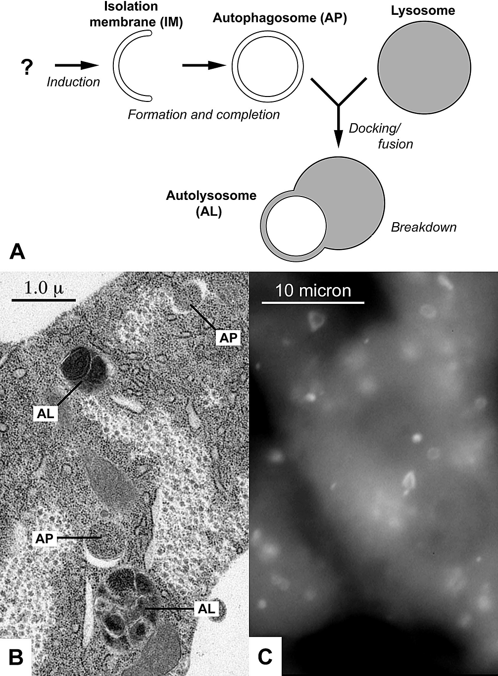
Autophagy (or autophagocytosis; from the Ancient Greek , , meaning "self-devouring" and , , meaning "hollow") is the natural, conserved degradation of the cell that removes unnecessary or dysfunctional components through a lysosome-dependent regulated mechanism. It allows the orderly degradation and recycling of cellular components. Although initially characterized as a primordial degradation pathway induced to protect against starvation, it has become increasingly clear that autophagy also plays a major role in the homeostasis of non-starved cells. Defects in autophagy have been linked to various human diseases, including neurodegeneration and cancer, and interest in modulating autophagy as a potential treatment for these diseases has grown rapidly. Four forms of autophagy have been identified: macroautophagy, microautophagy, chaperone-mediated autophagy (CMA), and crinophagy. In macroautophagy (the most thoroughly researched form of autophagy), cytoplasmic components (like mit ... [...More Info...] [...Related Items...] OR: [Wikipedia] [Google] [Baidu] |
Apoptosis
Apoptosis (from grc, ß╝ĆŽĆŽīŽĆŽäŽēŽā╬╣Žé, ap├│pt┼Źsis, 'falling off') is a form of programmed cell death that occurs in multicellular organisms. Biochemical events lead to characteristic cell changes (morphology) and death. These changes include blebbing, cell shrinkage, nuclear fragmentation, chromatin condensation, DNA fragmentation, and mRNA decay. The average adult human loses between 50 and 70 billion cells each day due to apoptosis. For an average human child between eight and fourteen years old, approximately twenty to thirty billion cells die per day. In contrast to necrosis, which is a form of traumatic cell death that results from acute cellular injury, apoptosis is a highly regulated and controlled process that confers advantages during an organism's life cycle. For example, the separation of fingers and toes in a developing human embryo occurs because cells between the digits undergo apoptosis. Unlike necrosis, apoptosis produces cell fragments called apoptotic ... [...More Info...] [...Related Items...] OR: [Wikipedia] [Google] [Baidu] |
Lipase
Lipase ( ) is a family of enzymes that catalyzes the hydrolysis of fats. Some lipases display broad substrate scope including esters of cholesterol, phospholipids, and of lipid-soluble vitamins and sphingomyelinases; however, these are usually treated separately from "conventional" lipases. Unlike esterases, which function in water, lipases "are activated only when adsorbed to an oilŌĆōwater interface". Lipases perform essential roles in digestion, transport and processing of dietary lipids in most, if not all, organisms. Structure and catalytic mechanism Classically, lipases catalyse the hydrolysis of triglycerides: :triglyceride + H2O ŌåÆ fatty acid + diacylglycerol :diacylglycerol + H2O ŌåÆ fatty acid + monacylglycerol :monacylglycerol + H2O ŌåÆ fatty acid + glycerol Lipases are serine hydrolases, i.e. they function by transesterification generating an acyl serine intermediate. Most lipases act at a specific position on the glycerol backbone of a lipid sub ... [...More Info...] [...Related Items...] OR: [Wikipedia] [Google] [Baidu] |
Hemolysin
Hemolysins or haemolysins are lipids and proteins that cause lysis of red blood cells by disrupting the cell membrane. Although the lytic activity of some microbe-derived hemolysins on red blood cells may be of great importance for nutrient acquisition, many hemolysins produced by pathogens do not cause significant destruction of red blood cells during infection. However, hemolysins are often capable of lysing red blood cells ''in vitro''. While most hemolysins are protein compounds, some are lipid biosurfactants. Properties Many bacteria produce hemolysins that can be detected in the laboratory. It is now believed that many clinically relevant fungi also produce hemolysins. Hemolysins can be identified by their ability to lyse red blood cells ''in vitro''. Not only are the erythrocytes affected by hemolysins, but there are also some effects among other blood cells, such as leucocytes (white blood cells). ''Escherichia coli'' hemolysin is potentially cytotoxic to monocytes ... [...More Info...] [...Related Items...] OR: [Wikipedia] [Google] [Baidu] |
Phospholipase
A phospholipase is an enzyme that hydrolyzes phospholipids into fatty acids and other lipophilic substances. Acids trigger the release of bound calcium from cellular stores and the consequent increase in free cytosolic Ca2+, an essential step in calcium signaling to regulate intracellular processes. There are four major classes, termed A, B, C, and D, which are distinguished by the type of reaction which they catalyze: *Phospholipase A **Phospholipase A1 ŌĆō cleaves the ''sn''-1 acyl chain (where ''sn'' refers to stereospecific numbering). **Phospholipase A2 ŌĆō cleaves the ''sn''-2 acyl chain, releasing arachidonic acid. * Phospholipase B ŌĆō cleaves both ''sn''-1 and ''sn''-2 acyl chains; this enzyme is also known as a lysophospholipase. *Phospholipase C ŌĆō cleaves before the phosphate, releasing diacylglycerol and a phosphate-containing head group. PLCs play a central role in signal transduction, releasing the second messenger inositol triphosphate. * Phospholipase D ŌĆ ... [...More Info...] [...Related Items...] OR: [Wikipedia] [Google] [Baidu] |


