|
Tuberculous Lymphadenitis
Tuberculous lymphadenitis (or tuberculous adenitis) is the most common form of tuberculosis infections that appears outside the lungs. Tuberculous lymphadenitis is a chronic, specific granulomatous inflammation of the lymph node with caseation necrosis, caused by infection with ''Mycobacterium tuberculosis'' or related bacteria. The characteristic morphological element is the tuberculous granuloma (caseating tubercule). This consists of giant multinucleated cells and ( Langhans cells), surrounded by epithelioid cells aggregates, T cell lymphocytes and fibroblasts. Granulomatous tubercules eventually develop central caseous necrosis and tend to become confluent, replacing the lymphoid tissue. Symptoms and signs In addition to swollen lymph nodes, called lymphadenitis, the person may experience mild fevers, not feel like eating, or lose weight. Cause It is usually caused by the most common cause of tuberculosis in the lungs, namely ''Mycobacterium tuberculosis''. It has sometime ... [...More Info...] [...Related Items...] OR: [Wikipedia] [Google] [Baidu] |
Infectious Disease (medical Specialty)
Infectious diseases or ID, also known as infectiology, is a medical specialty dealing with the diagnosis and treatment of infections. An infectious diseases specialist's practice consists of managing nosocomial ( healthcare-acquired) infections or community-acquired infections and is historically associated with hygiene, epidemiology, clinical microbiology, travel medicine and tropical medicine. Scope Infectious diseases specialists typically serve as consultants to other physicians in cases of complex infections, and often manage patients with HIV/AIDS and other forms of immunodeficiency. Although many common infections are treated by physicians without formal expertise in infectious diseases, specialists may be consulted for cases where an infection is difficult to diagnose or manage. They may also be asked to help determine the cause of a fever of unknown origin. Specialists in infectious diseases can practice both in hospitals (inpatient) and clinics (outpatient). In hospital ... [...More Info...] [...Related Items...] OR: [Wikipedia] [Google] [Baidu] |
Epithelioid Cell
According to a common point of view epithelioid cells (also called epithelioid histiocytes) are derivatives of activated macrophages resembling epithelial cells. Structure and function Structurally, epithelioid cells (when examined by light microscopy after stained with hematoxylin and eosin), are elongated, with finely granular, pale eosinophilic (pink) cytoplasm, and central, ovoid nuclei (oval or elongate), which are less dense than that of a lymphocyte. They have indistinct shape and often appear to merge into one another, forming aggregates known as giant cells. When examined by transmission electron microscopy in epithelioid cells in the field of Golgi lamellar complex are taped not only zonated, but also sleek vesicles with dense center, and also great many (more than 100) large granulas with diameters up to 340 nm and with finegranular matrix more light than in macrophage granulas, sometimes with perigranular halo. “The most prominent feature of these cells is the ... [...More Info...] [...Related Items...] OR: [Wikipedia] [Google] [Baidu] |
Antitubercular Agent
Tuberculosis management describes the techniques and procedures utilized for treating tuberculosis (TB). The medical standard for active TB is a short course treatment involving a combination of isoniazid, rifampicin (also known as Rifampin), pyrazinamide, and ethambutol for the first two months. During this initial period, Isoniazid is taken alongside pyridoxal phosphate to obviate peripheral neuropathy. Isoniazid is then taken coincident with rifampicin for the remaining four months of treatment. A patient is considered free of all living TB bacteria after six months. Latent tuberculosis or latent tuberculosis infection (LTBI) is treated with three to nine months of isoniazid alone. This long-term treatment often risks the development of hepatotoxicity. A combination of isoniazid plus rifampicin for a period of three to four months is shown to be an equally effective method for treating LTBI, while mitigating risks to hepatotoxicity. Treatment of LTBI is essential in preventi ... [...More Info...] [...Related Items...] OR: [Wikipedia] [Google] [Baidu] |
Biopsy
A biopsy is a medical test commonly performed by a surgeon, interventional radiologist, or an interventional cardiologist. The process involves extraction of sample cells or tissues for examination to determine the presence or extent of a disease. The tissue is then fixed, dehydrated, embedded, sectioned, stained and mounted before it is generally examined under a microscope by a pathologist; it may also be analyzed chemically. When an entire lump or suspicious area is removed, the procedure is called an excisional biopsy. An incisional biopsy or core biopsy samples a portion of the abnormal tissue without attempting to remove the entire lesion or tumor. When a sample of tissue or fluid is removed with a needle in such a way that cells are removed without preserving the histological architecture of the tissue cells, the procedure is called a needle aspiration biopsy. Biopsies are most commonly performed for insight into possible cancerous or inflammatory conditions. History T ... [...More Info...] [...Related Items...] OR: [Wikipedia] [Google] [Baidu] |
Collar Stud
A detachable collar is a shirt collar separate from the shirt, fastened to it by studs. The collar is usually made of a different fabric from the shirt, in which case it is almost always white, and, being unattached to the shirt, can be starched to a hard cardboard-like consistency. History The detachable collar was invented by Hannah Montague in Troy, New York, in 1827, after she snipped off the collar from one of her husband's shirts to wash it, and then sewed it back on. The Rev. Ebenezer Brown, a businessman in town, proceeded to commercialize it. The manufacture of detachable collars and the associated shirts became a significant industry in Troy. It was later that the benefit of being able to starch the collars became apparent, and for a short time, various other parts of the shirt, such as the front and cuffs, were also made detachable and treated to rigid stiffness. As more emphasis started to be placed on comfort in clothing this practice declined, and the stiff col ... [...More Info...] [...Related Items...] OR: [Wikipedia] [Google] [Baidu] |
Collar Bone
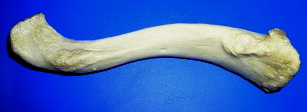
The clavicle, or collarbone, is a slender, S-shaped long bone approximately 6 inches (15 cm) long that serves as a strut between the shoulder blade and the sternum (breastbone). There are two clavicles, one on the left and one on the right. The clavicle is the only long bone in the body that lies horizontally. Together with the shoulder blade, it makes up the shoulder girdle. It is a palpable bone and, in people who have less fat in this region, the location of the bone is clearly visible. It receives its name from the Latin ''clavicula'' ("little key"), because the bone rotates along its axis like a key when the shoulder is abducted. The clavicle is the most commonly fractured bone. It can easily be fractured by impacts to the shoulder from the force of falling on outstretched arms or by a direct hit. Structure The collarbone is a thin doubly curved long bone that connects the arm to the trunk of the body. Located directly above the first rib, it acts as a strut to kee ... [...More Info...] [...Related Items...] OR: [Wikipedia] [Google] [Baidu] |
Mycobacterium Ulcerans
''Mycobacterium ulcerans'' is a species of bacteria found in various aquatic environments. The bacteria can infect humans and some other animals, causing persistent open wounds called Buruli ulcer. ''M. ulcerans'' is closely related to ''Mycobacterium marinum'', from which it evolved around one million years ago, and more distantly to the mycobacteria which cause tuberculosis and leprosy. Description ''M. ulcerans'' are rod-shaped bacteria. They appear purple ("Gram positive") under Gram stain and bright red ("acid fast") under Ziehl–Neelsen stain. On laboratory media, ''M. ulcerans'' grow slowly, forming small transparent colonies after four weeks. As colonies age, they develop irregular outlines and a rough, yellow surface. Taxonomy and evolution ''M. ulcerans'' is a species of mycobacteria within the phylum Actinomycetota. Within the genus ''Mycobacterium'', ''M. ulcerans'' is classified as both a "non-tuberculous mycobacterium" and a "slow-growing mycobacterium". ''M. ... [...More Info...] [...Related Items...] OR: [Wikipedia] [Google] [Baidu] |
Mycobacterium Bovis
''Mycobacterium bovis'' is a slow-growing (16- to 20-hour generation time) aerobic bacterium and the causative agent of tuberculosis in cattle (known as bovine TB). It is related to ''Mycobacterium tuberculosis'', the bacterium which causes tuberculosis in humans. ''M. bovis'' can jump the species barrier and cause tuberculosis-like infection in humans and other mammals. Bacterium morphology and staining The bacteria are curved or straight rods. They sometimes form filaments, which fragment into bacilli or cocci once disturbed. In tissues they form slender rods, straight or curved, or club-shaped. Short, relatively plump bacilli (rods) in tissue smears, large slender beaded rods in culture. They have no flagella or fimbria, and no capsule. ''Mycobacterium tuberculosis'' group bacteria are 1.0-4.0 µm long by 0.2-0.3 µm wide in tissues. In culture, they may appear as cocci, or as bacilli up to 6-8 µm long. The bacteria stain Gram-positive, acid-fast. The c ... [...More Info...] [...Related Items...] OR: [Wikipedia] [Google] [Baidu] |
Lymphadenitis
Lymphadenopathy or adenopathy is a disease of the lymph nodes, in which they are abnormal in size or consistency. Lymphadenopathy of an inflammatory type (the most common type) is lymphadenitis, producing swollen or enlarged lymph nodes. In clinical practice, the distinction between lymphadenopathy and lymphadenitis is rarely made and the words are usually treated as synonymous. Inflammation of the lymphatic vessels is known as lymphangitis. Infectious lymphadenitis affecting lymph nodes in the neck is often called scrofula. Lymphadenopathy is a common and nonspecific sign. Common causes include infections (from minor causes such as the common cold and post-vaccination swelling to serious ones such as HIV/AIDS), autoimmune diseases, and cancer. Lymphadenopathy is frequently idiopathic and self-limiting. Causes Lymph node enlargement is recognized as a common sign of infectious, autoimmune, or malignant disease. Examples may include: * Reactive: acute infection (''e.g.,'' ba ... [...More Info...] [...Related Items...] OR: [Wikipedia] [Google] [Baidu] |
Fibroblasts
A fibroblast is a type of biological cell that synthesizes the extracellular matrix and collagen, produces the structural framework ( stroma) for animal tissues, and plays a critical role in wound healing. Fibroblasts are the most common cells of connective tissue in animals. Structure Fibroblasts have a branched cytoplasm surrounding an elliptical, speckled nucleus having two or more nucleoli. Active fibroblasts can be recognized by their abundant rough endoplasmic reticulum. Inactive fibroblasts (called fibrocytes) are smaller, spindle-shaped, and have a reduced amount of rough endoplasmic reticulum. Although disjointed and scattered when they have to cover a large space, fibroblasts, when crowded, often locally align in parallel clusters. Unlike the epithelial cells lining the body structures, fibroblasts do not form flat monolayers and are not restricted by a polarizing attachment to a basal lamina on one side, although they may contribute to basal lamina components in s ... [...More Info...] [...Related Items...] OR: [Wikipedia] [Google] [Baidu] |
Lymphocytes
A lymphocyte is a type of white blood cell (leukocyte) in the immune system of most vertebrates. Lymphocytes include natural killer cells (which function in cell-mediated, cytotoxic innate immunity), T cells (for cell-mediated, cytotoxic adaptive immunity), and B cells (for humoral, antibody-driven adaptive immunity). They are the main type of cell found in lymph, which prompted the name "lymphocyte". Lymphocytes make up between 18% and 42% of circulating white blood cells. Types The three major types of lymphocyte are T cells, B cells and natural killer (NK) cells. Lymphocytes can be identified by their large nucleus. T cells and B cells T cells (thymus cells) and B cells ( bone marrow- or bursa-derived cells) are the major cellular components of the adaptive immune response. T cells are involved in cell-mediated immunity, whereas B cells are primarily responsible for humoral immunity (relating to antibodies). The function of T cells and B cells is to recognize specif ... [...More Info...] [...Related Items...] OR: [Wikipedia] [Google] [Baidu] |
Langhans Giant Cell
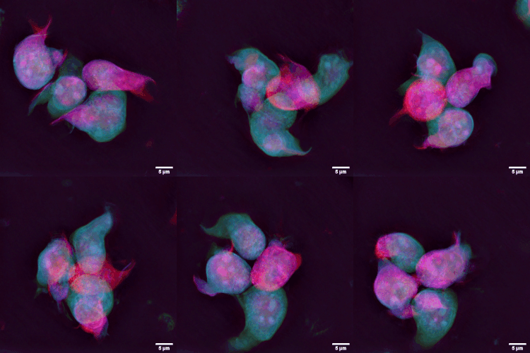
Langhans giant cells are large cells found in granulomatous conditions. They are formed by the fusion of epithelioid cells ( macrophages), and contain nuclei arranged in a horseshoe-shaped pattern in the cell periphery. Although traditionally their presence was associated with tuberculosis, they are not specific for tuberculosis or even for mycobacterial disease. In fact, they are found in nearly every form of granulomatous disease, regardless of etiology. Terminology Langhans giant cells are named after Theodor Langhans (1839–1915), a German pathologist. Causes In 2012, a research paper showed that when activated CD4+ T cells and monocytes are in close contact, interaction of CD40-CD40L between these two cells and subsequent IFNγ secretion by the T cells causes upregulation and secretion of fusion-related molecule DC-STAMP (dendritic cell-specific transmembrane protein) by the monocytes, which results in LGC formation. Clinical significance Langhans giant cells are oft ... [...More Info...] [...Related Items...] OR: [Wikipedia] [Google] [Baidu] |


.jpg)
