|
Transversus Thoracis
The transversus thoracis muscle (), also known as triangularis sterni, lies internal to the thoracic cage, anteriorly. It is usually a thin plane of muscular and tendinous fibers, however on athletic individuals it can be a thick 'slab of meat', situated upon the inner surface of the front wall of the chest. It is in the same layer as the subcostal muscles and the innermost intercostal muscles. Structure It arises on either side from the lower third of the posterior surface of the body of the sternum, from the posterior surface of the xiphoid process, and from the sternal ends of the costal cartilages of the lower three or four true ribs. Its fibers diverge upward and lateralward, to be inserted by slips into the lower borders and inner surfaces of the costal cartilages of the second, third, fourth, fifth, and sixth ribs. The lowest fibers of this muscle are horizontal in their direction, and are continuous with those of the transversus abdominis; the intermediate fibers ... [...More Info...] [...Related Items...] OR: [Wikipedia] [Google] [Baidu] |
Costal Cartilages
The costal cartilages are bars of hyaline cartilage that serve to prolong the ribs forward and contribute to the elasticity of the walls of the thorax. Costal cartilage is only found at the anterior ends of the ribs, providing medial extension. Differences from Ribs 1-12 The first seven pairs are connected with the sternum; the next three are each articulated with the lower border of the cartilage of the preceding rib; the last two have pointed extremities, which end in the wall of the abdomen. Like the ribs, the costal cartilages vary in their length, breadth, and direction. They increase in length from the first to the seventh, then gradually decrease to the twelfth. Their breadth, as well as that of the intervals between them, diminishes from the first to the last. They are broad at their attachments to the ribs, and taper toward their sternal extremities, excepting the first two, which are of the same breadth throughout, and the sixth, seventh, and eighth, which are enlarged ... [...More Info...] [...Related Items...] OR: [Wikipedia] [Google] [Baidu] |
Subcostal Muscles
Subcostal may refer to: * Subcostal nerve * Subcostal arteries The subcostal arteries, so named because they lie below the last ribs, constitute the lowest pair of branches derived from the thoracic aorta, and are in series with the intercostal arteries. Each passes along the lower border of the twelfth rib b ... * Subcostalis muscle {{disambig ... [...More Info...] [...Related Items...] OR: [Wikipedia] [Google] [Baidu] |
Thoracic Spinal Nerves
A spinal nerve is a mixed nerve, which carries motor, sensory, and autonomic signals between the spinal cord and the body. In the human body there are 31 pairs of spinal nerves, one on each side of the vertebral column. These are grouped into the corresponding cervical, thoracic, lumbar, sacral and coccygeal regions of the spine. There are eight pairs of cervical nerves, twelve pairs of thoracic nerves, five pairs of lumbar nerves, five pairs of sacral nerves, and one pair of coccygeal nerves. The spinal nerves are part of the peripheral nervous system. Structure Each spinal nerve is a mixed nerve, formed from the combination of nerve fibers from its dorsal and ventral roots. The dorsal root is the afferent sensory root and carries sensory information to the brain. The ventral root is the efferent motor root and carries motor information from the brain. The spinal nerve emerges from the spinal column through an opening (intervertebral foramen) between adjacent vertebra ... [...More Info...] [...Related Items...] OR: [Wikipedia] [Google] [Baidu] |
Intermediate Fibers
Intermediate fibers, also known as fast oxidative-glycolytic fibers, are fast twitch muscle fibers which have been converted via endurance training. These fibers are slightly larger in diameter, have more mitochondria as well as a greater blood supply and more endurance than typical fast twitch fibers. Most of the body's muscles are composed of these intermediate fibers. References * Visualizing Human Biology, Kathleen Anne Ireland, David J. Tenenbaum, raken jilani {{muscle-stub Muscular system ... [...More Info...] [...Related Items...] OR: [Wikipedia] [Google] [Baidu] |
Transversus Abdominis
The transverse abdominal muscle (TVA), also known as the transverse abdominis, transversalis muscle and transversus abdominis muscle, is a muscle layer of the anterior and lateral (front and side) abdominal wall which is deep to (layered below) the internal oblique muscle. It is thought by most fitness instructors to be a significant component of the core. Structure The transverse abdominal, so called for the direction of its fibers, is the innermost of the flat muscles of the abdomen. It is positioned immediately inside of the internal oblique muscle. The transverse abdominal arises as fleshy fibers, from the lateral third of the inguinal ligament, from the anterior three-fourths of the inner lip of the iliac crest, from the inner surfaces of the cartilages of the lower six ribs, interdigitating with the diaphragm, and from the thoracolumbar fascia. It ends anteriorly in a broad aponeurosis (the Spigelian fascia), the lower fibers of which curve inferomedially (medially an ... [...More Info...] [...Related Items...] OR: [Wikipedia] [Google] [Baidu] |
Second Rib
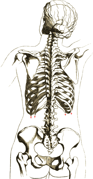
The rib cage, as an enclosure that comprises the ribs, vertebral column and sternum in the thorax of most vertebrates, protects vital organs such as the heart, lungs and great vessels. The sternum, together known as the thoracic cage, is a semi-rigid bony and cartilaginous structure which surrounds the thoracic cavity and supports the shoulder girdle to form the core part of the human skeleton. A typical human thoracic cage consists of 12 pairs of ribs and the adjoining costal cartilages, the sternum (along with the manubrium and xiphoid process), and the 12 thoracic vertebrae articulating with the ribs. Together with the skin and associated fascia and muscles, the thoracic cage makes up the thoracic wall and provides attachments for extrinsic skeletal muscles of the neck, upper limbs, upper abdomen and back. The rib cage intrinsically holds the muscles of respiration ( diaphragm, intercostal muscles, etc.) that are crucial for active inhalation and forced exhalation, and ther ... [...More Info...] [...Related Items...] OR: [Wikipedia] [Google] [Baidu] |
Human Sternum
The sternum or breastbone is a long flat bone located in the central part of the chest. It connects to the ribs via cartilage and forms the front of the rib cage, thus helping to protect the heart, lungs, and major blood vessels from injury. Shaped roughly like a necktie, it is one of the largest and longest flat bones of the body. Its three regions are the manubrium, the body, and the xiphoid process. The word "sternum" originates from the Ancient Greek στέρνον (stérnon), meaning "chest". Structure The sternum is a narrow, flat bone, forming the middle portion of the front of the chest. The top of the sternum supports the clavicles (collarbones) and its edges join with the costal cartilages of the first two pairs of ribs. The inner surface of the sternum is also the attachment of the sternopericardial ligaments. Its top is also connected to the sternocleidomastoid muscle. The sternum consists of three main parts, listed from the top: * Manubrium * Body (g ... [...More Info...] [...Related Items...] OR: [Wikipedia] [Google] [Baidu] |
Innermost Intercostal
The innermost intercostal muscle is a layer of intercostal muscles. It may also be called the intima of the internal intercostal muscles. It is the deepest muscular layer of the thorax, with muscle fibres running vertically (in parallel with the internal intercostal muscles). It is present only in the middle of each intercostal space, and often not present higher up the rib cage. It lies deep to the plane that contains the intercostal nerves and intercostal vessels, and the internal intercostal muscles The internal intercostal muscles (intercostales interni) are a group of skeletal muscles located between the ribs. They are eleven in number on either side. They commence anteriorly at the sternum, in the intercostal spaces between the cartilage .... The diaphragm is continuous with the innermost intercostal muscle. Additional images File:Innermost intercostal muscles animation.gif, Innermost intercostal muscle (shown in red). Animation. File:1114 Thorax zoom.png, A cutout of ... [...More Info...] [...Related Items...] OR: [Wikipedia] [Google] [Baidu] |
Thoracic Cage
The rib cage, as an enclosure that comprises the ribs, vertebral column and sternum in the thorax of most vertebrates, protects vital organs such as the heart, lungs and great vessels. The sternum, together known as the thoracic cage, is a semi-rigid bony and cartilaginous structure which surrounds the thoracic cavity and supports the shoulder girdle to form the core part of the human skeleton. A typical human thoracic cage consists of 12 pairs of ribs and the adjoining costal cartilages, the sternum (along with the manubrium and xiphoid process), and the 12 thoracic vertebrae articulating with the ribs. Together with the skin and associated fascia and muscles, the thoracic cage makes up the thoracic wall and provides attachments for extrinsic skeletal muscles of the neck, upper limbs, upper abdomen and back. The rib cage intrinsically holds the muscles of respiration ( diaphragm, intercostal muscles, etc.) that are crucial for active inhalation and forced exhalation, and the ... [...More Info...] [...Related Items...] OR: [Wikipedia] [Google] [Baidu] |
True Ribs
The rib cage, as an enclosure that comprises the ribs, vertebral column and sternum in the thorax of most vertebrates, protects vital organs such as the heart, lungs and great vessels. The sternum, together known as the thoracic cage, is a semi-rigid bony and cartilaginous structure which surrounds the thoracic cavity and supports the shoulder girdle to form the core part of the human skeleton. A typical human thoracic cage consists of 12 pairs of ribs and the adjoining costal cartilages, the sternum (along with the manubrium and xiphoid process), and the 12 thoracic vertebrae articulating with the ribs. Together with the skin and associated fascia and muscles, the thoracic cage makes up the thoracic wall and provides attachments for extrinsic skeletal muscles of the neck, upper limbs, upper abdomen and back. The rib cage intrinsically holds the muscles of respiration (diaphragm, intercostal muscles, etc.) that are crucial for active inhalation and forced exhalation, and ther ... [...More Info...] [...Related Items...] OR: [Wikipedia] [Google] [Baidu] |
Intercostal Nerves
The intercostal nerves are part of the somatic nervous system, and arise from the anterior rami of the thoracic spinal nerves from T1 to T11. The intercostal nerves are distributed chiefly to the thoracic pleura and abdominal peritoneum, and differ from the anterior rami of the other spinal nerves in that each pursues an independent course without plexus formation. The first two nerves supply fibers to the upper limb and thorax; the next four distribute to the walls of the thorax; the lower five supply the walls of the thorax and abdomen. The 7th intercostal nerve end at the xyphoid process of the sternum. The 10th intercostal nerve terminates at the navel. The 12th ( subcostal) thoracic is distributed to the walls of the abdomen and groin. Each of these fibers contains around 1300 axons. Unlike the nerves from the autonomic nervous system that innervate the visceral pleura of the thoracic cavity, the intercostal nerves arise from the somatic nervous system. This enables them ... [...More Info...] [...Related Items...] OR: [Wikipedia] [Google] [Baidu] |
Intercostal Arteries
The intercostal arteries are a group of arteries that supply the area between the ribs ("costae"), called the intercostal space. The highest intercostal artery (supreme intercostal artery or superior intercostal artery) is an artery in the human body that usually gives rise to the first and second posterior intercostal arteries, which supply blood to their corresponding intercostal space. It usually arises from the costocervical trunk, which is a branch of the subclavian artery. Some anatomists may contend that there is no supreme intercostal artery, only a supreme intercostal vein. The anterior intercostal branches of internal thoracic artery supply the upper five or six intercostal spaces. The internal thoracic artery (previously called as internal mammary artery) then divides into the superior epigastric artery and musculophrenic artery. The latter gives out the remaining anterior intercostal branches. Two in number in each space, these small vessels pass lateralward, ... [...More Info...] [...Related Items...] OR: [Wikipedia] [Google] [Baidu] |


