|
Transverse Temporal Gyri
The transverse temporal gyri, also called Heschl's gyri () or Heschl's convolutions, are gyri found in the area of primary auditory cortex buried within the lateral sulcus of the human brain, occupying Brodmann areas 41 and 42. Transverse temporal gyri are superior to and separated from the planum temporale (cortex involved in language production) by Heschl's sulcus. Transverse temporal gyri are found in varying numbers in both the right and left hemispheres of the brain and one study found that this number is not related to the hemisphere or dominance of hemisphere studied in subjects. Transverse temporal gyri can be viewed in the sagittal plane as either an omega shape (if one gyrus is present) or a heart shape (if two gyri and a sulcus are present). Transverse temporal gyri are the first cortical structures to process incoming auditory information. Anatomically, the transverse temporal gyri are distinct in that they run mediolaterally (toward the center of the brain), rather ... [...More Info...] [...Related Items...] OR: [Wikipedia] [Google] [Baidu] |
Brain
A brain is an organ that serves as the center of the nervous system in all vertebrate and most invertebrate animals. It is located in the head, usually close to the sensory organs for senses such as vision. It is the most complex organ in a vertebrate's body. In a human, the cerebral cortex contains approximately 14–16 billion neurons, and the estimated number of neurons in the cerebellum is 55–70 billion. Each neuron is connected by synapses to several thousand other neurons. These neurons typically communicate with one another by means of long fibers called axons, which carry trains of signal pulses called action potentials to distant parts of the brain or body targeting specific recipient cells. Physiologically, brains exert centralized control over a body's other organs. They act on the rest of the body both by generating patterns of muscle activity and by driving the secretion of chemicals called hormones. This centralized control allows rapid and coordinated responses ... [...More Info...] [...Related Items...] OR: [Wikipedia] [Google] [Baidu] |
Planum Temporale
The planum temporale is the cortical area just posterior to the auditory cortex (Heschl's gyrus) within the Sylvian fissure. It is a triangular region which forms the heart of Wernicke's area, one of the most important functional areas for language. Original studies on this area found that the planum temporale was one of the most asymmetric regions in the brain, with this area being up to ten times larger in the left cerebral hemisphere than the right. Location The planum temporale makes up the superior surface of the superior temporal gyrus to the parietal lobe. The posterior extent of the planum temporale has been variably defined, which has led to disputes to estimates of size and degree of asymmetry. Asymmetry The planum temporale shows a significant asymmetry. In 65% of all individuals the left planum temporale appears to be more developed, while the right planum temporale is more developed in only 11%. In some people’s brains, the planum temporale is more than five times ... [...More Info...] [...Related Items...] OR: [Wikipedia] [Google] [Baidu] |
Superior Temporal Gyrus
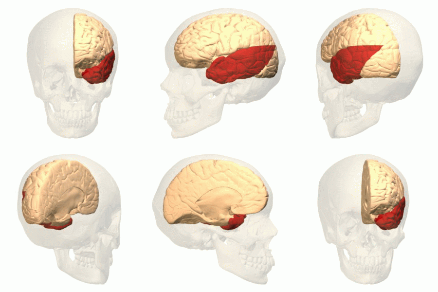
The superior temporal gyrus (STG) is one of three (sometimes two) gyri in the temporal lobe of the human brain, which is located laterally to the head, situated somewhat above the external ear. The superior temporal gyrus is bounded by: * the lateral sulcus above; * the superior temporal sulcus (not always present or visible) below; * an imaginary line drawn from the preoccipital notch to the lateral sulcus posteriorly. The superior temporal gyrus contains several important structures of the brain, including: * Brodmann areas 41 and 42, marking the location of the auditory cortex, the cortical region responsible for the sensation of sound; * Wernicke's area, Brodmann 22p, an important region for the processing of speech so that it can be understood as language. The superior temporal gyrus contains the auditory cortex, which is responsible for processing sounds. Specific sound frequencies map precisely onto the auditory cortex. This auditory (or tonotopic) map is similar ... [...More Info...] [...Related Items...] OR: [Wikipedia] [Google] [Baidu] |
Event-related Potential
An event-related potential (ERP) is the measured brain response that is the direct result of a specific sensory, cognitive, or motor event. More formally, it is any stereotyped electrophysiological response to a stimulus. The study of the brain in this way provides a noninvasive means of evaluating brain functioning. ERPs are measured by means of electroencephalography (EEG). The magnetoencephalography (MEG) equivalent of ERP is the ERF, or event-related field. Evoked potentials and induced potentials are subtypes of ERPs. History With the discovery of the electroencephalogram (EEG) in 1924, Hans Berger revealed that one could measure the electrical activity of the human brain by placing electrodes on the scalp and amplifying the signal. Changes in voltage can then be plotted over a period of time. He observed that the voltages could be influenced by external events that stimulated the senses. The EEG proved to be a useful source in recording brain activity over the ... [...More Info...] [...Related Items...] OR: [Wikipedia] [Google] [Baidu] |
Functional Magnetic Resonance Imaging
Functional magnetic resonance imaging or functional MRI (fMRI) measures brain activity by detecting changes associated with blood flow. This technique relies on the fact that cerebral blood flow and neuronal activation are coupled. When an area of the brain is in use, blood flow to that region also increases. The primary form of fMRI uses the blood-oxygen-level dependent (BOLD) contrast, discovered by Seiji Ogawa in 1990. This is a type of specialized brain and body scan used to map neural activity in the brain or spinal cord of humans or other animals by imaging the change in blood flow (hemodynamic response) related to energy use by brain cells. Since the early 1990s, fMRI has come to dominate brain mapping research because it does not involve the use of injections, surgery, the ingestion of substances, or exposure to ionizing radiation. This measure is frequently corrupted by noise from various sources; hence, statistical procedures are used to extract the underlying signal. ... [...More Info...] [...Related Items...] OR: [Wikipedia] [Google] [Baidu] |
Richard L
Richard is a male given name. It originates, via Old French, from Old Frankish and is a compound of the words descending from Proto-Germanic ''*rīk-'' 'ruler, leader, king' and ''*hardu-'' 'strong, brave, hardy', and it therefore means 'strong in rule'. Nicknames include "Richie", "Dick", "Dickon", " Dickie", " Rich", "Rick", " Rico", " Ricky", and more. Richard is a common English, German and French male name. It's also used in many more languages, particularly Germanic, such as Norwegian, Danish, Swedish, Icelandic, and Dutch, as well as other languages including Irish, Scottish, Welsh and Finnish. Richard is cognate with variants of the name in other European languages, such as the Swedish "Rickard", the Catalan "Ricard" and the Italian "Riccardo", among others (see comprehensive variant list below). People named Richard Multiple people with the same name * Richard Andersen (other) * Richard Anderson (other) * Richard Cartwright (other) ... [...More Info...] [...Related Items...] OR: [Wikipedia] [Google] [Baidu] |
Cerebral Cortex
The cerebral cortex, also known as the cerebral mantle, is the outer layer of neural tissue of the cerebrum of the brain in humans and other mammals. The cerebral cortex mostly consists of the six-layered neocortex, with just 10% consisting of allocortex. It is separated into two cortices, by the longitudinal fissure that divides the cerebrum into the left and right cerebral hemispheres. The two hemispheres are joined beneath the cortex by the corpus callosum. The cerebral cortex is the largest site of neural integration in the central nervous system. It plays a key role in attention, perception, awareness, thought, memory, language, and consciousness. The cerebral cortex is part of the brain responsible for cognition. In most mammals, apart from small mammals that have small brains, the cerebral cortex is folded, providing a greater surface area in the confined volume of the cranium. Apart from minimising brain and cranial volume, cortical folding is crucial f ... [...More Info...] [...Related Items...] OR: [Wikipedia] [Google] [Baidu] |
Brodmann Areas 41 And 42
Brodmann areas 41 and 42 are parts of the primary auditory cortex. Brodmann area 41 is also known as the anterior transverse temporal area 41 (H). It is a cytoarchitectonic division of the cerebral cortex occupying the anterior transverse temporal gyrus (H) in the bank of the lateral sulcus on the dorsal surface of the temporal lobe. Brodmann area 41 is bounded medially by the parainsular area 52 (H) and laterally by the posterior transverse temporal area 42 (H) (Brodmann-1909). Brodmann area 42 is also known as the posterior transverse temporal area 42 (H), and is also a subdivision of the temporal lobe. Brodmann area 42 is bounded medially by the anterior transverse temporal area 41 (H) and laterally by the superior temporal area 22 (Brodmann-1909). Function Brodmann areas 41 and 42 are parts of the primary auditory cortex. This is the first cortical destination of auditory information stemming from the thalamus. Neural activity in this brain part corresponds most strong ... [...More Info...] [...Related Items...] OR: [Wikipedia] [Google] [Baidu] |
Temporal Lobe
The temporal lobe is one of the four major lobes of the cerebral cortex in the brain of mammals. The temporal lobe is located beneath the lateral fissure on both cerebral hemispheres of the mammalian brain. The temporal lobe is involved in processing sensory input into derived meanings for the appropriate retention of visual memory, language comprehension, and emotion association. ''Temporal'' refers to the head's temples. Structure The temporal lobe consists of structures that are vital for declarative or long-term memory. Declarative (denotative) or explicit memory is conscious memory divided into semantic memory (facts) and episodic memory (events). Medial temporal lobe structures that are critical for long-term memory include the hippocampus, along with the surrounding hippocampal region consisting of the perirhinal, parahippocampal, and entorhinal neocortical regions. The hippocampus is critical for memory formation, and the surrounding medial temporal cortex is ... [...More Info...] [...Related Items...] OR: [Wikipedia] [Google] [Baidu] |
Human Brain
The human brain is the central organ (anatomy), organ of the human nervous system, and with the spinal cord makes up the central nervous system. The brain consists of the cerebrum, the brainstem and the cerebellum. It controls most of the activities of the human body, body, processing, integrating, and coordinating the information it receives from the Sensory nervous system, sense organs, and making decisions as to the instructions sent to the rest of the body. The brain is contained in, and protected by, the neurocranium, skull bones of the human head, head. The cerebrum, the largest part of the human brain, consists of two cerebral hemispheres. Each hemisphere has an inner core composed of white matter, and an outer surface – the cerebral cortex – composed of grey matter. The cortex has an outer layer, the neocortex, and an inner allocortex. The neocortex is made up of six Cerebral cortex#Layers of neocortex, neuronal layers, while the allocortex has three or four. Each ... [...More Info...] [...Related Items...] OR: [Wikipedia] [Google] [Baidu] |
Lateral Sulcus
In neuroanatomy, the lateral sulcus (also called Sylvian fissure, after Franciscus Sylvius, or lateral fissure) is one of the most prominent features of the human brain. The lateral sulcus is a deep fissure in each hemisphere that separates the frontal and parietal lobes from the temporal lobe. The insular cortex lies deep within the lateral sulcus. Anatomy The lateral sulcus divides both the frontal lobe and parietal lobe above from the temporal lobe below. It is in both hemispheres of the brain. The lateral sulcus is one of the earliest-developing sulci of the human brain. It first appears around the fourteenth gestational week. The insular cortex lies deep within the lateral sulcus. The lateral sulcus has a number of side branches. Two of the most prominent and most regularly found are the ascending (also called vertical) ramus and the horizontal ramus of the lateral fissure, which subdivide the inferior frontal gyrus. The lateral sulcus also contains the transverse tempor ... [...More Info...] [...Related Items...] OR: [Wikipedia] [Google] [Baidu] |


