|
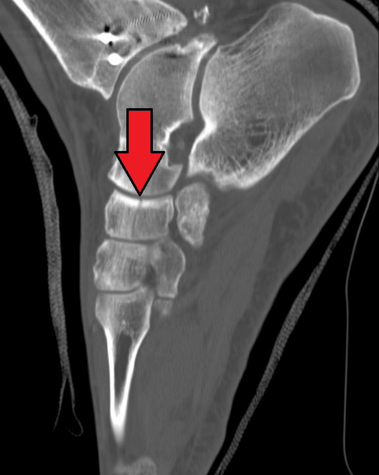
Tibialis Posterior
The tibialis posterior muscle is the most central of all the leg muscles, and is located in the deep posterior compartment of the leg. It is the key stabilizing muscle of the lower leg. Structure The tibialis posterior muscle originates on the inner posterior border of the fibula laterally. It is also attached to the interosseous membrane medially, which attaches to the tibia and fibula. The tendon of the tibialis posterior muscle (sometimes called the posterior tibial tendon) descends posterior to the medial malleolus. It terminates by dividing into plantar, main, and recurrent components. The main portion inserts into the tuberosity of the navicular bone. The smaller portion inserts into the plantar surface of the medial cuneiform. The plantar portion inserts into the bases of the second, third and fourth metatarsals, the intermediate and lateral cuneiforms and the cuboid. The recurrent portion inserts into the sustentaculum tali of the calcaneus. Blood is supplied to the m ... [...More Info...] [...Related Items...] OR: [Wikipedia] [Google] [Baidu] |
Tibia
The tibia (; ), also known as the shinbone or shankbone, is the larger, stronger, and anterior (frontal) of the two bones in the leg below the knee in vertebrates (the other being the fibula, behind and to the outside of the tibia); it connects the knee with the ankle. The tibia is found on the medial side of the leg next to the fibula and closer to the median plane. The tibia is connected to the fibula by the interosseous membrane of leg, forming a type of fibrous joint called a syndesmosis with very little movement. The tibia is named for the flute ''tibia''. It is the second largest bone in the human body, after the femur. The leg bones are the strongest long bones as they support the rest of the body. Structure In human anatomy, the tibia is the second largest bone next to the femur. As in other vertebrates the tibia is one of two bones in the lower leg, the other being the fibula, and is a component of the knee and ankle joints. The ossification or formation of the bone ... [...More Info...] [...Related Items...] OR: [Wikipedia] [Google] [Baidu] |
Navicular Bone
The navicular bone is a small bone found in the feet of most mammals. Human anatomy The navicular bone in humans is one of the tarsal bones, found in the foot. Its name derives from the human bone's resemblance to a small boat, caused by the strongly concave proximal articular surface. The term ''navicular bone'' or ''hand navicular bone'' was formerly used for the scaphoid bone, one of the carpal bones of the wrist. The navicular bone in humans is located on the medial side of the foot, and articulates proximally with the talus, distally with the three cuneiform bones, and laterally with the cuboid. It is the last of the foot bones to start ossification and does not tend to do so until the end of the third year in girls and the beginning of the fourth year in boys, although a large range of variation has been reported. The tibialis posterior is the only muscle that attaches to the navicular bone. The main portion of the muscle inserts into the tuberosity of the navi ... [...More Info...] [...Related Items...] OR: [Wikipedia] [Google] [Baidu] |
Exercise
Exercise is a body activity that enhances or maintains physical fitness and overall health and wellness. It is performed for various reasons, to aid growth and improve strength, develop muscles and the cardiovascular system, hone athletic skills, weight loss or maintenance, improve health, or simply for enjoyment. Many individuals choose to exercise outdoors where they can congregate in groups, socialize, and improve well-being as well as mental health. In terms of health benefits, the amount of recommended exercise depends upon the goal, the type of exercise, and the age of the person. Even doing a small amount of exercise is healthier than doing none. Classification Physical exercises are generally grouped into three types, depending on the overall effect they have on the human body: * Aerobic exercise is any physical activity that uses large muscle groups and causes the body to use more oxygen than it would while resting. The goal of aerobic exercise is to increa ... [...More Info...] [...Related Items...] OR: [Wikipedia] [Google] [Baidu] |
Eversion (kinesiology)
Motion, the process of movement, is described using specific anatomical terms. Motion includes movement of organs, joints, limbs, and specific sections of the body. The terminology used describes this motion according to its direction relative to the anatomical position of the body parts involved. Anatomists and others use a unified set of terms to describe most of the movements, although other, more specialized terms are necessary for describing unique movements such as those of the hands, feet, and eyes. In general, motion is classified according to the anatomical plane it occurs in. ''Flexion'' and ''extension'' are examples of ''angular'' motions, in which two axes of a joint are brought closer together or moved further apart. ''Rotational'' motion may occur at other joints, for example the shoulder, and are described as ''internal'' or ''external''. Other terms, such as ''elevation'' and ''depression'', describe movement above or below the horizontal plane. Many anatomica ... [...More Info...] [...Related Items...] OR: [Wikipedia] [Google] [Baidu] |
Valgus Deformity
A valgus deformity is a condition in which the bone segment distal Standard anatomical terms of location are used to unambiguously describe the anatomy of animals, including humans. The terms, typically derived from Latin or Greek roots, describe something in its standard anatomical position. This position pro ... to a joint is angled outward, that is, angled laterally, away from the body's midline. The opposite deformation, where the twist or angulation is directed medially, toward the center of the body, is called varus deformity, varus. Common causes of valgus knee (genu valgum or "knock-knee") in adults include arthritis of the knee and traumatic injuries. Knee arthritis with valgus knee Rheumatoid knee commonly presents as valgus knee. Osteoarthritis knee may also sometimes present with valgus deformity though varus deformity is common. Total knee arthroplasty (TKA) to correct valgus deformity is surgically difficult and requires specialized implants called Knee repl ... [...More Info...] [...Related Items...] OR: [Wikipedia] [Google] [Baidu] |
Flat Feet
Flat feet (also called pes planus or fallen arches) is a postural deformity in which the arches of the foot collapse, with the entire sole of the foot coming into complete or near-complete contact with the ground. Sometimes children are born with flat feet (congenital). There is a functional relationship between the structure of the arch of the foot and the biomechanics of the lower leg. The arch provides an elastic, springy connection between the forefoot and the hind foot so that a majority of the forces incurred during weight bearing on the foot can be dissipated before the force reaches the long bones of the leg and thigh. In pes planus, the head of the talus bone is displaced medially and distal from the navicular bone. As a result, the Plantar calcaneonavicular ligament (spring ligament) and the tendon of the tibialis posterior muscle are stretched to the extent that the individual with pes planus loses the function of the medial longitudinal arch (MLA). If the MLA is ... [...More Info...] [...Related Items...] OR: [Wikipedia] [Google] [Baidu] |
Arches Of The Foot
The arches of the foot, formed by the tarsus (skeleton), tarsal and metatarsal bones, strengthened by ligaments and tendons, allow the foot to support the weight of the body in the erect posture with the least weight. They are categorized as longitudinal and transverse arches. Structure Longitudinal arches The longitudinal arches of the foot can be divided into medial and lateral arches. Medial arch The medial arch is higher than the lateral longitudinal arch. It is made up by the calcaneus, the talus bone, talus, the navicular, the three Cuneiform (anatomy), cuneiforms (medial, intermediate, and lateral), and the first, second, and third metatarsals. Its summit is at the superior articular surface of the talus, and its two extremities or piers, on which it rests in standing, are the tuberosity on the plantar surface of the calcaneus posteriorly and the heads of the first, second, and third metatarsal bones anteriorly. The chief characteristic of this arch is its elasticity, ... [...More Info...] [...Related Items...] OR: [Wikipedia] [Google] [Baidu] |
Tibial Nerve
The tibial nerve is a branch of the sciatic nerve. The tibial nerve passes through the popliteal fossa to pass below the arch of soleus. Structure Popliteal fossa The tibial nerve is the larger terminal branch of the sciatic nerve with root values of L4, L5, S1, S2, and S3. It lies superficial (or posterior) to the popliteal vessels, extending from the superior angle to the inferior angle of the popliteal fossa, crossing the popliteal vessels from lateral to medial side. It gives off branches as shown below: * Muscular branches - Muscular branches arise from the distal part of the popliteal fossa. It supplies the medial and lateral heads of gastrocnemius, soleus, plantaris and popliteus muscles. Nerve to popliteus crosses the popliteus muscle, runs downwards and laterally, winds around the lower border of the popliteus to supply the deep (or anterior) surface of the popliteus. This nerve also supplies the tibialis posterior muscle, superior tibiofibular joint, tibia bone, intero ... [...More Info...] [...Related Items...] OR: [Wikipedia] [Google] [Baidu] |
Posterior Tibial Artery
The posterior tibial artery of the lower limb is an artery that carries blood to the posterior compartment of the leg and plantar surface of the foot. It branches from the popliteal artery via the tibial-fibular trunk. Structure The posterior tibial artery arises from the popliteal artery in the popliteal fossa. It is accompanied by a deep vein, the posterior tibial vein, along its course. It passes just posterior to the medial malleolus of the tibia, but anterior to the Achilles tendon. It passes into the foot deep to the flexor retinaculum of the foot. It runs through the tarsal tunnel. Branches The posterior tibial artery gives rise to: * medial plantar artery. * lateral plantar artery. * fibular artery, which is said to rise from the bifurcation of the tibial-fibular trunk and the posterior tibial artery. * calcaneal branch to the medial aspect of the calcaneus. Function The posterior tibial artery supplies oxygenated blood to the posterior compartment of the leg and t ... [...More Info...] [...Related Items...] OR: [Wikipedia] [Google] [Baidu] |
Calcaneus
In humans and many other primates, the calcaneus (; from the Latin ''calcaneus'' or ''calcaneum'', meaning heel) or heel bone is a bone of the tarsus of the foot which constitutes the heel. In some other animals, it is the point of the hock. Structure In humans, the calcaneus is the largest of the tarsal bones and the largest bone of the foot. Its long axis is pointed forwards and laterally. The talus bone, calcaneus, and navicular bone are considered the proximal row of tarsal bones. In the calcaneus, several important structures can be distinguished:Platzer (2004), p 216 There is a large calcaneal tuberosity located posteriorly on plantar surface with medial and lateral tubercles on its surface. Besides, there is another peroneal tubecle on its lateral surface. On its lower edge on either side are its lateral and medial processes (serving as the origins of the abductor hallucis and abductor digiti minimi). The Achilles tendon is inserted into a roughened area on its superio ... [...More Info...] [...Related Items...] OR: [Wikipedia] [Google] [Baidu] |
Sustentaculum Tali
In humans and many other primates, the calcaneus (; from the Latin ''calcaneus'' or ''calcaneum'', meaning heel) or heel bone is a bone of the tarsus of the foot which constitutes the heel. In some other animals, it is the point of the hock. Structure In humans, the calcaneus is the largest of the tarsal bones and the largest bone of the foot. Its long axis is pointed forwards and laterally. The talus bone, calcaneus, and navicular bone are considered the proximal row of tarsal bones. In the calcaneus, several important structures can be distinguished:Platzer (2004), p 216 There is a large calcaneal tuberosity located posteriorly on plantar surface with medial and lateral tubercles on its surface. Besides, there is another peroneal tubecle on its lateral surface. On its lower edge on either side are its lateral and medial processes (serving as the origins of the abductor hallucis and abductor digiti minimi). The Achilles tendon is inserted into a roughened area on its supe ... [...More Info...] [...Related Items...] OR: [Wikipedia] [Google] [Baidu] |
Cuboid Bone
In the human body, the cuboid bone is one of the seven tarsal bones of the foot. Structure The cuboid bone is the most lateral of the bones in the distal row of the tarsus. It is roughly cubical in shape, and presents a prominence in its inferior (or plantar) surface, the tuberosity of the cuboid. The bone provides a groove where the tendon of the peroneus longus muscle passes to reach its insertion in the first metatarsal and medial cuneiform bones. Surfaces The dorsal surface, directed upward and lateralward, is rough, for the attachment of ligaments. The plantar surface presents in front a deep groove, the peroneal sulcus, which runs obliquely forward and medialward; it lodges the tendon of the peroneus longus, and is bounded behind by a prominent ridge, to which the long plantar ligament is attached. The ridge ends laterally in an eminence, the tuberosity, the surface of which presents an oval facet; on this facet glides the sesamoid bone or cartilage frequently found ... [...More Info...] [...Related Items...] OR: [Wikipedia] [Google] [Baidu] |


