|
Thyroid Papillary Carcinomas
Papillary thyroid cancer or papillary thyroid carcinoma is the most common type of thyroid cancer Thyroid cancer is cancer that develops from the tissues of the thyroid gland. It is a disease in which cells grow abnormally and have the potential to spread to other parts of the body. Symptoms can include swelling or a lump in the neck. C ..., representing 75 percent to 85 percent of all thyroid cancer cases.Chapter 20 in: 8th edition. It occurs more frequently in women and presents in the 20–55 year age group. It is also the predominant cancer type in children with thyroid cancer, and in patients with thyroid cancer who have had previous radiation to the head and neck. It is often well-cellular differentiation, differentiated, slow-growing, and localized, although it can metastasis, metastasize. Diagnosis Papillary thyroid carcinoma is usually discovered on routine examination as an asymptomatic thyroid nodule that appears as a neck mass. In some instances, the mass may ... [...More Info...] [...Related Items...] OR: [Wikipedia] [Google] [Baidu] |
Pap Stain
Papanicolaou stain (also Papanicolaou's stain and Pap stain) is a multichromatic (multicolored) Cytopathology, cytological staining technique developed by Georgios_Papanikolaou, George Papanicolaou in 1942. The Papanicolaou stain is one of the most widely used stains in cytology, where it is used to aid pathologists in making a diagnosis. Although most notable for its use in the detection of cervical cancer in the Pap test or Pap smear, it is also used to stain non-gynecological specimen preparations from a variety of bodily secretions and from fine needle aspiration, small needle biopsies of organs and tissues. Papanicolaou published three formulations of this stain in 1942, 1954, and 1960. Usage Pap staining is used to differentiate cells in smear preparations (in which samples are spread or smeared onto a glass microscope slide) from various body fluid, bodily secretions and biopsies, needle biopsies; the specimens may include gynecological smears (Pap smears), sputum, brushi ... [...More Info...] [...Related Items...] OR: [Wikipedia] [Google] [Baidu] |
Papillary Carcinoma Of The Thyroid
Papilla (Latin, 'nipple') or papillae may refer to: In animals * Papilla (fish anatomy), in the mouth of fish * Basilar papilla, a sensory organ of lizards, amphibians and fish * Dental papilla, in a developing tooth * Dermal papillae, part of the skin * Major duodenal papilla, in the duodenum * Minor duodenal papilla, in the duodenum * Genital papilla, a feature of the external genitalia of some animals * Interdental papilla, part of the gums * Lacrimal papilla, on the bottom eyelid * Lingual papillae, small structures on the upper surface of the tongue * Renal papilla, part of the kidney In plants and fungi * Papilla (mycology), a nipple-shaped protrusion in the center of the cap * Stigmatic papilla, part of the stigma (botany) See also * * * Blister, a small pocket of body fluid within the upper layers of the skin * Papillary muscle, a muscle in the heart * Papilloma, a benign epithelial tumor * Papule A papule is a small, well-defined bump in the skin. It may have ... [...More Info...] [...Related Items...] OR: [Wikipedia] [Google] [Baidu] |
Micrograph
A micrograph or photomicrograph is a photograph or digital image taken through a microscope or similar device to show a magnified image of an object. This is opposed to a macrograph or photomacrograph, an image which is also taken on a microscope but is only slightly magnified, usually less than 10 times. Micrography is the practice or art of using microscopes to make photographs. A micrograph contains extensive details of microstructure. A wealth of information can be obtained from a simple micrograph like behavior of the material under different conditions, the phases found in the system, failure analysis, grain size estimation, elemental analysis and so on. Micrographs are widely used in all fields of microscopy. Types Photomicrograph A light micrograph or photomicrograph is a micrograph prepared using an optical microscope, a process referred to as ''photomicroscopy''. At a basic level, photomicroscopy may be performed simply by connecting a camera to a microscope, th ... [...More Info...] [...Related Items...] OR: [Wikipedia] [Google] [Baidu] |
Mitochondria
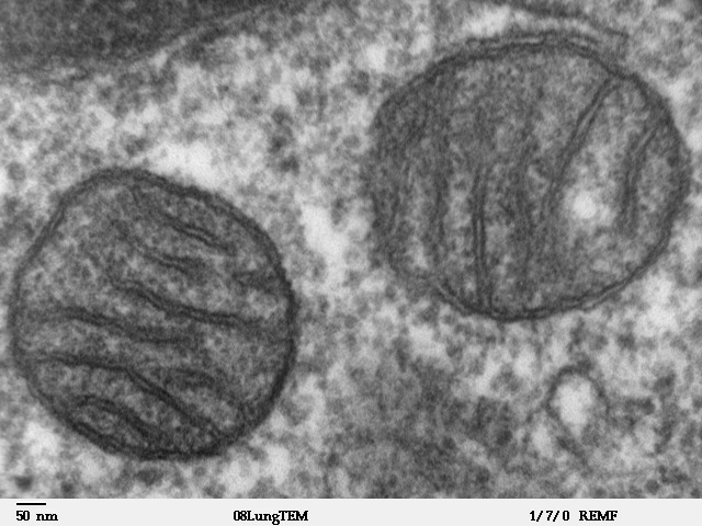
A mitochondrion (; ) is an organelle found in the Cell (biology), cells of most Eukaryotes, such as animals, plants and Fungus, fungi. Mitochondria have a double lipid bilayer, membrane structure and use aerobic respiration to generate adenosine triphosphate (ATP), which is used throughout the cell as a source of chemical energy. They were discovered by Albert von Kölliker in 1857 in the voluntary muscles of insects. The term ''mitochondrion'' was coined by Carl Benda in 1898. The mitochondrion is popularly nicknamed the "powerhouse of the cell", a phrase coined by Philip Siekevitz in a 1957 article of the same name. Some cells in some multicellular organisms lack mitochondria (for example, mature mammalian red blood cells). A large number of unicellular organisms, such as microsporidia, parabasalids and diplomonads, have reduced or transformed their mitochondria into mitosome, other structures. One eukaryote, ''Monocercomonoides'', is known to have completely lost its mitocho ... [...More Info...] [...Related Items...] OR: [Wikipedia] [Google] [Baidu] |
Snowflake
A snowflake is a single ice crystal that has achieved a sufficient size, and may have amalgamated with others, which falls through the Earth's atmosphere as snow.Knight, C.; Knight, N. (1973). Snow crystals. Scientific American, vol. 228, no. 1, pp. 100–107.Hobbs, P.V. 1974. Ice Physics. Oxford: Clarendon Press. Each flake nucleates around a dust particle in supersaturated air masses by attracting Supercooling, supercooled cloud water droplets, which freezing, freeze and accrete in crystal form. Complex shapes Emergence, emerge as the flake moves through differing temperature and humidity zones in the atmosphere, such that individual snowflakes differ in detail from one another, but may be categorized in eight broad classifications and at least 80 individual variants. The main constituent shapes for ice crystals, from which combinations may occur, are needle, column, plate, and rime. Snow appears white in color despite being made of clear ice. This is due to diffuse reflection ... [...More Info...] [...Related Items...] OR: [Wikipedia] [Google] [Baidu] |
Lungs
The lungs are the primary organs of the respiratory system in humans and most other animals, including some snails and a small number of fish. In mammals and most other vertebrates, two lungs are located near the backbone on either side of the heart. Their function in the respiratory system is to extract oxygen from the air and transfer it into the bloodstream, and to release carbon dioxide from the bloodstream into the atmosphere, in a process of gas exchange. Respiration is driven by different muscular systems in different species. Mammals, reptiles and birds use their different muscles to support and foster breathing. In earlier tetrapods, air was driven into the lungs by the pharyngeal muscles via buccal pumping, a mechanism still seen in amphibians. In humans, the main muscle of respiration that drives breathing is the diaphragm. The lungs also provide airflow that makes vocal sounds including human speech possible. Humans have two lungs, one on the left and one on the ... [...More Info...] [...Related Items...] OR: [Wikipedia] [Google] [Baidu] |
Blood Vessels
The blood vessels are the components of the circulatory system that transport blood throughout the human body. These vessels transport blood cells, nutrients, and oxygen to the tissues of the body. They also take waste and carbon dioxide away from the tissues. Blood vessels are needed to sustain life, because all of the body's tissues rely on their functionality. There are five types of blood vessels: the arteries, which carry the blood away from the heart; the arterioles; the capillaries, where the exchange of water and chemicals between the blood and the tissues occurs; the venules; and the veins, which carry blood from the capillaries back towards the heart. The word ''vascular'', meaning relating to the blood vessels, is derived from the Latin ''vas'', meaning vessel. Some structures – such as cartilage, the epithelium, and the lens and cornea of the eye – do not contain blood vessels and are labeled ''avascular''. Etymology * artery: late Middle English; from Latin ' ... [...More Info...] [...Related Items...] OR: [Wikipedia] [Google] [Baidu] |
Lymphatics
The lymphatic vessels (or lymph vessels or lymphatics) are thin-walled vessels (tubes), structured like blood vessels, that carry lymph. As part of the lymphatic system, lymph vessels are complementary to the cardiovascular system. Lymph vessels are lined by endothelial cells, and have a thin layer of smooth muscle, and adventitia that binds the lymph vessels to the surrounding tissue. Lymph vessels are devoted to the propulsion of the lymph from the lymph capillaries, which are mainly concerned with the absorption of interstitial fluid from the tissues. Lymph capillaries are slightly bigger than their counterpart capillaries of the vascular system. Lymph vessels that carry lymph to a lymph node are called afferent lymph vessels, and those that carry it from a lymph node are called efferent lymph vessels, from where the lymph may travel to another lymph node, may be returned to a vein, or may travel to a larger lymph duct. Lymph ducts drain the lymph into one of the subclavian ve ... [...More Info...] [...Related Items...] OR: [Wikipedia] [Google] [Baidu] |
Noninvasive Follicular Thyroid Neoplasm With Papillary-like Nuclear Features
Noninvasive follicular thyroid neoplasm with papillary-like nuclear features (NIFTP) is an indolent thyroid tumor that was previously classified as an encapsulated follicular variant of papillary thyroid carcinoma, necessitating a new classification as it was recognized that encapsulated tumors without invasion have an indolent behavior, and may be over-treated if classified as a type of cancer. Signs and symptoms The clinical presentation of the patients is identical to other thyroid tumors, where there is usually a painless, asymptomatic, mobile thyroid gland nodule or enlargement. Depending on the size, additional symptoms of hoarseness, difficulty swallowing, or other compression symptoms may be experienced. In nearly all cases, the patients do not have any thyroid hormone dysfunction (hyperthyroidism or hypothyroidism – respectively, excessive or low hormone levels). Genetics This tumor shows a very high association with other follicular-pattern tumors, with RAS mutations ... [...More Info...] [...Related Items...] OR: [Wikipedia] [Google] [Baidu] |
Hematogenous
Bloodstream infections (BSIs), which include bacteremias when the infections are bacterial and fungemias when the infections are fungal, are infections present in the blood. Blood is normally a sterile environment, so the detection of microbes in the blood (most commonly accomplished by blood cultures) is always abnormal. A bloodstream infection is different from sepsis, which is the host response to bacteria. Bacteria can enter the bloodstream as a severe complication of infections (like pneumonia or meningitis), during surgery (especially when involving mucous membranes such as the gastrointestinal tract), or due to catheters and other foreign bodies entering the arteries or veins (including during intravenous drug abuse). Transient bacteremia can result after dental procedures or brushing of teeth. Bacteremia can have several important health consequences. The immune response to the bacteria can cause sepsis and septic shock, which has a high mortality rate. Bacteria can al ... [...More Info...] [...Related Items...] OR: [Wikipedia] [Google] [Baidu] |
Psammoma
A psammoma body is a round collection of calcium, seen microscopically. The term is derived from the Greek word ψάμμος (''psámmos''), meaning "sand". Cause Psammoma bodies are associated with the papillary (nipple-like) histomorphology and are thought to arise from, # Infarction and calcification of papillae tips. # Calcification of intralymphatic tumor thrombi. Association with lesions Psammoma bodies are commonly seen in certain tumors such as: * Papillary thyroid carcinoma * Papillary renal cell carcinoma * Ovarian papillary serous cystadenoma and cystadenocarcinoma * Endometrial adenocarcinomas (Papillary serous carcinoma ~3%-4%) * Meningiomas, in the central nervous system * Peritoneal and Pleural Mesothelioma * Somatostatinoma (pancreas) * Prolactinoma of the pituitary *Glucagonoma * Micropapillary subtype of Lung Adenocarcinoma Benign lesions Psammoma bodies may be seen in: * Endosalpingiosis * Psammomatous melanotic schwannoma * Melanocytic nevusRapini, Rona ... [...More Info...] [...Related Items...] OR: [Wikipedia] [Google] [Baidu] |
Staining
Staining is a technique used to enhance contrast in samples, generally at the microscopic level. Stains and dyes are frequently used in histology (microscopic study of biological tissues), in cytology (microscopic study of cells), and in the medical fields of histopathology, hematology, and cytopathology that focus on the study and diagnoses of diseases at the microscopic level. Stains may be used to define biological tissues (highlighting, for example, muscle fibers or connective tissue), cell populations (classifying different blood cells), or organelles within individual cells. In biochemistry, it involves adding a class-specific ( DNA, proteins, lipids, carbohydrates) dye to a substrate to qualify or quantify the presence of a specific compound. Staining and fluorescent tagging can serve similar purposes. Biological staining is also used to mark cells in flow cytometry, and to flag proteins or nucleic acids in gel electrophoresis. Light microscopes are used for viewin ... [...More Info...] [...Related Items...] OR: [Wikipedia] [Google] [Baidu] |
.jpg)


.jpg)


