|
Thoracic Aorta
The descending thoracic aorta is a part of the aorta located in the thorax. It is a continuation of the aortic arch. It is located within the posterior mediastinal cavity, but frequently bulges into the left pleural cavity. The descending thoracic aorta begins at the lower border of the fourth thoracic vertebra and ends in front of the lower border of the twelfth thoracic vertebra, at the aortic hiatus in the diaphragm where it becomes the abdominal aorta. At its commencement, it is situated on the left of the vertebral column; it approaches the median line as it descends; and, at its termination, lies directly in front of the column. The descending thoracic aorta has a curved shape that faces forward, and has small branches. It has a radius of approximately 1.16 cm. Structure The descending thoracic aorta is part of the aorta, which has different parts named according to their structure or location. The descending thoracic aorta is a continuation of the descending aorta ... [...More Info...] [...Related Items...] OR: [Wikipedia] [Google] [Baidu] |
Descending Aorta
In human anatomy, the descending aorta is part of the aorta, the largest artery in the body. The descending aorta begins at the aortic arch and runs down through the chest and abdomen. The descending aorta anatomically consists of two portions or segments, the thoracic and the abdominal aorta, in correspondence with the two great cavities of the trunk in which it is situated. Within the abdomen, the descending aorta branches into the two common iliac arteries which serve the pelvis and eventually legs. The ductus arteriosus connects to the junction between the pulmonary artery and the descending aorta in foetal life. This artery later regresses as the ligamentum arteriosum. See also *Abbott artery Abbott's artery describes an anomalous artery that arises from the posteromedial aspect of the proximal part of the descending aorta. Normally a minor congenital abnormality, its presence is important during surgical repair of coarctation of the ... References External links ... [...More Info...] [...Related Items...] OR: [Wikipedia] [Google] [Baidu] |
Aortic Valve
The aortic valve is a valve in the heart of humans and most other animals, located between the left ventricle and the aorta. It is one of the four valves of the heart and one of the two semilunar valves, the other being the pulmonary valve. The aortic valve normally has three cusps or leaflets, although in 1–2% of the population it is found to congenitally have two leaflets. The aortic valve is the last structure in the heart the blood travels through before stopping the flow through the systemic circulation. Structure The aortic valve normally has three cusps however there is some discrepancy in their naming. They may be called the left coronary, right coronary and non-coronary cusp. Some sources also advocate they be named as a left, right and posterior cusp. Anatomists have traditionally named them the left posterior (origin of left coronary), anterior (origin of the right coronary) and right posterior. The three cusps, when the valve is closed, contain a sinus called an a ... [...More Info...] [...Related Items...] OR: [Wikipedia] [Google] [Baidu] |
Pleura
The pulmonary pleurae (''sing.'' pleura) are the two opposing layers of serous membrane overlying the lungs and the inside of the surrounding chest walls. The inner pleura, called the visceral pleura, covers the surface of each lung and dips between the lobes of the lung as ''fissures'', and is formed by the invagination of lung buds into each thoracic sac during embryonic development. The outer layer, called the parietal pleura, lines the inner surfaces of the thoracic cavity on each side of the mediastinum, and can be subdivided into ''mediastinal'' (covering the side surfaces of the fibrous pericardium, oesophagus and thoracic aorta), ''diaphragmatic'' (covering the upper surface of the diaphragm), ''costal'' (covering the inside of rib cage) and cervical (covering the underside of the suprapleural membrane) pleurae. The visceral and the mediastinal parietal pleurae are connected at the root of the lung ("hilum") through a smooth fold known as ''pleural reflections'', and ... [...More Info...] [...Related Items...] OR: [Wikipedia] [Google] [Baidu] |
Thoracic Duct
In human anatomy, the thoracic duct is the larger of the two lymph ducts of the lymphatic system. It is also known as the ''left lymphatic duct'', ''alimentary duct'', ''chyliferous duct'', and ''Van Hoorne's canal''. The other duct is the right lymphatic duct. The thoracic duct carries chyle, a liquid containing both lymph and emulsified fats, rather than pure lymph. It also collects most of the lymph in the body other than from the right thorax, arm, head, and neck (which are drained by the right lymphatic duct). The thoracic duct usually starts from the level of the twelfth thoracic vertebra (T12) and extends to the root of the neck. It drains into the systemic (blood) circulation at the junction of the left subclavian and internal jugular veins, at the commencement of the brachiocephalic vein. When the duct ruptures, the resulting flood of liquid into the pleural cavity is known as chylothorax. Structure In adults, the thoracic duct is typically 38–45 cm in length an ... [...More Info...] [...Related Items...] OR: [Wikipedia] [Google] [Baidu] |
Azygos Veins
The azygos vein is a vein running up the right side of the thoracic vertebral column draining itself towards the superior vena cava. It connects the systems of superior vena cava and inferior vena cava and can provide an alternative path for blood to the right atrium when either of the venae cavae is blocked. Structure The azygos vein transports deoxygenated blood from the posterior walls of the thorax and abdomen into the superior vena cava. It is formed by the union of the ascending lumbar veins with the right subcostal veins at the level of the 12th thoracic vertebra, ascending to the right of the descending aorta and thoracic duct, passing behind the right crus of diaphragm, anterior to the vertebral bodies of T12 to T5 and right posterior intercostal arteries. At the level of T4 vertebrae, it arches over the root of the right lung from behind to the front to join the superior vena cava. The trachea and oesophagus is located medially to the arch of the azygous vein. The ... [...More Info...] [...Related Items...] OR: [Wikipedia] [Google] [Baidu] |
Hemiazygos Vein
The hemiazygos vein (vena azygos minor inferior) is a vein running superiorly in the lower thoracic region, just to the left side of the vertebral column. Structure The hemiazygos vein and the accessory hemiazygos vein, when taken together, essentially serve as the left-sided equivalent of the azygos vein. That is, the azygos vein serves to drain most of the posterior intercostal veins on the right side of the body, and the hemiazygos vein and the accessory hemiazygos vein drain most of the posterior intercostal veins on the left side of the body. Specifically, the hemiazygos vein mirrors the bottom part of the azygos vein. The structure of the hemiazygos vein is often variable. It usually begins in the left ascending lumbar vein or renal vein, and passes upward through the left crus of the diaphragm to enter the thorax. It continues ascending on the left side of the vertebral column, and around the level of the ninth thoracic vertebra, it passes rightward across the column, behin ... [...More Info...] [...Related Items...] OR: [Wikipedia] [Google] [Baidu] |
Vertebral Column
The vertebral column, also known as the backbone or spine, is part of the axial skeleton. The vertebral column is the defining characteristic of a vertebrate in which the notochord (a flexible rod of uniform composition) found in all chordata, chordates has been replaced by a segmented series of bone: vertebrae separated by intervertebral discs. Individual vertebrae are named according to their region and position, and can be used as anatomical landmarks in order to guide procedures such as Lumbar puncture, lumbar punctures. The vertebral column houses the spinal canal, a cavity that encloses and protects the spinal cord. There are about 50,000 species of animals that have a vertebral column. The human vertebral column is one of the most-studied examples. Many different diseases in humans can affect the spine, with spina bifida and scoliosis being recognisable examples. The general structure of human vertebrae is fairly typical of that found in mammals, reptiles, and birds. Th ... [...More Info...] [...Related Items...] OR: [Wikipedia] [Google] [Baidu] |
Upper Limb
The upper limbs or upper extremities are the forelimbs of an upright-postured tetrapod vertebrate, extending from the scapulae and clavicles down to and including the digits, including all the musculatures and ligaments involved with the shoulder, elbow, wrist and knuckle joints. In humans, each upper limb is divided into the arm, forearm and hand, and is primarily used for climbing, lifting and manipulating objects. Definition In formal usage, the term "arm" only refers to the structures from the shoulder to the elbow, explicitly excluding the forearm, and thus "upper limb" and "arm" are not synonymous. However, in casual usage, the terms are often used interchangeably. The term "upper arm" is redundant in anatomy, but in informal usage is used to distinguish between the two terms. Structure In the human body the muscles of the upper limb can be classified by origin, topography, function, or innervation. While a grouping by innervation reveals embryological and phylogenet ... [...More Info...] [...Related Items...] OR: [Wikipedia] [Google] [Baidu] |
Neck
The neck is the part of the body on many vertebrates that connects the head with the torso. The neck supports the weight of the head and protects the nerves that carry sensory and motor information from the brain down to the rest of the body. In addition, the neck is highly flexible and allows the head to turn and flex in all directions. The structures of the human neck are anatomically grouped into four compartments; vertebral, visceral and two vascular compartments. Within these compartments, the neck houses the cervical vertebrae and cervical part of the spinal cord, upper parts of the respiratory and digestive tracts, endocrine glands, nerves, arteries and veins. Muscles of the neck are described separately from the compartments. They bound the neck triangles. In anatomy, the neck is also called by its Latin names, or , although when used alone, in context, the word ''cervix'' more often refers to the uterine cervix, the neck of the uterus. Thus the adjective ''cervical'' ma ... [...More Info...] [...Related Items...] OR: [Wikipedia] [Google] [Baidu] |
Head
A head is the part of an organism which usually includes the ears, brain, forehead, cheeks, chin, eyes, nose, and mouth, each of which aid in various sensory functions such as sight, hearing, smell, and taste. Some very simple animals may not have a head, but many bilaterally symmetric forms do, regardless of size. Heads develop in animals by an evolutionary trend known as cephalization. In bilaterally symmetrical animals, nervous tissue concentrate at the anterior region, forming structures responsible for information processing. Through biological evolution, sense organs and feeding structures also concentrate into the anterior region; these collectively form the head. Human head The human head is an anatomical unit that consists of the Human skull, skull, hyoid bone and cervical vertebrae. The term "skull" collectively denotes the mandible (lower jaw bone) and the cranium (upper portion of the skull that houses the brain). Sculptures of human heads are general ... [...More Info...] [...Related Items...] OR: [Wikipedia] [Google] [Baidu] |
Subclavian Artery
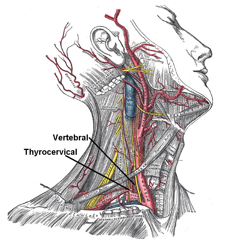
In human anatomy, the subclavian arteries are paired major arteries of the upper thorax, below the clavicle. They receive blood from the aortic arch. The left subclavian artery supplies blood to the left arm and the right subclavian artery supplies blood to the right arm, with some branches supplying the head and thorax. On the left side of the body, the subclavian comes directly off the aortic arch, while on the right side it arises from the relatively short brachiocephalic artery when it bifurcates into the subclavian and the right common carotid artery. The usual branches of the subclavian on both sides of the body are the vertebral artery, the internal thoracic artery, the thyrocervical trunk, the costocervical trunk and the dorsal scapular artery, which may branch off the transverse cervical artery, which is a branch of the thyrocervical trunk. The subclavian becomes the axillary artery at the lateral border of the first rib. Structure From its origin, the subclavian artery t ... [...More Info...] [...Related Items...] OR: [Wikipedia] [Google] [Baidu] |
Common Carotid Artery
In anatomy, the left and right common carotid arteries (carotids) ( in Merriam-Webster Online Dictionary '.) are that supply the head and neck with ; they divide in the neck to form the and |
_Victoria_blue-HE.jpg)


