|
Tetracosactide
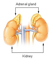
Adrenocorticotropic hormone is used as a medication and as Medical test#By method, diagnostic agent in the ACTH stimulation test. The form that is purified from pig pituitary glands is known as corticotropin is a medication and naturally occurring Peptide, polypeptide tropic hormone produced and secreted by the Anterior pituitary, anterior pituitary gland. The form that is made synthetically is tetracosactide, also known as synacthen, tetracosactrin and cosyntropin. It consists of the first 24 (of a total of 39) amino acids of ACTH and retains full function of the parent peptide. Tetracosactide stimulates the release of corticosteroids such as cortisol from the adrenal glands, and is used for the ACTH stimulation test to assess adrenal gland function. Medical uses Both corticotropin and tetracosactide have been used for diagnostic purposes to determine adrenocortical insufficiency, particularly in Addison's disease, via the ACTH stimulation test. However, as of 2015 the ... [...More Info...] [...Related Items...] OR: [Wikipedia] [Google] [Baidu] [Amazon] |
Addison's Disease
Addison's disease, also known as primary adrenal insufficiency, is a rare long-term endocrine disorder characterized by inadequate production of the steroid hormones cortisol and aldosterone by the two outer layers of the cells of the adrenal glands ( adrenal cortex), causing adrenal insufficiency. Symptoms generally develop slowly and insidiously and may include abdominal pain and gastrointestinal abnormalities, weakness, and weight loss. Darkening of the skin in certain areas may also occur. Under certain circumstances, an adrenal crisis may occur with low blood pressure, vomiting, lower back pain, and loss of consciousness. Mood changes may also occur. Rapid onset of symptoms indicates acute adrenal failure, which is a clinical emergency. An adrenal crisis can be triggered by stress, such as from an injury, surgery, or infection. Addison's disease arises when the adrenal gland does not produce sufficient amounts of the steroid hormones cortisol and (sometimes) a ... [...More Info...] [...Related Items...] OR: [Wikipedia] [Google] [Baidu] [Amazon] |
ACTH Stimulation Test
The ACTH test (also called the cosyntropin, tetracosactide, or Synacthen test) is a medical test usually requested and interpreted by endocrinologists to assess the functioning of the adrenal glands' stress response by measuring the adrenal response to adrenocorticotropic hormone (ACTH; corticotropin) or another corticotropic agent such as tetracosactide (cosyntropin, tetracosactrin; Synacthen) or alsactide (Synchrodyn). ACTH is a hormone produced in the anterior pituitary gland that stimulates the adrenal glands to release cortisol, dehydroepiandrosterone (DHEA), dehydroepiandrosterone sulfate (DHEA-S), and aldosterone. During the test, a small amount of synthetic ACTH is injected, and the amount of cortisol (and sometimes aldosterone) that the adrenals produce in response is measured. This test may cause mild side effects in some individuals. This test is used to diagnose or exclude primary and secondary adrenal insufficiency, Addison's disease, and related conditio ... [...More Info...] [...Related Items...] OR: [Wikipedia] [Google] [Baidu] [Amazon] |
Medical Test
A medical test is a medical procedure performed to detect, diagnose, or monitor diseases, disease processes, susceptibility, or to determine a course of treatment. Medical tests such as, physical and visual exams, diagnostic imaging, genetic testing, chemical and cellular analysis, relating to clinical chemistry and molecular diagnostics, are typically performed in a medical setting. Types of tests By purpose Medical tests can be classified by their purposes, including diagnosis, screening or monitoring. Diagnostic A diagnostic test is a procedure performed to confirm or determine the presence of disease in an individual suspected of having a disease, usually following the report of symptoms, or based on other medical test results. This includes posthumous diagnosis. Examples of such tests are: * Using nuclear medicine to examine a patient suspected of having a lymphoma. * Measuring the blood sugar in a person suspected of having diabetes mellitus after periods of in ... [...More Info...] [...Related Items...] OR: [Wikipedia] [Google] [Baidu] [Amazon] |
Edema
Edema (American English), also spelled oedema (British English), and also known as fluid retention, swelling, dropsy and hydropsy, is the build-up of fluid in the body's tissue (biology), tissue. Most commonly, the legs or arms are affected. Symptoms may include skin that feels tight, the area feeling heavy, and joint stiffness. Other symptoms depend on the underlying cause. Causes may include Chronic venous insufficiency, venous insufficiency, heart failure, kidney problems, hypoalbuminemia, low protein levels, liver problems, deep vein thrombosis, infections, kwashiorkor, angioedema, certain medications, and lymphedema. It may also occur in immobile patients (stroke, spinal cord injury, aging), or with temporary immobility such as prolonged sitting or standing, and during menstruation or pregnancy. The condition is more concerning if it starts suddenly, or pain or shortness of breath is present. Treatment depends on the underlying cause. If the underlying mechanism involve ... [...More Info...] [...Related Items...] OR: [Wikipedia] [Google] [Baidu] [Amazon] |
Sarcoidosis
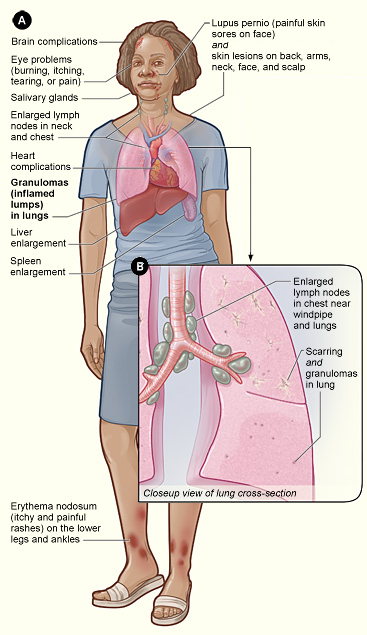
Sarcoidosis (; also known as Besnier–Boeck–Schaumann disease) is a disease involving abnormal collections of White blood cell, inflammatory cells that form lumps known as granulomata. The disease usually begins in the lungs, skin, or lymph nodes. Less commonly affected are the eyes, liver, heart, and Human brain, brain, though any Organ (anatomy), organ can be affected. The signs and symptoms depend on the organ involved. Often, no symptoms or only mild symptoms are seen. When it affects the lungs, wheezing, coughing, shortness of breath, or chest pain may occur. Some may have Löfgren syndrome with fever, Bilateral hilar lymphadenopathy, enlarged hilar lymph nodes, arthritis, and a rash known as erythema nodosum. The cause of sarcoidosis is unknown. Some believe it may be due to an immune reaction to a trigger such as an infection or chemicals in those who are genetically predisposed. Those with affected family members are at greater risk. Diagnosis is partly based on signs ... [...More Info...] [...Related Items...] OR: [Wikipedia] [Google] [Baidu] [Amazon] |
Chorioretinitis
Chorioretinitis is an inflammation of the choroid (thin pigmented vascular coat of the eye) and retina of the eye. It is a form of posterior uveitis. Inflammation of these layers can lead to vision-threatening complications. If only the choroid is inflamed, not the retina, the condition is termed choroiditis. The ophthalmologist's goal in treating these potentially blinding conditions is to eliminate the inflammation and minimize the potential risk of therapy to the patient. Symptoms Symptoms may include the presence of floating black spots, blurred vision, pain or redness in the eye, sensitivity to light, or excessive tearing. Causes Chorioretinitis is often caused by toxoplasmosis and cytomegalovirus infections (mostly seen in immunodeficient subjects such as people with HIV/AIDS or on immunosuppressant drugs). Congenital toxoplasmosis via transplacental transmission can also lead to sequelae such as chorioretinitis along with hydrocephalus and cerebral calcifications. Other ... [...More Info...] [...Related Items...] OR: [Wikipedia] [Google] [Baidu] [Amazon] |
Optic Neuritis
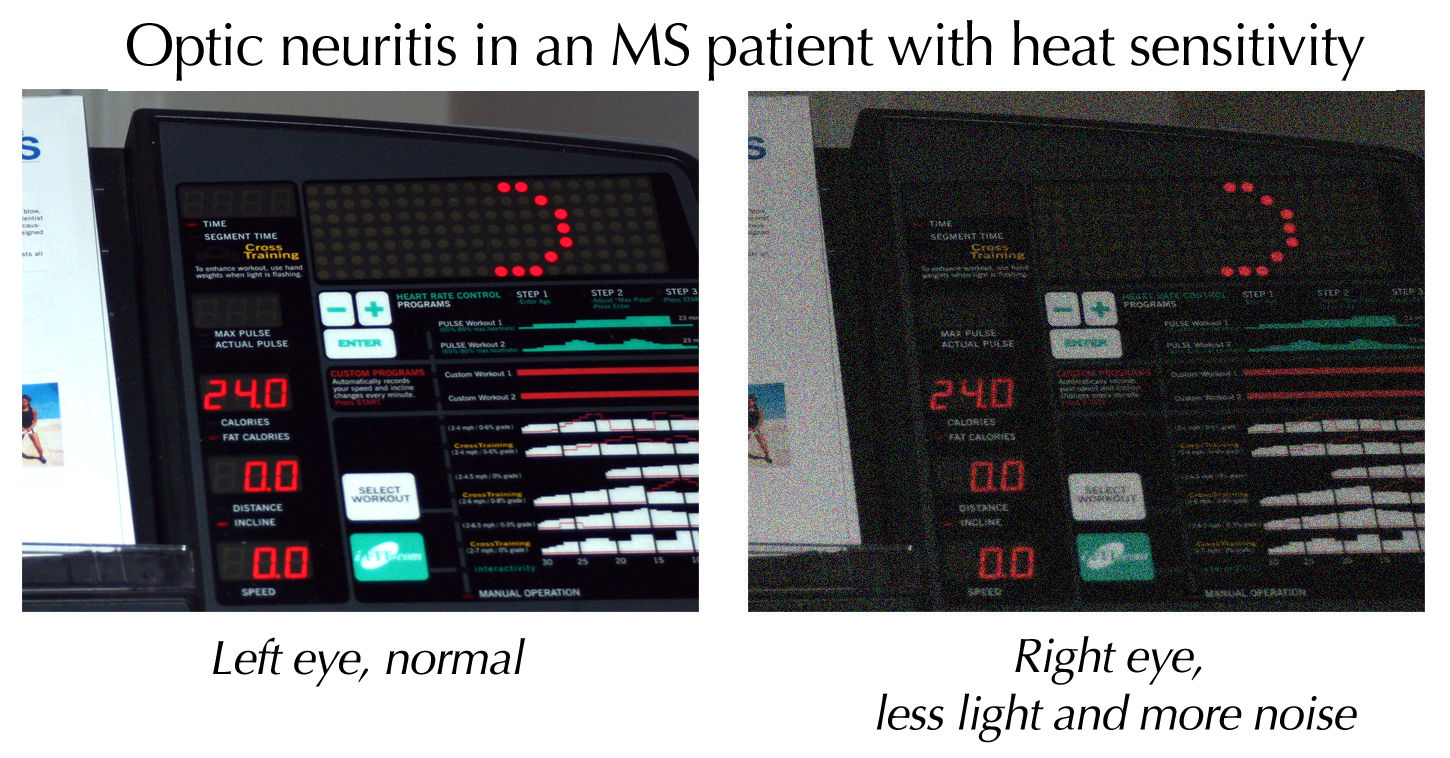
Optic neuritis (ON) is a debilitating condition that is defined as inflammation of cranial nerve II which results in disruption of the neurologic pathways that allow visual sensory information received by the retina to be able to be transmitted to the visual cortex of the brain. This disorder of the optic nerve may arise through various pathophysiologic mechanisms, such as through Demyelinating disease, demyelination or inflammation, leading to partial or total loss of vision. Optic neuritis may be a result of standalone idiopathic disease, but is often a manifestation that occurs secondary to an underlying disease. Signs of ON classically present as sudden-onset visual impairment in one or both eyes that can range in severity from mild visual blurring to complete blindness in the affected eye(s). Although pain is typically considered a hallmark feature of optic neuritis, the absence of pain does not preclude a diagnosis or consideration of ON as some patients may report painlessne ... [...More Info...] [...Related Items...] OR: [Wikipedia] [Google] [Baidu] [Amazon] |
Choroiditis
Chorioretinitis is an inflammation of the choroid (thin pigmented vascular coat of the eye) and retina of the eye. It is a form of posterior uveitis. Inflammation of these layers can lead to vision-threatening complications. If only the choroid is inflamed, not the retina, the condition is termed choroiditis. The ophthalmologist's goal in treating these potentially blinding conditions is to eliminate the inflammation and minimize the potential risk of therapy to the patient. Symptoms Symptoms may include the presence of floating black spots, blurred vision, pain or redness in the eye, sensitivity to light, or excessive tearing. Causes Chorioretinitis is often caused by toxoplasmosis and cytomegalovirus infections (mostly seen in immunodeficient subjects such as people with HIV/AIDS or on immunosuppressant drugs). Congenital toxoplasmosis via transplacental transmission can also lead to sequelae such as chorioretinitis along with hydrocephalus and cerebral calcifications. Other ... [...More Info...] [...Related Items...] OR: [Wikipedia] [Google] [Baidu] [Amazon] |
Uveitis
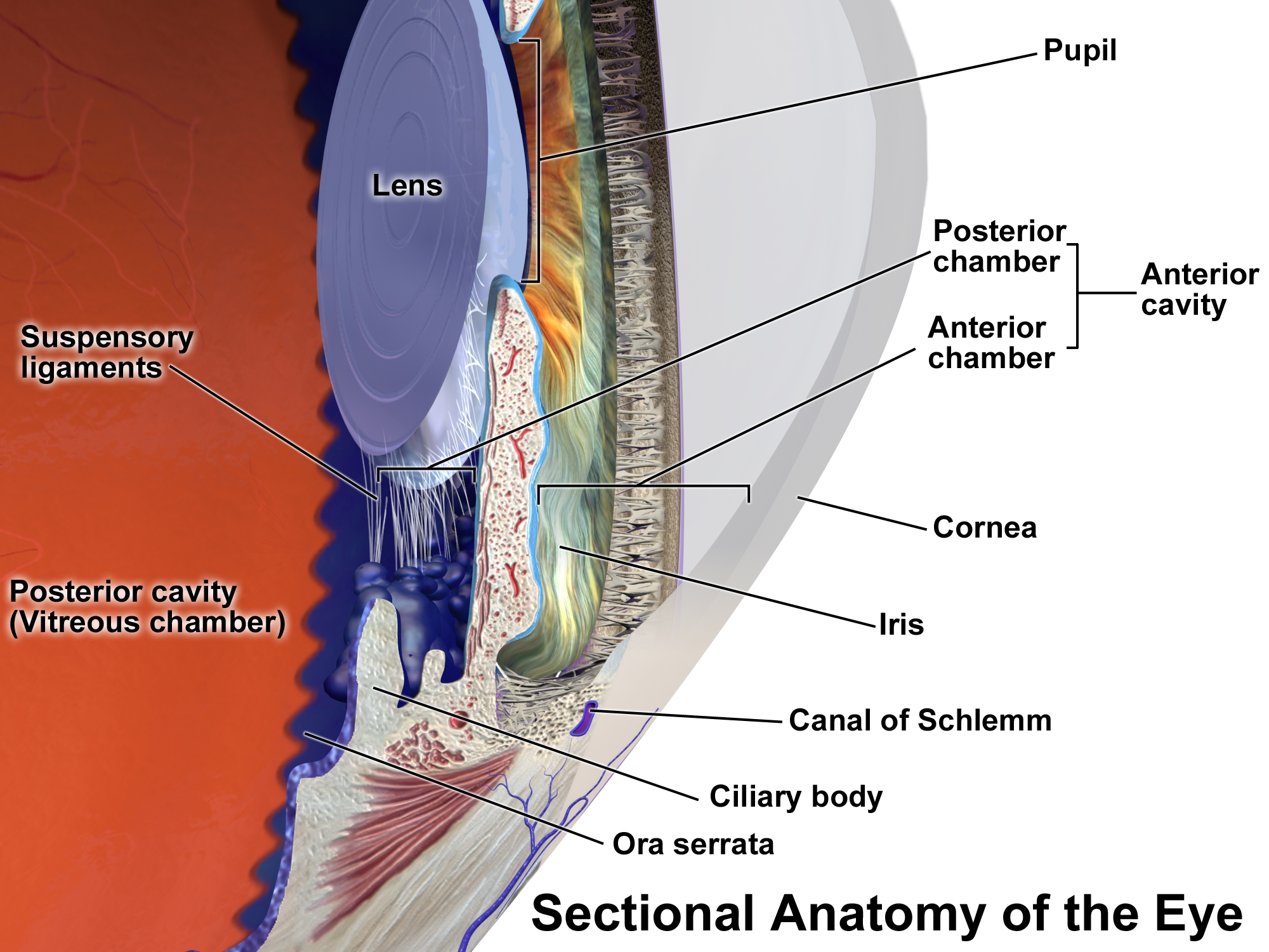
Uveitis () is inflammation of the uvea, the pigmented layer of the eye between the inner retina and the outer fibrous layer composed of the sclera and cornea. The uvea consists of the middle layer of pigmented vascular structures of the eye and includes the iris, ciliary body, and choroid. Uveitis is described anatomically, by the part of the eye affected, as anterior, intermediate or posterior, or panuveitic if all parts are involved. Anterior uveitis ( iridocyclitis) is the most common, with the incidence of uveitis overall affecting approximately 1:4500, most commonly those between the ages of 20–60. Symptoms include eye pain, eye redness, floaters and blurred vision, and ophthalmic examination may show dilated ciliary blood vessels and the presence of cells in the anterior chamber. Uveitis may arise spontaneously, have a genetic component, or be associated with an autoimmune disease or infection. While the eye is a relatively protected environment, its immune mecha ... [...More Info...] [...Related Items...] OR: [Wikipedia] [Google] [Baidu] [Amazon] |
Iridocyclitis
Uveitis () is inflammation of the uvea, the pigmented layer of the eye between the inner retina and the outer fibrous layer composed of the sclera and cornea. The uvea consists of the middle layer of pigmented vascular structures of the eye and includes the iris, ciliary body, and choroid. Uveitis is described anatomically, by the part of the eye affected, as anterior, intermediate or posterior, or panuveitic if all parts are involved. Anterior uveitis ( iridocyclitis) is the most common, with the incidence of uveitis overall affecting approximately 1:4500, most commonly those between the ages of 20–60. Symptoms include eye pain, eye redness, floaters and blurred vision, and ophthalmic examination may show dilated ciliary blood vessels and the presence of cells in the anterior chamber. Uveitis may arise spontaneously, have a genetic component, or be associated with an autoimmune disease or infection. While the eye is a relatively protected environment, its immune mechanisms ... [...More Info...] [...Related Items...] OR: [Wikipedia] [Google] [Baidu] [Amazon] |
Keratitis
Keratitis is a condition in which the human eye, eye's cornea, the clear dome on the front surface of the eye, becomes inflammation, inflamed. The condition is often marked by moderate to intense pain and usually involves any of the following symptoms: pain, impaired eyesight, photophobia (light sensitivity), Red eye (medicine), red eye and a 'gritty' sensation. Diagnosis of infectious keratitis is usually made clinically based on the signs and symptoms as well as eye examination, but corneal scrapings may be obtained and evaluated using microbiological culture or other testing to identify the causative pathogen. Classification (by chronicity) Acute * Acute epithelial keratitis * Nummular keratitis * Interstitial keratitis * Disciform keratitis Chronic * Neurotrophic keratitis * Mucous plaque keratitis Classification (infective) Viral The most common causes of viral keratitis include herpes simplex virus (HSV) and varicella zoster virus (VZV), which cause herpes of the eye ... [...More Info...] [...Related Items...] OR: [Wikipedia] [Google] [Baidu] [Amazon] |


