|
Tectorial Membrane
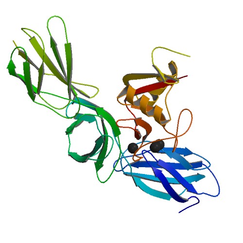
The tectoria membrane (TM) is one of two acellular membranes in the cochlea of the inner ear, the other being the basilar membrane (BM). "Tectorial" in anatomy means forming a cover. The TM is located above the spiral limbus and the spiral organ of Corti and extends along the longitudinal length of the cochlea parallel to the BM. Radially the TM is divided into three zones, the limbal, middle and marginal zones. Of these the limbal zone is the thinnest (transversally) and overlies the auditory teeth of Huschke with its inside edge attached to the spiral limbus. The marginal zone is the thickest (transversally) and is divided from the middle zone by Hensen's Stripe. It overlies the sensory inner hair cells and electrically-motile outer hair cells of the organ of Corti and during acoustic stimulation stimulates the inner hair cells through fluid coupling, and the outer hair cells via direct connection to their tallest stereocilia. Structure The TM is a gel-like structure containing ... [...More Info...] [...Related Items...] OR: [Wikipedia] [Google] [Baidu] |
Spiral Organ Of Corti
The organ of Corti, or spiral organ, is the receptor organ for hearing and is located in the mammalian cochlea. This highly varied strip of epithelial cells allows for transduction of auditory signals into nerve impulses' action potential. Transduction occurs through vibrations of structures in the inner ear causing displacement of cochlear fluid and movement of hair cells at the organ of Corti to produce electrochemical signals.The Ear Pujol, R., Irving, S., 2013 Italian anatomist (1822–1876) discovered the organ of Corti in 1851. The structure evolved from the [...More Info...] [...Related Items...] OR: [Wikipedia] [Google] [Baidu] |
Cochlea
The cochlea is the part of the inner ear involved in hearing. It is a spiral-shaped cavity in the bony labyrinth, in humans making 2.75 turns around its axis, the modiolus. A core component of the cochlea is the Organ of Corti, the sensory organ of hearing, which is distributed along the partition separating the fluid chambers in the coiled tapered tube of the cochlea. The name cochlea derives . Structure The cochlea (plural is cochleae) is a spiraled, hollow, conical chamber of bone, in which waves propagate from the base (near the middle ear and the oval window) to the apex (the top or center of the spiral). The spiral canal of the cochlea is a section of the bony labyrinth of the inner ear that is approximately 30 mm long and makes 2 turns about the modiolus. The cochlear structures include: * Three ''scalae'' or chambers: ** the vestibular duct or ''scala vestibuli'' (containing perilymph), which lies superior to the cochlear duct and abuts the oval window ** the ty ... [...More Info...] [...Related Items...] OR: [Wikipedia] [Google] [Baidu] |
Basilar Membrane
The basilar membrane is a stiff structural element within the cochlea of the inner ear which separates two liquid-filled tubes that run along the coil of the cochlea, the scala media and the scala tympani. The basilar membrane moves up and down in response to incoming sound waves, which are converted to traveling waves on the basilar membrane. Structure The basilar membrane is a pseudo-resonant structure that, like the strings on an instrument, varies in width and stiffness. But unlike the parallel strings of a guitar, the basilar membrane is not a discrete set of resonant structures, but a single structure with varying width, stiffness, mass, damping, and duct dimensions along its length. The motion of the basilar membrane is generally described as a traveling wave. The properties of the membrane at a given point along its length determine its characteristic frequency (CF), the frequency at which it is most sensitive to sound vibrations. The basilar membrane is widest (0.42– ... [...More Info...] [...Related Items...] OR: [Wikipedia] [Google] [Baidu] |
Spiral Limbus
The osseous spiral lamina consists of two plates of bone, and between these are the canals for the transmission of the filaments of the acoustic nerve. On the upper plate of that part of the lamina which is outside the vestibular membrane, the periosteum The periosteum is a membrane that covers the outer surface of all bones, except at the articular surfaces (i.e. the parts within a joint space) of long bones. Endosteum lines the inner surface of the medullary cavity of all long bones. Structure ... is thickened to form the limbus spiralis (or limbus laminæ spiralis), this ends externally in a concavity, the sulcus spiralis internus, which represents, on section, the form of the letter C. References External links * http://www.anatomyatlases.org/MicroscopicAnatomy/Section16/Plate16311.shtml * http://www.med.uiuc.edu/histo/small/atlas/objects/103.htm Ear {{Anatomy-stub ... [...More Info...] [...Related Items...] OR: [Wikipedia] [Google] [Baidu] |
Auditory Teeth Of Huschke
Auditory means of or relating to the process of hearing: * Auditory system, the neurological structures and pathways of sound perception ** Auditory bulla, part of auditory system found in mammals other than primates ** Auditory nerve, also known as the cochlear nerve is one of two parts of a cranial nerve ** Auditory ossicles, three bones in the middle ear that transmit sounds * Hearing (sense), the auditory sense, the sense by which sound is perceived * Ear, the auditory end organ * Cochlea, the auditory branch of the inner ear * Sound, the physical signal perceived by the auditory system * External auditory meatus, the ear canal * Primary auditory cortex, the part of the higher-level of the brain that serves hearing * Auditory agnosia * Auditory exclusion, a form of temporary hearing loss under high stress * Auditory feedback, an aid to control speech production and singing * Auditory hallucination, perceiving sounds without auditory stimulus * Auditory illusion, sound trick ... [...More Info...] [...Related Items...] OR: [Wikipedia] [Google] [Baidu] |
Inner Hair Cells
Hair cells are the sensory receptors of both the auditory system and the vestibular system in the ears of all vertebrates, and in the lateral line organ of fishes. Through mechanotransduction, hair cells detect movement in their environment. In mammals, the auditory hair cells are located within the spiral organ of Corti on the thin basilar membrane in the cochlea of the inner ear. They derive their name from the tufts of stereocilia called ''hair bundles'' that protrude from the apical surface of the cell into the fluid-filled cochlear duct. The stereocilia number from 50-100 in each cell while being tightly packed together and decrease in size the further away they are located from the kinocilium. The hair bundles are arranged as stiff columns that move at their base in response to stimuli applied to the tips. Mammalian cochlear hair cells are of two anatomically and functionally distinct types, known as outer, and inner hair cells. Damage to these hair cells results in ... [...More Info...] [...Related Items...] OR: [Wikipedia] [Google] [Baidu] |
Outer Hair Cells
Hair cells are the sensory receptors of both the auditory system and the vestibular system in the ears of all vertebrates, and in the lateral line organ of fishes. Through mechanotransduction, hair cells detect movement in their environment. In mammals, the auditory hair cells are located within the spiral organ of Corti on the thin basilar membrane in the cochlea of the inner ear. They derive their name from the tufts of stereocilia called ''hair bundles'' that protrude from the apical surface of the cell into the fluid-filled cochlear duct. The stereocilia number from 50-100 in each cell while being tightly packed together and decrease in size the further away they are located from the kinocilium. The hair bundles are arranged as stiff columns that move at their base in response to stimuli applied to the tips. Mammalian cochlear hair cells are of two anatomically and functionally distinct types, known as outer, and inner hair cells. Damage to these hair cells results ... [...More Info...] [...Related Items...] OR: [Wikipedia] [Google] [Baidu] |
Collagen
Collagen () is the main structural protein in the extracellular matrix found in the body's various connective tissues. As the main component of connective tissue, it is the most abundant protein in mammals, making up from 25% to 35% of the whole-body protein content. Collagen consists of amino acids bound together to form a triple helix of elongated fibril known as a collagen helix. It is mostly found in connective tissue such as cartilage, bones, tendons, ligaments, and skin. Depending upon the degree of mineralization, collagen tissues may be rigid (bone) or compliant (tendon) or have a gradient from rigid to compliant (cartilage). Collagen is also abundant in corneas, blood vessels, the gut, intervertebral discs, and the dentin in teeth. In muscle tissue, it serves as a major component of the endomysium. Collagen constitutes one to two percent of muscle tissue and accounts for 6% of the weight of the skeletal muscle tissue. The fibroblast is the most common cell that crea ... [...More Info...] [...Related Items...] OR: [Wikipedia] [Google] [Baidu] |
Glycoprotein
Glycoproteins are proteins which contain oligosaccharide chains covalently attached to amino acid side-chains. The carbohydrate is attached to the protein in a cotranslational or posttranslational modification. This process is known as glycosylation. Secreted extracellular proteins are often glycosylated. In proteins that have segments extending extracellularly, the extracellular segments are also often glycosylated. Glycoproteins are also often important integral membrane proteins, where they play a role in cell–cell interactions. It is important to distinguish endoplasmic reticulum-based glycosylation of the secretory system from reversible cytosolic-nuclear glycosylation. Glycoproteins of the cytosol and nucleus can be modified through the reversible addition of a single GlcNAc residue that is considered reciprocal to phosphorylation and the functions of these are likely to be an additional regulatory mechanism that controls phosphorylation-based signalling. In contrast, ... [...More Info...] [...Related Items...] OR: [Wikipedia] [Google] [Baidu] |
Proteoglycan
Proteoglycans are proteins that are heavily glycosylated. The basic proteoglycan unit consists of a "core protein" with one or more covalently attached glycosaminoglycan (GAG) chain(s). The point of attachment is a serine (Ser) residue to which the glycosaminoglycan is joined through a tetrasaccharide bridge (e.g. chondroitin sulfate- GlcA- Gal-Gal- Xyl-PROTEIN). The Ser residue is generally in the sequence -Ser-Gly-X-Gly- (where X can be any amino acid residue but proline), although not every protein with this sequence has an attached glycosaminoglycan. The chains are long, linear carbohydrate polymers that are negatively charged under physiological conditions due to the occurrence of sulfate and uronic acid groups. Proteoglycans occur in connective tissue. Types Proteoglycans are categorized by their relative size (large and small) and the nature of their glycosaminoglycan chains. Types include: Certain members are considered members of the "small leucine-rich proteoglyc ... [...More Info...] [...Related Items...] OR: [Wikipedia] [Google] [Baidu] |
TECTA
Alpha-tectorin is a protein that in humans is encoded by the ''TECTA'' gene. The tectorial membrane is an apical extracellular matrix (aECM) of the inner ear that contacts the stereocilia bundles of specialized sensory hair cells. Sound induces movement of these hair cells relative to the tectorial membrane, deflects the stereocilia, and leads to fluctuations in hair-cell membrane potential, transducing sound into electrical signals. Alpha-tectorin is one of the major noncollagenous components of the tectorial membrane. Mutations in the TECTA gene have been shown to be responsible for autosomal dominant In genetics, dominance is the phenomenon of one variant (allele) of a gene on a chromosome masking or overriding the effect of a different variant of the same gene on the other copy of the chromosome. The first variant is termed dominant and t ... nonsyndromic hearing impairment and a recessive form of sensorineural pre-lingual non-syndromic deafness. References Further ... [...More Info...] [...Related Items...] OR: [Wikipedia] [Google] [Baidu] |
Anisotropic
Anisotropy () is the property of a material which allows it to change or assume different properties in different directions, as opposed to isotropy. It can be defined as a difference, when measured along different axes, in a material's physical or mechanical properties (absorbance, refractive index, conductivity, tensile strength, etc.). An example of anisotropy is light coming through a polarizer. Another is wood, which is easier to split along its grain than across it. Fields of interest Computer graphics In the field of computer graphics, an anisotropic surface changes in appearance as it rotates about its geometric normal, as is the case with velvet. Anisotropic filtering (AF) is a method of enhancing the image quality of textures on surfaces that are far away and steeply angled with respect to the point of view. Older techniques, such as bilinear and trilinear filtering, do not take into account the angle a surface is viewed from, which can result in aliasing or bl ... [...More Info...] [...Related Items...] OR: [Wikipedia] [Google] [Baidu] |


