|
Trabecular Cartilage
Trabecular cartilages (trabeculae cranii, sometimes simply trabeculae, prechordal cartilages) are paired, rod-shaped cartilages, which develop in the head of the vertebrate embryo. They are the primordia of the anterior part of the cranial base, and are derived from the cranial neural crest cells. Structure The trabecular cartilages generally appear as a paired, rod-shaped cartilages at the ventral side of the forebrain and lateral side of the adenohypophysis in the vertebrate embryo. During development, their anterior ends fuse and form the ''trabecula communis''. Their posterior ends fuse with the caudal-most parachordal cartilages. Development Most skeletons are of mesodermal origin in vertebrates. Especially axial skeletal elements, such as the vertebrae, are derived from the paraxial mesoderm (e.g., somites), which is regulated by molecular signals from the notochord. Trabecular cartilages, however, originate from the neural crest, and since they are located anterior to t ... [...More Info...] [...Related Items...] OR: [Wikipedia] [Google] [Baidu] |
Head
A head is the part of an organism which usually includes the ears, brain, forehead, cheeks, chin, eyes, nose, and mouth, each of which aid in various sensory functions such as sight, hearing, smell, and taste. Some very simple animals may not have a head, but many bilaterally symmetric forms do, regardless of size. Heads develop in animals by an evolutionary trend known as cephalization. In bilaterally symmetrical animals, nervous tissue concentrate at the anterior region, forming structures responsible for information processing. Through biological evolution, sense organs and feeding structures also concentrate into the anterior region; these collectively form the head. Human head The human head is an anatomical unit that consists of the Human skull, skull, hyoid bone and cervical vertebrae. The term "skull" collectively denotes the mandible (lower jaw bone) and the cranium (upper portion of the skull that houses the brain). Sculptures of human heads are general ... [...More Info...] [...Related Items...] OR: [Wikipedia] [Google] [Baidu] |
Mesenchyme
Mesenchyme () is a type of loosely organized animal embryonic connective tissue of undifferentiated cells that give rise to most tissues, such as skin, blood or bone. The interactions between mesenchyme and epithelium help to form nearly every organ in the developing embryo. Vertebrates Structure Mesenchyme is characterized morphologically by a prominent ground substance matrix containing a loose aggregate of reticular fibers and unspecialized mesenchymal stem cells. Mesenchymal cells can migrate easily (in contrast to epithelial cells, which lack mobility), are organized into closely adherent sheets, and are polarized in an apical-basal orientation. Development The mesenchyme originates from the mesoderm. From the mesoderm, the mesenchyme appears as an embryologically primitive "soup". This "soup" exists as a combination of the mesenchymal cells plus serous fluid plus the many different tissue proteins. Serous fluid is typically stocked with the many serous elements, such a ... [...More Info...] [...Related Items...] OR: [Wikipedia] [Google] [Baidu] |
Gavin De Beer
Sir Gavin Rylands de Beer (1 November 1899 – 21 June 1972) was a British evolutionary embryologist, known for his work on heterochrony as recorded in his 1930 book ''Embryos and Ancestors''. He was director of the Natural History Museum, London, president of the Linnean Society of London, and a winner of the Royal Society's Darwin Medal for his studies on evolution. Biography Born on 1 November 1899 in Malden, Surrey (now part of London), de Beer spent most of his childhood in France, where he was educated at the Parisian École Pascal. During this time, he also visited Switzerland, a country with which he remained fascinated for the rest of his life. His education continued at Harrow and Magdalen College, Oxford, where he graduated with a degree in zoology in 1921, after a pause to serve in the First World War in the Grenadier Guards and the Army Education Corps. In 1923 he was made a fellow of Merton College, Oxford, and began to teach at the university's zoology depart ... [...More Info...] [...Related Items...] OR: [Wikipedia] [Google] [Baidu] |
Edwin Stephen Goodrich
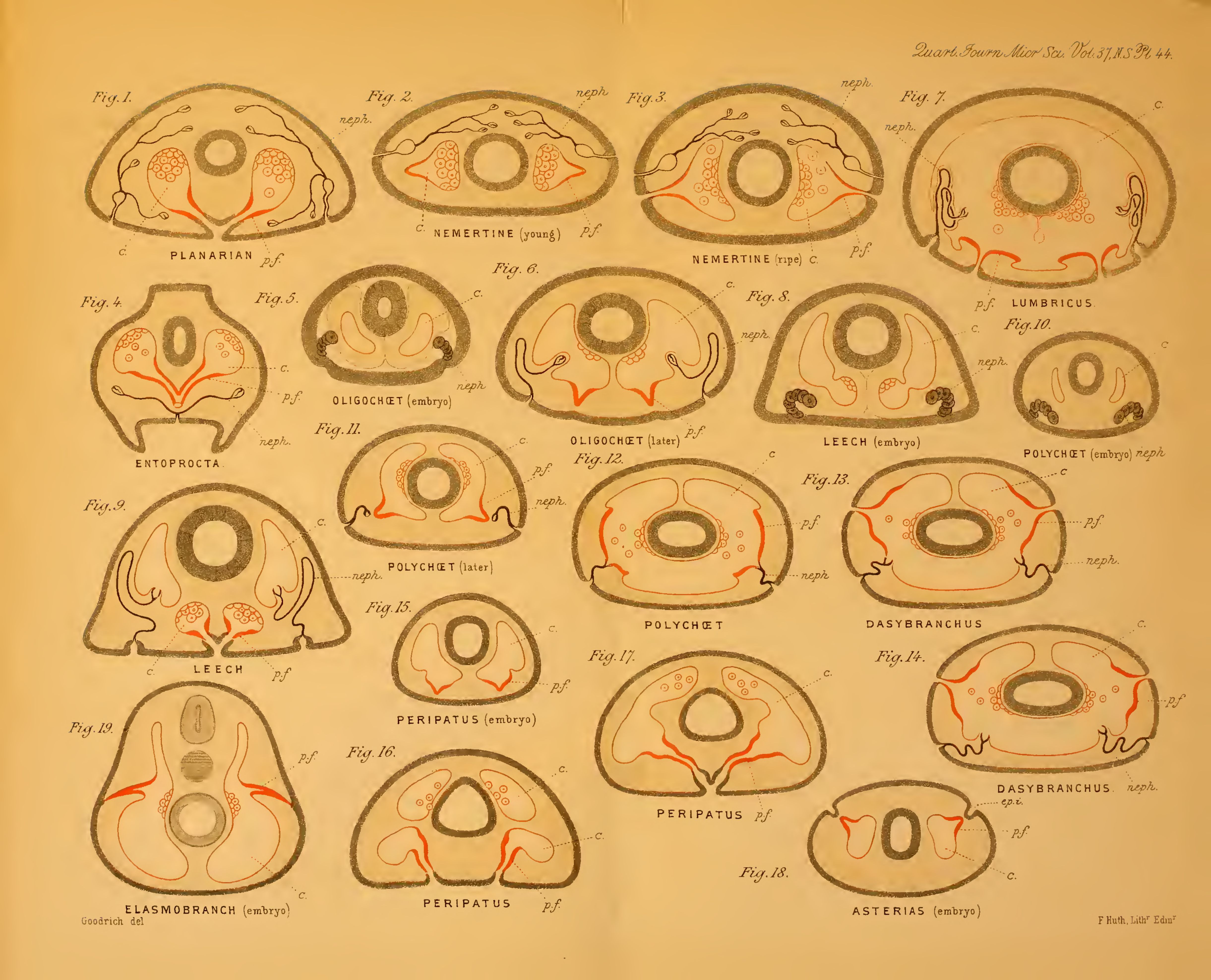
Edwin Stephen Goodrich FRS (Weston-super-Mare, 21 June 1868 – Oxford, 6 January 1946), was an English zoologist, specialising in comparative anatomy, embryology, palaeontology, and evolution. He held the Linacre Chair of Zoology in the University of Oxford from 1921 to 1946. He served as editor of the ''Quarterly Journal of Microscopical Science'' from 1920 until his death. Life Goodrich's father died when he was only two weeks old, and his mother took her children to live with her mother at Pau, France, where he attended the local English school and a French lycée. In 1888 he entered the Slade School of Art at University College London; there he met E. Ray Lankester, who interested him in zoology. On coming to Oxford from London, Goodrich entered Merton College, Oxford as an undergraduate in 1891 and, while acting as assistant to Lankester, read for the final honour school in Zoology; he was awarded the Rolleston Memorial Prize in 1894 and graduated with first-class hon ... [...More Info...] [...Related Items...] OR: [Wikipedia] [Google] [Baidu] |
Mandibular Arch
The pharyngeal arches, also known as visceral arches'','' are structures seen in the embryonic development of vertebrates that are recognisable precursors for many structures. In fish, the arches are known as the branchial arches, or gill arches. In the human embryo, the arches are first seen during the fourth week of development. They appear as a series of outpouchings of mesoderm on both sides of the developing pharynx. The vasculature of the pharyngeal arches is known as the aortic arches. In fish, the branchial arches support the gills. Structure In vertebrates, the pharyngeal arches are derived from all three germ layers (the primary layers of cells that form during embryogenesis). Neural crest cells enter these arches where they contribute to features of the skull and facial skeleton such as bone and cartilage. However, the existence of pharyngeal structures before neural crest cells evolved is indicated by the existence of neural crest-independent mechanisms of pharyng ... [...More Info...] [...Related Items...] OR: [Wikipedia] [Google] [Baidu] |
Pharyngeal Arches
The pharyngeal arches, also known as visceral arches'','' are structures seen in the embryonic development of vertebrates that are recognisable precursors for many structures. In fish, the arches are known as the branchial arches, or gill arches. In the human embryo, the arches are first seen during the fourth week of development. They appear as a series of outpouchings of mesoderm on both sides of the developing pharynx. The vasculature of the pharyngeal arches is known as the aortic arches. In fish, the branchial arches support the gills. Structure In vertebrates, the pharyngeal arches are derived from all three germ layers (the primary layers of cells that form during embryogenesis). Neural crest cells enter these arches where they contribute to features of the skull and facial skeleton such as bone and cartilage. However, the existence of pharyngeal structures before neural crest cells evolved is indicated by the existence of neural crest-independent mechanisms of pharyng ... [...More Info...] [...Related Items...] OR: [Wikipedia] [Google] [Baidu] |
Splanchnocranium
The splanchnocranium (or visceral skeleton) is the portion of the cranium that is derived from pharyngeal arches. ''Splanchno'' indicates to the gut because the face forms around the mouth, which is an end of the gut. The splanchnocranium consists of cartilage and endochondral bone. In mammals, the splanchnocranium comprises the three ear ossicles (i.e., incus, malleus, and stapes), as well as the alisphenoid, the styloid process, the hyoid apparatus, and the thyroid cartilage. In other tetrapods, such as amphibians and reptiles, homologous bones to those of mammals, such as the quadrate, articular, columella, and entoglossus are part of the splanchnocranium. See also * Dermatocranium * Endocranium The endocranium in comparative anatomy is a part of the skull base in vertebrates and it represents the basal, inner part of the cranium. The term is also applied to the outer layer of the dura mater in human anatomy. Structure Structurally, t ... References {{anatomy-stub ... [...More Info...] [...Related Items...] OR: [Wikipedia] [Google] [Baidu] |
Thomas Henry Huxley
Thomas Henry Huxley (4 May 1825 – 29 June 1895) was an English biologist and anthropologist specialising in comparative anatomy. He has become known as "Darwin's Bulldog" for his advocacy of Charles Darwin's theory of evolution. The stories regarding Huxley's famous 1860 Oxford evolution debate with Samuel Wilberforce were a key moment in the wider acceptance of evolution and in his own career, although some historians think that the surviving story of the debate is a later fabrication. Huxley had been planning to leave Oxford on the previous day, but, after an encounter with Robert Chambers, the author of '' Vestiges'', he changed his mind and decided to join the debate. Wilberforce was coached by Richard Owen, against whom Huxley also debated about whether humans were closely related to apes. Huxley was slow to accept some of Darwin's ideas, such as gradualism, and was undecided about natural selection, but despite this he was wholehearted in his public support of Darw ... [...More Info...] [...Related Items...] OR: [Wikipedia] [Google] [Baidu] |
Grass Snake
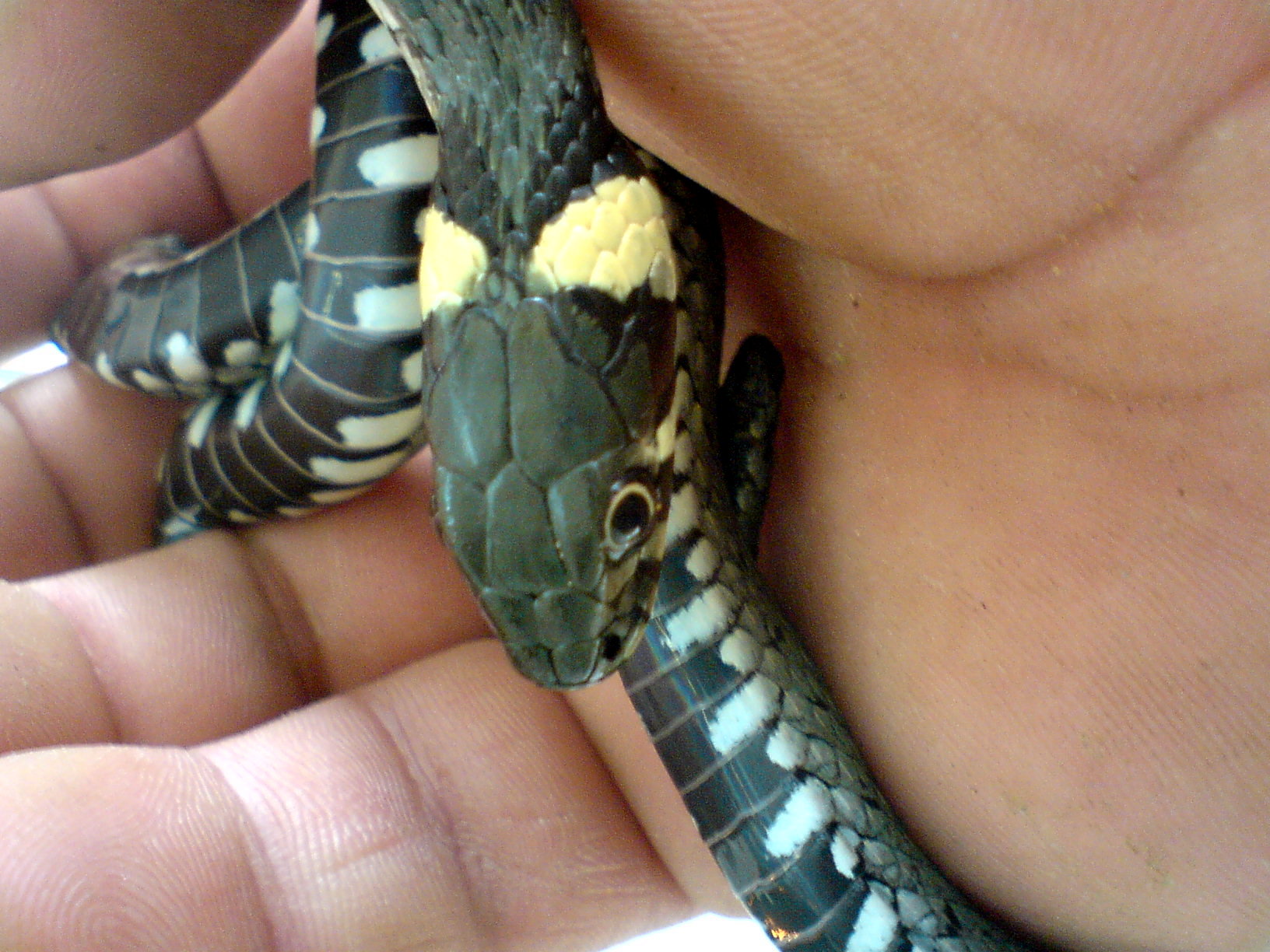
The grass snake (''Natrix natrix''), sometimes called the ringed snake or water snake, is a Eurasian non-venomous colubrid snake. It is often found near water and feeds almost exclusively on amphibians. Subspecies Many subspecies are recognized, including: ''Natrix natrix helvetica'' ( Lacépède, 1789) was formerly treated as a subspecies, but following genetic analysis it was recognised in August 2017 as a separate species, ''Natrix helvetica'', the barred grass snake. Four other subspecies were transferred from ''N. natrix'' to ''N. helvetica'', becoming ''N. helvetica cettii'', ''N. helvetica corsa'', ''N. helvetica lanzai'' and ''N. helvetica sicula''. Description The grass snake is typically dark green or brown in colour with a characteristic yellow or whitish collar behind the head, which explains the alternative name ringed snake. The colour may also range from grey to black, with darker colours being more prevalent in colder regions, ... [...More Info...] [...Related Items...] OR: [Wikipedia] [Google] [Baidu] |
Gnathostomata
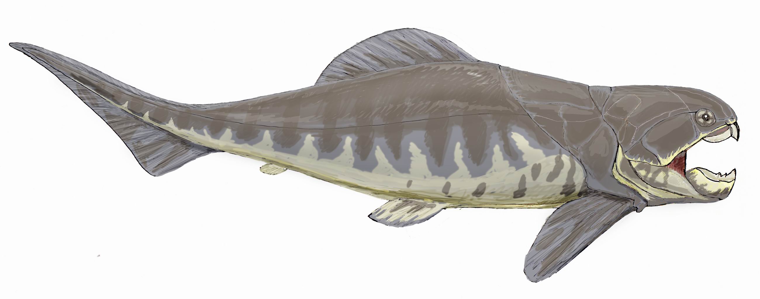
Gnathostomata (; from Greek: (') "jaw" + (') "mouth") are the jawed vertebrates. Gnathostome diversity comprises roughly 60,000 species, which accounts for 99% of all living vertebrates, including humans. In addition to opposing jaws, living gnathostomes have true teeth (a characteristic which has subsequently been lost in some), paired appendages (pectoral and pelvic fins, arms, legs, wings, etc.), the elastomeric protein of elastin, and a horizontal semicircular canal of the inner ear, along with physiological and cellular anatomical characters such as the myelin sheaths of neurons, and an adaptive immune system that has the discrete lymphoid organs of spleen and thymus, and uses V(D)J recombination to create antigen recognition sites, rather than using genetic recombination in the variable lymphocyte receptor gene. It is now assumed that Gnathostomata evolved from ancestors that already possessed a pair of both pectoral and pelvic fins. Until recently these ancestors, know ... [...More Info...] [...Related Items...] OR: [Wikipedia] [Google] [Baidu] |


