|
Thoracic Duct
In human anatomy, the thoracic duct (also known as the ''left lymphatic duct'', ''alimentary duct'', ''chyliferous duct'', and ''Van Hoorne's canal'') is the larger of the two lymph ducts of the lymphatic system (the other being the right lymphatic duct). The thoracic duct usually begins from the upper aspect of the cisterna chyli, passing out of the abdomen through the aortic hiatus into first the posterior mediastinum and then the superior mediastinum, extending as high up as the root of the neck before descending to drain into the systemic (blood) circulation at the venous angle. The thoracic duct carries chyle, a liquid containing both lymph and emulsified fats, rather than pure lymph. It also collects most of the lymph in the body other than from the right thorax, arm, head, and neck (which are drained by the right lymphatic duct). When the duct ruptures, the resulting flood of liquid into the pleural cavity is known as chylothorax. Structure In adults, the thoraci ... [...More Info...] [...Related Items...] OR: [Wikipedia] [Google] [Baidu] |
Cisterna Chyli
The cisterna chyli or receptaculum chyli (chy·li pronounced: ˈkī-ˌlī) is a dilated sac at the lower end of the thoracic duct in most mammals into which lymph from the intestinal trunk and two lumbar lymphatic trunks flow. It receives fatty chyle from the intestines and thus acts as a conduit for the lipid products of digestion. It is the most common drainage trunk of most of the body's lymphatics. The cisterna chyli is a retroperitoneal structure. Structure In humans, the cisterna chyli is located posterior to the abdominal aorta on the anterior aspect of the bodies of the first and second lumbar vertebrae (L1 and L2). There it forms the beginning of the primary lymph vessel, the thoracic duct, which transports lymph and chyle from the abdomen via the aortic opening of the diaphragm up to the junction of left subclavian vein and internal jugular veins. Other animals In dogs, the cisterna chyli is located to the left and often ventral to the aorta; in cats it is left a ... [...More Info...] [...Related Items...] OR: [Wikipedia] [Google] [Baidu] |
Neck
The neck is the part of the body in many vertebrates that connects the head to the torso. It supports the weight of the head and protects the nerves that transmit sensory and motor information between the brain and the rest of the body. Additionally, the neck is highly flexible, allowing the head to turn and move in all directions. Anatomically, the human neck is divided into four compartments: vertebral, visceral, and two vascular compartments. Within these compartments, the neck houses the cervical vertebrae, the cervical portion of the spinal cord, upper parts of the respiratory and digestive tracts, endocrine glands, nerves, arteries and veins. The muscles of the neck, which are separate from the compartments, form the boundaries of the neck triangles. In anatomy, the neck is also referred to as the or . However, when the term ''cervix'' is used alone, it often refers to the uterine cervix, the neck of the uterus. Therefore, the adjective ''cervical'' ... [...More Info...] [...Related Items...] OR: [Wikipedia] [Google] [Baidu] |
Brachiocephalic Vein
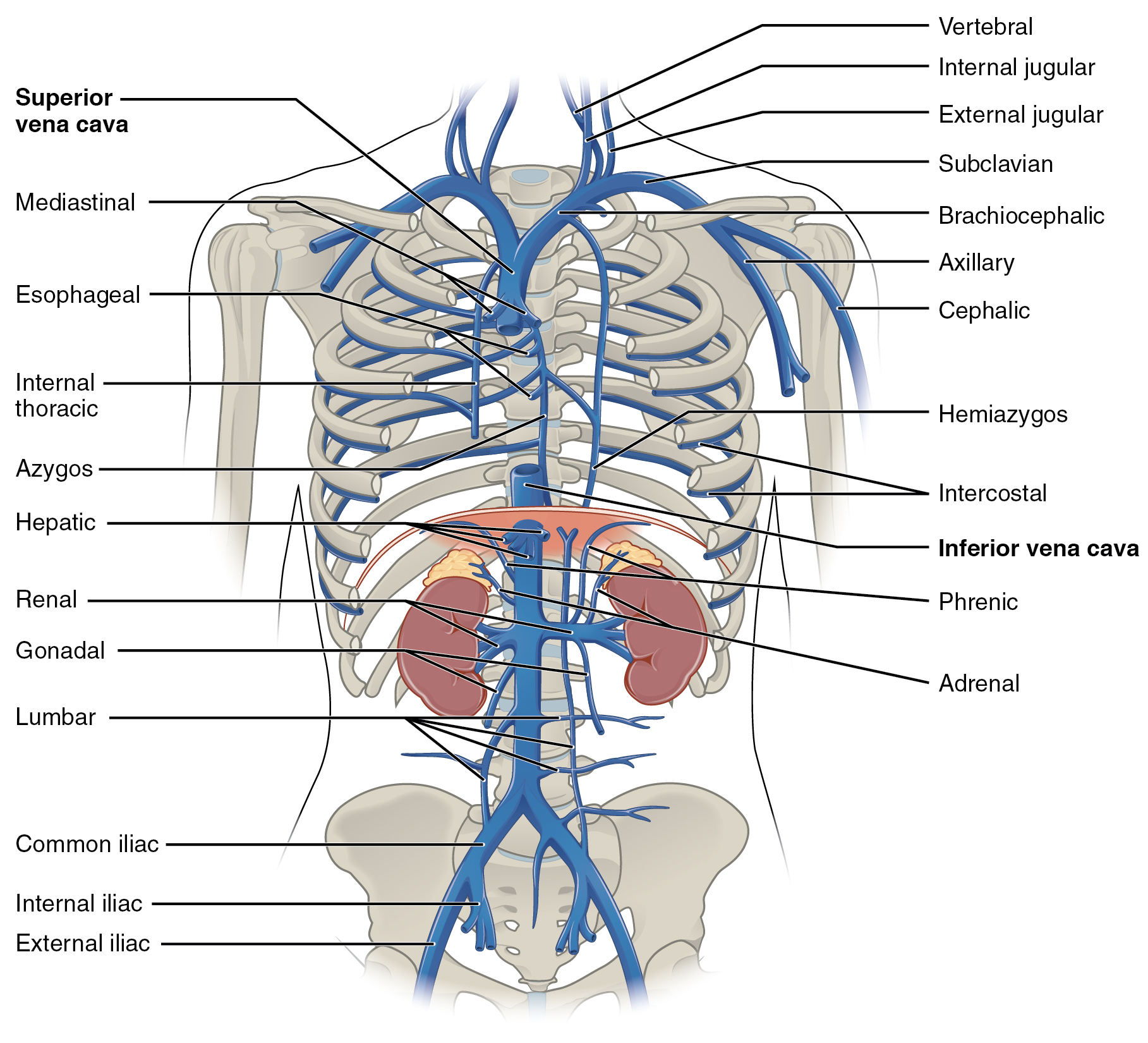
The left and right brachiocephalic veins (previously called innominate veins) are major veins in the Thorax, upper chest, formed by the union of the ipsilateral internal jugular vein and subclavian vein (the so-called venous angle) behind the sternoclavicular joint. The left brachiocephalic vein is more than twice the length of the right brachiocephalic vein. These veins merge to form the superior vena cava, a great vessel, posterior to the junction of the first costal cartilage with the Manubrium, manubrium of the sternum. The brachiocephalic veins are the major veins returning blood to the superior vena cava. Left and right veins Left brachiocephalic vein The left brachiocephalic vein is about 6cm, more than twice the length of the right brachiocephalic vein. and is formed by the confluence of the left subclavian vein, subclavian and left internal jugular veins. In addition the left vein receives drainage from the following tributaries: * The left vertebral vein, internal thor ... [...More Info...] [...Related Items...] OR: [Wikipedia] [Google] [Baidu] |
Lumbar Trunk
The lumbar trunks are formed by the union of the efferent vessels from the lateral aortic lymph nodes. They receive the lymph from the lower limbs, from the walls and viscera of the pelvis, from the kidneys and suprarenal glands and the deep lymphatics of the greater part of the abdominal wall. Ultimately, the lumbar trunks empty into the cisterna chyli, a dilatation at the beginning of the thoracic duct In human anatomy, the thoracic duct (also known as the ''left lymphatic duct'', ''alimentary duct'', ''chyliferous duct'', and ''Van Hoorne's canal'') is the larger of the two lymph ducts of the lymphatic system (the other being the right lymph .... References External links Overview at uams.edu Lymphatics of the torso {{lymphatic-stub ... [...More Info...] [...Related Items...] OR: [Wikipedia] [Google] [Baidu] |
Phrenic Nerve
The phrenic nerve is a mixed nerve that originates from the C3–C5 spinal nerves in the neck. The nerve is important for breathing because it provides exclusive motor control of the diaphragm, the primary muscle of respiration. In humans, the right and left phrenic nerves are primarily supplied by the C4 spinal nerve, but there is also a contribution from the C3 and C5 spinal nerves. From its origin in the neck, the nerve travels downward into the chest to pass between the heart and lungs towards the diaphragm. In addition to motor fibers, the phrenic nerve contains sensory fibers, which receive input from the central tendon of the diaphragm and the mediastinal pleura, as well as some sympathetic nerve fibers. Although the nerve receives contributions from nerve roots of the cervical plexus and the brachial plexus, it is usually considered separate from either plexus. The name of the nerve comes from Ancient Greek ''phren'' 'diaphragm'. Structure The phrenic nerve or ... [...More Info...] [...Related Items...] OR: [Wikipedia] [Google] [Baidu] |
Thyrocervical Trunk
The thyrocervical trunks are very small arteries of the neck arising from the subclavian arteries, lateral to the vertebral arteries. They divide into branches: the inferior thyroid artery, suprascapular artery, and the transverse cervical artery. The thyrocervical trunks supply the thyroid gland and some scapular muscles. Structure The thyrocervical trunk is a branch of the subclavian artery. It arises from the first portion of this vessel, between the origin of the subclavian artery and the inner border of the anterior scalene muscle. It is located distally to the vertebral artery and proximally to the costocervical trunk. It is short and wide artery. Branches The thyrocervical trunk soon divides into branches: the inferior thyroid artery, the suprascapular artery, and the transverse cervical artery. The transverse cervical artery is present in about 2/3 of cases. In a third of cases the superficial cervical artery and the dorsal scapular artery arise as the transve ... [...More Info...] [...Related Items...] OR: [Wikipedia] [Google] [Baidu] |
Carotid Sheath
The carotid sheath is a condensation of the deep cervical fascia enveloping multiple vital neurovascular structures of the neck, including the common and internal carotid arteries, the internal jugular vein, the vagus nerve (CN X), and ansa cervicalis. The carotid sheath helps protects the structures contained therein. Anatomy One carotid sheath is situated on each side of the neck, extending between the base of the skull superiorly and the thorax inferiorly. Superiorly, the carotid sheath encircles the margins of the carotid canal and jugular foramen. Inferiorly, it terminates at the arch of the aorta; it is continuous inferiorly with the axillary sheath at the venous angle. Its inferior end occurs at the level of the first rib and sternum inferiorly (varying between the levels of C7 and T4). Structure The carotid sheath is a fibrous connective tissue formation surrounding several important structures of the neck. It is thicker around the arteries than around the ... [...More Info...] [...Related Items...] OR: [Wikipedia] [Google] [Baidu] |
Vagus Nerve
The vagus nerve, also known as the tenth cranial nerve (CN X), plays a crucial role in the autonomic nervous system, which is responsible for regulating involuntary functions within the human body. This nerve carries both sensory and motor fibers and serves as a major pathway that connects the brain to various organs, including the heart, lungs, and digestive tract. As a key part of the parasympathetic nervous system, the vagus nerve helps regulate essential involuntary functions like heart rate, breathing, and digestion. By controlling these processes, the vagus nerve contributes to the body's "rest and digest" response, helping to calm the body after stress, lower heart rate, improve digestion, and maintain homeostasis. The vagus nerve consists of two branches: the right and left vagus nerves. In the neck, the right vagus nerve contains approximately 105,000 fibers, while the left vagus nerve has about 87,000 fibers, according to one source. However, other sources report sl ... [...More Info...] [...Related Items...] OR: [Wikipedia] [Google] [Baidu] |
Common Carotid Artery
In anatomy, the left and right common carotid arteries (carotids) () are artery, arteries that supply the head and neck with oxygenated blood; they divide in the neck to form the external carotid artery, external and internal carotid artery, internal carotid arteries. Structure The common carotid arteries are present on the left and right sides of the body. These arteries originate from different arteries but follow symmetrical courses. The right common carotid originates in the neck from the brachiocephalic trunk; the left from the aortic arch in the thorax. These split into the external and internal carotid arteries at the upper border of the thyroid cartilage, at around the level of the fourth cervical vertebra. The left common carotid artery can be thought of as having two parts: a thoracic (chest) part and a cervical (neck) part. The right common carotid originates in or close to the neck and contains only a small thoracic portion. There are studies in the bioengineering l ... [...More Info...] [...Related Items...] OR: [Wikipedia] [Google] [Baidu] |
Alar Fascia
The alar fascia a portion of prevertebral fascia that may or may not be considered a distinct anatomical structure. When acknowledged, it is described as anterior to the prevertebral fascia. Anatomy Superiorly, it extends to the base of the skull; inferiorly, it extends to the second thoracic vertebra. Inferiorly, it unites with the visceral fascia of the neck. Anatomical relations The alar fascia represents the posterior boundary of the retropharyngeal space The retropharyngeal space (abbreviated as "RPS") is a potential space and deep compartment of the head and neck situated posterior to the pharynx. The RPS is bounded anteriorly by the buccopharyngeal fascia, posteriorly by the alar fascia, and l .... Research In 2015, the anatomy of the alar fascia was revisited using dissection in conjunction with E12 plastination. The authors revealed that the alar fascia originated as a well defined midline structure at the level of C1 and does not reach the base of the skull. I ... [...More Info...] [...Related Items...] OR: [Wikipedia] [Google] [Baidu] |
Thoracic Aorta
The thoracic aorta is a part of the aorta located in the thorax. It is a continuation of the aortic arch. It is located within the posterior mediastinal cavity, but frequently bulges into the left pleural cavity. The descending thoracic aorta begins at the lower border of the fourth thoracic vertebra and ends in front of the lower border of the twelfth thoracic vertebra, at the aortic hiatus in the diaphragm where it becomes the abdominal aorta. At its commencement, it is situated on the left of the vertebral column; it approaches the median line as it descends; and, at its termination, lies directly in front of the column. The thoracic aorta has a curved shape that faces forward, and has small branches. It has a radius of approximately 1.16 cm. Structure The thoracic aorta is part of the descending aorta, which has different parts named according to their structure or location. The thoracic aorta is a continuation of the descending aorta and becomes the abdominal aorta ... [...More Info...] [...Related Items...] OR: [Wikipedia] [Google] [Baidu] |
Posterior Mediastinum
Posterior may refer to: * Posterior (anatomy), the end of an organism opposite to anterior ** Buttocks, as a euphemism * Posterior horn (other) * Posterior probability The posterior probability is a type of conditional probability that results from updating the prior probability with information summarized by the likelihood via an application of Bayes' rule. From an epistemological perspective, the posteri ..., the conditional probability that is assigned when the relevant evidence is taken into account * Posterior tense, a relative future tense {{disambiguation ... [...More Info...] [...Related Items...] OR: [Wikipedia] [Google] [Baidu] |


