|
Teres Minor
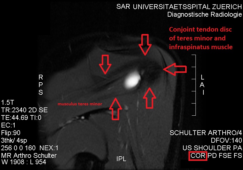
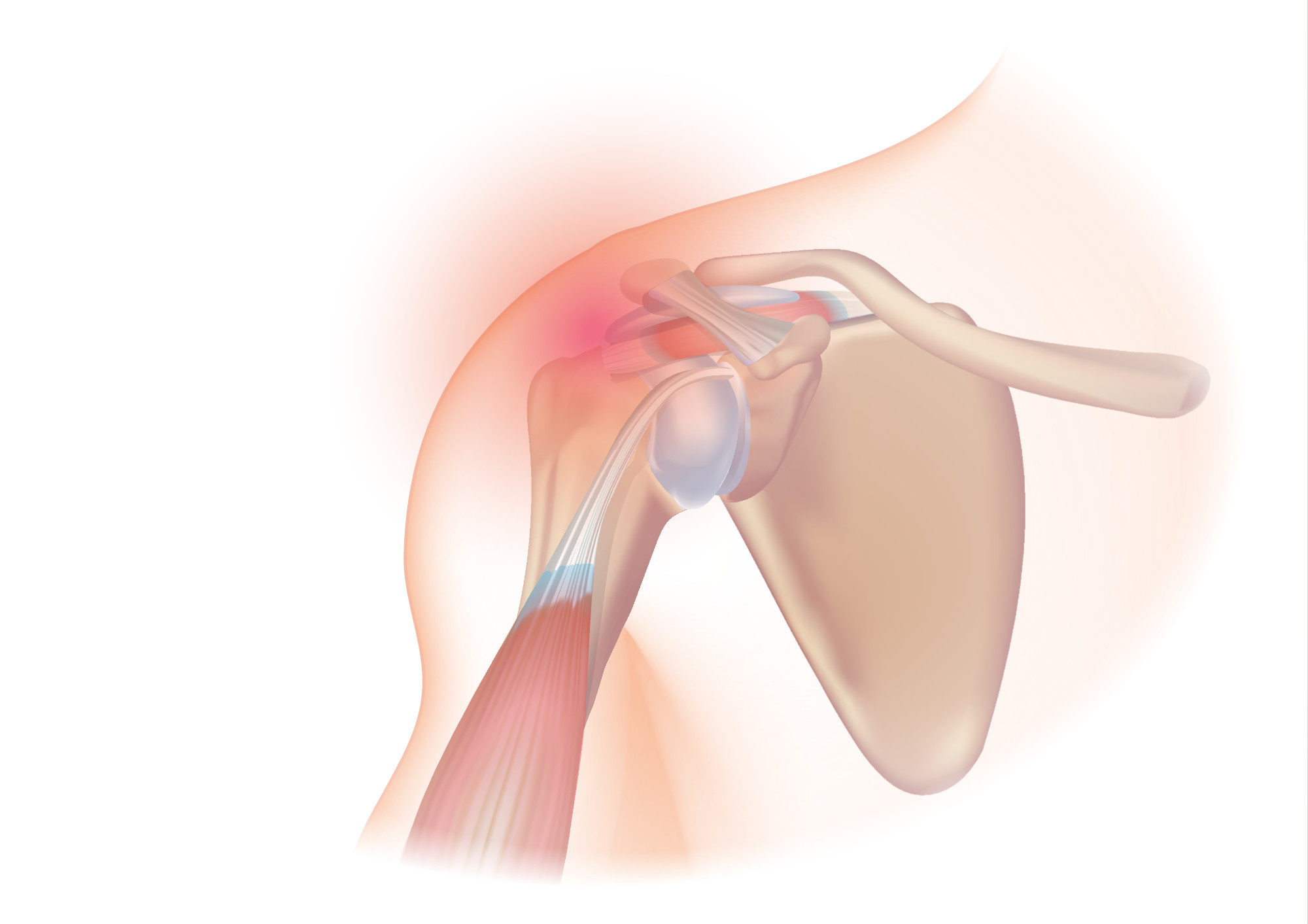
The teres minor (Latin ''teres'' meaning 'rounded') is a narrow, elongated muscle of the rotator cuff. The muscle originates from the lateral border and adjacent posterior surface of the corresponding right or left scapula and inserts at both the greater tubercle of the humerus and the posterior surface of the joint capsule. The primary function of the teres minor is to modulate the action of the deltoid, preventing the humeral head from sliding upward as the arm is abducted. It also functions to rotate the humerus laterally. The teres minor is innervated by the axillary nerve. Structure It arises from the dorsal surface of the axillary border of the scapula for the upper two-thirds of its extent, and from two aponeurotic laminae, one of which separates it from the infraspinatus muscle, the other from the teres major muscle. Its fibers run obliquely upwards and laterally; the upper ones end in a tendon which is inserted into the lowest of the three impressions on the greater tub ... [...More Info...] [...Related Items...] OR: [Wikipedia] [Google] [Baidu] |
Scapula
The scapula (: scapulae or scapulas), also known as the shoulder blade, is the bone that connects the humerus (upper arm bone) with the clavicle (collar bone). Like their connected bones, the scapulae are paired, with each scapula on either side of the body being roughly a mirror image of the other. The name derives from the Classical Latin word for trowel or small shovel, which it was thought to resemble. In compound terms, the prefix omo- is used for the shoulder blade in medical terminology. This prefix is derived from ὦμος (ōmos), the Ancient Greek word for shoulder, and is cognate with the Latin , which in Latin signifies either the shoulder or the upper arm bone. The scapula forms the back of the shoulder girdle. In humans, it is a flat bone, roughly triangular in shape, placed on a posterolateral aspect of the thoracic cage. Structure The scapula is a thick, flat bone lying on the thoracic wall that provides an attachment for three groups of muscles: intrinsic, e ... [...More Info...] [...Related Items...] OR: [Wikipedia] [Google] [Baidu] |
Aponeurotic
An aponeurosis (; : aponeuroses) is a flattened tendon by which muscle attaches to bone or fascia. Aponeuroses exhibit an ordered arrangement of collagen fibres, thus attaining high tensile strength in a particular direction while being vulnerable to tensional or shear forces in other directions. They have a shiny, whitish-silvery color, are histologically similar to tendons, and are very sparingly supplied with blood vessels and nerves. When dissected, aponeuroses are papery and peel off by sections. The primary regions with thick aponeuroses are in the ventral abdominal region, the dorsal lumbar region, the ventriculus in birds, and the palmar (palms) and plantar (soles) regions. Anatomy Anterior abdominal aponeuroses The anterior abdominal aponeuroses are located just superficial to the rectus abdominis muscle. It has for its borders the external oblique, pectoralis muscles, and the latissimus dorsi. Posterior lumbar aponeuroses The posterior lumbar aponeuroses ar ... [...More Info...] [...Related Items...] OR: [Wikipedia] [Google] [Baidu] |
Cysts
A cyst is a closed Wikt:sac, sac, having a distinct Cell envelope, envelope and cell division, division compared with the nearby Biological tissue, tissue. Hence, it is a cluster of Cell (biology), cells that have grouped together to form a sac (like the manner in which water molecules group together to form a bubble); however, the distinguishing aspect of a cyst is that the cells forming the "shell" of such a sac are distinctly abnormal (in both appearance and behaviour) when compared with all surrounding cells for that given location. A cyst may contain air, fluids, or semi-solid material. A collection of pus is called an abscess, not a cyst. Once formed, a cyst may resolve on its own. When a cyst fails to resolve, it may need to be removed surgically, but that would depend upon its type and location. Cancer-related cysts are formed as a defense mechanism for the body following the development of mutations that lead to an uncontrolled cellular division. Once that mutation has o ... [...More Info...] [...Related Items...] OR: [Wikipedia] [Google] [Baidu] |
Paresthesia
Paresthesia is a sensation of the skin that may feel like numbness (''hypoesthesia''), tingling, pricking, chilling, or burning. It can be temporary or Chronic condition, chronic and has many possible underlying causes. Paresthesia is usually painless and can occur anywhere on the body, but most commonly in the arms and legs. The most familiar kind of paresthesia is the sensation known as ''pins and needles'' after having a limb "fall asleep" (''obdormition''). A less common kind is formication, the sensation of insects crawling on the skin. Causes Transient Paresthesias of the hands, feet, legs, and arms are common transient symptoms. The briefest electric shock type of paresthesia can be caused by tweaking the ulnar nerve near the elbow; this phenomenon is colloquially known as bumping one's "funny bone". Similar brief shocks can be experienced when any other nerve is tweaked (e.g. a pinched neck nerve may cause a brief shock-like paresthesia toward the scalp). In the older a ... [...More Info...] [...Related Items...] OR: [Wikipedia] [Google] [Baidu] |
Posterior Humeral Circumflex Artery
The posterior humeral circumflex artery (posterior circumflex artery or posterior circumflex humeral artery) arises from the third part of the axillary artery at the distal border of the subscapularis. Anatomy Course and relations It passes posteriorward with the axillary nerve through the quadrangular space. It winds laterally around the surgical neck of the humerus. Distribution It is distributed to the shoulder joint, teres major, teres minor, deltoid, and (long and lateral heads of) triceps brachii. Anastomoses It forms anastomoses with the anterior humeral circumflex artery, (deltoid branch of) profunda brachii artery, (acromial branches of) suprascapular artery The suprascapular artery is a branch of the thyrocervical trunk on the neck. Structure At first, it passes downward and laterally across the scalenus anterior and phrenic nerve, being covered by the sternocleidomastoid muscle; it then crosses t ..., (acromial branches of) and thoracoacromial artery. ... [...More Info...] [...Related Items...] OR: [Wikipedia] [Google] [Baidu] |
Quadrangular Space
The quadrangular space, also known as the quadrilateral space (of Velpeau) and the foramen humerotricipitale, is one of the three spaces in the axillary space. The other two spaces are: triangular space and triangular interval. Structure The quadrangular space is one of the three spaces in the axillary space. Boundaries The quadrangular space is defined by: - "Scapular Region: Quadrangular Space of Scapular Region" * ''above/superior:'' teres minor muscle. * ''below/inferior:'' teres major muscle. * ''medially:'' long head of the triceps brachii muscle (lateral margin). * ''laterally:'' surgical neck of the humerus. * ''anteriorly:'' subscapularis muscle. Contents The quadrangular space transmits the axillary nerve, the posterior humeral circumflex artery and the posterior circumflex humeral vein. Clinical significance The quadrangular space is a clinically important anatomic space in the arm as it provides the anterior regions of the axilla a passageway to the poste ... [...More Info...] [...Related Items...] OR: [Wikipedia] [Google] [Baidu] |
Rotator Cuff Tear
Rotator cuff tendinopathy is a process of senescence. The pathophysiology is mucoid degeneration. Most people develop rotator cuff tendinopathy within their lifetime. As part of rotator cuff tendinopathy, the tendon can thin and develop a defect. This defect is often referred to as a rotator cuff tear. Acute, traumatic rupture of the rotator cuff tendons can also occur, but is less common. Traumatic rupture of the rotator cuff usually involves the tendons of more than one muscle. Rotator cuff tendinopathy is, by far, the most common reason people seek care for shoulder pain. Pain related to rotator cuff tendinopathy is typically on the front side of the shoulder, down to the elbow, and worse reaching up or back. Diagnosis is based on symptoms and physical examination, examination. Medical imaging is used mostly to plan surgery and is not needed for diagnosis. Treatment may include pain medication such as NSAIDs and specific exercises. It is recommended that people who are una ... [...More Info...] [...Related Items...] OR: [Wikipedia] [Google] [Baidu] |
Atrophy
Atrophy is the partial or complete wasting away of a part of the body. Causes of atrophy include mutations (which can destroy the gene to build up the organ), malnutrition, poor nourishment, poor circulatory system, circulation, loss of hormone, hormonal support, loss of nerve supply to the target Organ (anatomy), organ, excessive amount of apoptosis of cells, and disuse or lack of exercise or disease intrinsic to the tissue itself. In medical practice, hormonal and nerve inputs that maintain an organ or body part are said to have ''trophic'' effects. A diminished muscular trophic condition is designated as ''atrophy''. Atrophy is reduction in size of cell, organ or tissue, after attaining its normal mature growth. In contrast, hypoplasia is the reduction in the cellular numbers of an organ, or tissue that has not attained normal maturity. Atrophy is the general physiological process of reabsorption and breakdown of biological tissue, tissues, involving apoptosis. When it occurs ... [...More Info...] [...Related Items...] OR: [Wikipedia] [Google] [Baidu] |
Posterior Deltoid
The deltoid muscle is the muscle forming the rounded contour of the human shoulder. It is also known as the 'common shoulder muscle', particularly in other animals such as the domestic cat. Anatomically, the deltoid muscle is made up of three distinct sets of muscle fibers, namely the # anterior or clavicular part (pars clavicularis) ( More commonly known as the front delt.) # posterior or scapular part (pars scapularis) ( More commonly known as the rear delt.) # intermediate or acromial part (pars acromialis) ( More commonly known as the side delt) The deltoid's fibres are pennate muscle. However, electromyography suggests that it consists of at least seven groups that can be independently coordinated by the nervous system. It was previously called the deltoideus (plural ''deltoidei'') and the name is still used by some anatomists. It is called so because it is in the shape of the Greek capital letter delta (Δ). Deltoid is also further shortened in slang as "delt". A study of ... [...More Info...] [...Related Items...] OR: [Wikipedia] [Google] [Baidu] |
Head Of The Humerus
The humerus (; : humeri) is a long bone in the arm that runs from the shoulder to the elbow. It connects the scapula and the two bones of the lower arm, the radius and ulna, and consists of three sections. The humeral upper extremity consists of a rounded head, a narrow neck, and two short processes (tubercles, sometimes called tuberosities). The body is cylindrical in its upper portion, and more prismatic below. The lower extremity consists of 2 epicondyles, 2 processes ( trochlea and capitulum), and 3 fossae ( radial fossa, coronoid fossa, and olecranon fossa). As well as its true anatomical neck, the constriction below the greater and lesser tubercles of the humerus is referred to as its surgical neck due to its tendency to fracture, thus often becoming the focus of surgeons. Etymology The word "humerus" is derived from Late Latin , from Latin , meaning upper arm, shoulder, and is linguistically related to Gothic (shoulder) and Greek . Structure Upper extremity The u ... [...More Info...] [...Related Items...] OR: [Wikipedia] [Google] [Baidu] |
Infraspinatus
In human anatomy, the infraspinatus muscle is a thick triangular muscle, which occupies the chief part of the infraspinatous fossa.''Gray's Anatomy'', see infobox. As one of the four muscles of the rotator cuff, the main function of the infraspinatus is to externally rotate the humerus and stabilize the shoulder joint. Structure It attaches medially to the infraspinous fossa of the scapula and laterally to the middle facet of the greater tubercle of the humerus. The muscle arises by fleshy fibers from the medial two-thirds of the infraspinatous fossa, and by tendinous fibers from the ridges on its surface; it also arises from the infraspinatous fascia which covers it, and separates it from the teres major and teres minor. The fibers converge to a tendon, which glides over the lateral border of the spine of the scapula and passing across the posterior part of the capsule of the shoulder-joint, is inserted into the middle impression on the greater tubercle of the humerus. The t ... [...More Info...] [...Related Items...] OR: [Wikipedia] [Google] [Baidu] |
Pseudoganglion
A ganglion (: ganglia) is a group of neuron cell bodies in the peripheral nervous system. In the somatic nervous system, this includes dorsal root ganglia and trigeminal ganglia among a few others. In the autonomic nervous system, there are both sympathetic and parasympathetic ganglia which contain the cell bodies of postganglionic sympathetic and parasympathetic neurons respectively. A pseudoganglion looks like a ganglion, but only has nerve fibers and has no nerve cell bodies. Structure Ganglia are primarily made up of somata and dendritic structures, which are bundled or connected. Ganglia often interconnect with other ganglia to form a complex system of ganglia known as a plexus. Ganglia provide relay points and intermediary connections between different neurological structures in the body, such as the peripheral and central nervous systems. Among vertebrates there are three major groups of ganglia: * Dorsal root ganglia (also known as the spinal ganglia) contain the ... [...More Info...] [...Related Items...] OR: [Wikipedia] [Google] [Baidu] |


