|
Teres Major
The teres major muscle is a muscle of the upper limb. It attaches to the scapula and the humerus and is one of the seven scapulohumeral muscles. It is a thick but somewhat flattened muscle. The teres major muscle (from Latin ''teres'', meaning "rounded") is positioned above the latissimus dorsi muscle and assists in the extension and medial rotation of the humerus. This muscle is commonly confused as a rotator cuff muscle, but it is not because it does not attach to the capsule of the shoulder joint, unlike the teres minor muscle for example. Structure The teres major muscle originates on the dorsal surface of the inferior angle and the lower part of the lateral border of the scapula. The fibers of teres major insert into the medial lip of the intertubercular sulcus of the humerus. Relations The tendon, at its insertion, lies behind that of the latissimus dorsi, from which it is separated by a bursa, the two tendons being, however, united along their lower borders for a ... [...More Info...] [...Related Items...] OR: [Wikipedia] [Google] [Baidu] |
Upper Extremity
The upper limbs or upper extremities are the forelimbs of an upright-postured tetrapod vertebrate, extending from the scapulae and clavicles down to and including the digits, including all the musculatures and ligaments involved with the shoulder, elbow, wrist and knuckle joints. In humans, each upper limb is divided into the arm, forearm and hand, and is primarily used for climbing, lifting and manipulating objects. Definition In formal usage, the term "arm" only refers to the structures from the shoulder to the elbow, explicitly excluding the forearm, and thus "upper limb" and "arm" are not synonymous. However, in casual usage, the terms are often used interchangeably. The term "upper arm" is redundant in anatomy, but in informal usage is used to distinguish between the two terms. Structure In the human body the muscles of the upper limb can be classified by origin, topography, function, or innervation. While a grouping by innervation reveals embryological and phylogeneti ... [...More Info...] [...Related Items...] OR: [Wikipedia] [Google] [Baidu] |
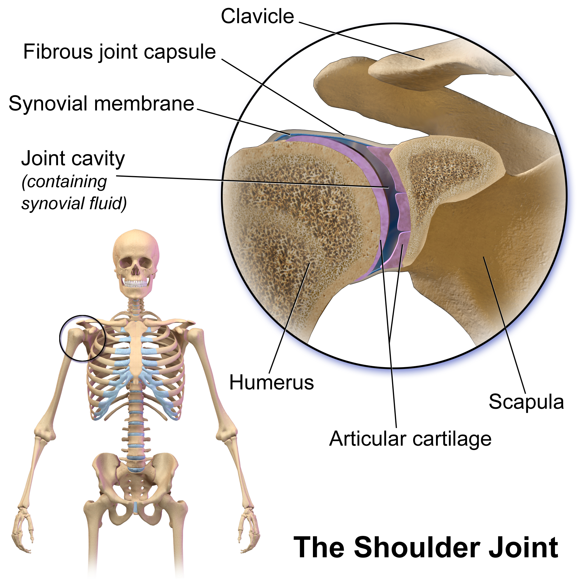
Shoulder Joint
The shoulder joint (or glenohumeral joint from Greek ''glene'', eyeball, + -''oid'', 'form of', + Latin ''humerus'', shoulder) is structurally classified as a synovial ball-and-socket joint and functionally as a diarthrosis and multiaxial joint. It involves an articulation between the glenoid fossa of the scapula (shoulder blade) and the head of the humerus (upper arm bone). Due to the very loose joint capsule that gives a limited interface of the humerus and scapula, it is the most mobile joint of the human body. Structure The shoulder joint is a ball-and-socket joint between the scapula and the humerus. The socket of the glenoid fossa of the scapula is itself quite shallow, but it is made deeper by the addition of the glenoid labrum. The glenoid labrum is a ring of cartilaginous fibre attached to the circumference of the cavity. This ring is continuous with the tendon of the biceps brachii above. Spaces Significant joint spaces are: * The normal glenohumeral space is 4� ... [...More Info...] [...Related Items...] OR: [Wikipedia] [Google] [Baidu] |
Brachial Plexus
The brachial plexus is a network () of nerves formed by the anterior rami of the lower four cervical nerves and first thoracic nerve ( C5, C6, C7, C8, and T1). This plexus extends from the spinal cord, through the cervicoaxillary canal in the neck, over the first rib, and into the armpit, it supplies afferent and efferent nerve fibers the to chest, shoulder, arm, forearm, and hand. Structure The brachial plexus is divided into five ''roots'', three ''trunks'', six ''divisions'' (three anterior and three posterior), three ''cords'', and five ''branches''. There are five "terminal" branches and numerous other "pre-terminal" or "collateral" branches, such as the subscapular nerve, the thoracodorsal nerve, and the long thoracic nerve, that leave the plexus at various points along its length. A common structure used to identify part of the brachial plexus in cadaver dissections is the M or W shape made by the musculocutaneous nerve, lateral cord, median nerve, medial cord, and ... [...More Info...] [...Related Items...] OR: [Wikipedia] [Google] [Baidu] |
Posterior Cord
The posterior cord is a part of the brachial plexus The brachial plexus is a network () of nerves formed by the anterior rami of the lower four cervical nerves and first thoracic nerve ( C5, C6, C7, C8, and T1). This plexus extends from the spinal cord, through the cervicoaxillary canal in th .... It consists of contributions from all of the roots of the brachial plexus. The posterior cord gives rise to the following nerves: Additional images File:PLEXUS BRACHIALIS.jpg, Brachial plexus File:Slide12OOO.JPG, Posterior cord File:Slide1SSS.JPG, Posterior cord File:Slide1cord.JPG, Brachial plexus.Deep dissection. File:Slide1ecc.JPG, Brachial plexus.Deep dissection.Anterolateral view References MBBS resources http://mbbsbasic.googlepages.com/ External links * - "Axilla, dissection, anterior view" Nerves of the upper limb {{neuroscience-stub ... [...More Info...] [...Related Items...] OR: [Wikipedia] [Google] [Baidu] |
Upper Subscapular Nerve
The upper (superior) subscapular nerve is the first branch of the posterior cord of the brachial plexus. The upper subscapular nerve contains axons from the ventral rami of the C5 and C6 cervical spinal nerves. It innervates the superior portion of the subscapularis muscle. The inferior portion of the subscapularis is innervated by the lower subscapular nerve. Structure The axons which form the upper subscapular nerve travel from the ventral rami of C5 and C6. They join at the upper trunk and move through its posterior division to form the posterior cord, along with the other two posterior divisions of the middle and lower trunks. The axons then branch from the posterior cord and form the upper subscapular nerve. Function The upper subscapular nerve innervates the superior portion of the subscapularis muscle The subscapularis is a large triangular muscle which fills the subscapular fossa and inserts into the lesser tubercle of the humerus and the front of the cap ... [...More Info...] [...Related Items...] OR: [Wikipedia] [Google] [Baidu] |
Thoracodorsal Nerve
The thoracodorsal nerve is a nerve present in humans and other animals, also known as the middle subscapular nerve or the long subscapular nerve. It supplies the latissimus dorsi muscle. Structure The thoracodorsal nerve arises from the brachial plexus. It derives its fibers from the sixth, seventh, and eighth cervical nerves. It is derived from their ventral rami, in spite of the fact that the latissimus dorsi is found in the back. The thoracodorsal nerve is a branch of the posterior cord of the brachial plexus, and is made up of fibres from the posterior divisions of all three trunks of the brachial plexus. It follows the course of the subscapular artery, along the posterior wall of the axilla to the latissimus dorsi muscle, in which it may be traced as far as the lower border of the muscle. Function The thoracodorsal nerve innervates the latissimus dorsi muscle on its deep surface. Clinical Significance The latissimus dorsi is occasionally used for transplantation, a ... [...More Info...] [...Related Items...] OR: [Wikipedia] [Google] [Baidu] |
Lower Subscapular Nerve
The lower subscapular nerve, also known as the inferior subscapular nerve, is the third branch of the posterior cord of the brachial plexus. It innervates the inferior portion of the subscapularis muscle and the teres major muscle. Structure The lower subscapular nerve contains axons from the ventral rami of the C5 and C6 cervical spinal nerves. It is the third branch of the posterior cord of the brachial plexus. It gives branches to 2 muscles: * subscapularis muscle. It usually gives 4 branches to innervate the subscapularis, and can give up to 8 branches. * teres major muscle. Function The lower subscapular nerve innervates the subscapularis muscle and the teres major muscle. These muscles medially rotate and adduct the humerus The humerus (; ) is a long bone in the arm that runs from the shoulder to the elbow. It connects the scapula and the two bones of the lower arm, the radius and ulna, and consists of three sections. The humeral upper extremity consists of a roun ... [...More Info...] [...Related Items...] OR: [Wikipedia] [Google] [Baidu] |
Axillary Space
The axillary spaces are anatomic spaces. through which axillary contents leave the axilla. They consist of the quadrangular space, triangular space, and triangular interval. It is bounded by teres major, teres minor, medial border of the humerus, and long head of triceps brachii. They should not be confused with the true "axillary space" within the borders of the axilla. Structure Axilla The true axilla is a conical space with its apex at the Cervico-axillary Canal, Base at the axillary fascia and skin of the armpit. When viewed in an axillary plane (axillary cut), it is more triangle with: Medial Wall: Serratus Anterior, Anterior Wall: pectoral muscles, Posterior Wall: subscapularis muscle, where the "apex" of the triangle is the humerus Quadrangular space This space is in the posterior wall of the axilla. It is a quadrangular space bounded laterally by surgical neck of the humerus, medially by long head of triceps brachii and inferiorly by teres major. It is bounded su ... [...More Info...] [...Related Items...] OR: [Wikipedia] [Google] [Baidu] |
Lesser Tubercle
The lesser tubercle of the humerus, although smaller, is more prominent than the greater tubercle: it is situated in front, and is directed medially and anteriorly. The projection of the lesser tubercle is anterior from the junction that is found between the Anatomical neck of humerus, anatomical neck and the Humerus, shaft of the humerus and easily identified due to the Bicipital groove, intertubercular sulcus (Bicipital groove). Above and in front it presents an impression for the insertion of the tendon of the subscapularis. Additional images File:Gray326.png, The left shoulder and acromioclavicular joints, and the proper ligaments of the scapula. File:Human arm bones diagram.svg, Human arm bones diagram References External links * * * Diagram at uwlax.edu Bones of the upper limb Humerus {{musculoskeletal-stub ... [...More Info...] [...Related Items...] OR: [Wikipedia] [Google] [Baidu] |
Bursa (anatomy)
( grc-gre, Προῦσα, Proûsa, Latin: Prusa, ota, بورسه, Arabic:بورصة) is a city in northwestern Turkey and the administrative center of Bursa Province. The fourth-most populous city in Turkey and second-most populous in the Marmara Region, Bursa is one of the industrial centers of the country. Most of Turkey's automotive production takes place in Bursa. As of 2019, the Metropolitan Province was home to 3,056,120 inhabitants, 2,161,990 of whom lived in the 3 city urban districts ( Osmangazi, Yildirim and Nilufer) plus Gursu and Kestel, largely conurbated. Bursa was the first major and second overall capital of the Ottoman State between 1335 and 1363. The city was referred to as (, meaning "God's Gift" in Ottoman Turkish, a name of Persian origin) during the Ottoman period, while a more recent nickname is ("") in reference to the parks and gardens located across its urban fabric, as well as to the vast and richly varied forests of the surrounding re ... [...More Info...] [...Related Items...] OR: [Wikipedia] [Google] [Baidu] |
Latissimus Dorsi
The latissimus dorsi () is a large, flat muscle on the back that stretches to the sides, behind the arm, and is partly covered by the trapezius on the back near the midline. The word latissimus dorsi (plural: ''latissimi dorsorum'') comes from Latin and means "broadest uscleof the back", from "latissimus" ( la, broadest)' and "dorsum" ( la, back). The pair of muscles are commonly known as "lats", especially among bodybuilders. The latissimus dorsi is the largest muscle in the upper body. The latissimus dorsi is responsible for extension, adduction, transverse extension also known as horizontal abduction (or horizontal extension), flexion from an extended position, and (medial) internal rotation of the shoulder joint. It also has a synergistic role in extension and lateral flexion of the lumbar spine. Due to bypassing the scapulothoracic joints and attaching directly to the spine, the actions the latissimi dorsi have on moving the arms can also influence the movement of the s ... [...More Info...] [...Related Items...] OR: [Wikipedia] [Google] [Baidu] |
Tendon
A tendon or sinew is a tough, high-tensile-strength band of dense fibrous connective tissue that connects muscle to bone. It is able to transmit the mechanical forces of muscle contraction to the skeletal system without sacrificing its ability to withstand significant amounts of tension. Tendons are similar to ligaments; both are made of collagen. Ligaments connect one bone to another, while tendons connect muscle to bone. Structure Histologically, tendons consist of dense regular connective tissue. The main cellular component of tendons are specialized fibroblasts called tendon cells (tenocytes). Tenocytes synthesize the extracellular matrix of tendons, abundant in densely packed collagen fibers. The collagen fibers are parallel to each other and organized into tendon fascicles. Individual fascicles are bound by the endotendineum, which is a delicate loose connective tissue containing thin collagen fibrils and elastic fibres. Groups of fascicles are bounded by the epit ... [...More Info...] [...Related Items...] OR: [Wikipedia] [Google] [Baidu] |


